Biological effects of radiation
Levels of radiation damage
- Every exposure to ionising radiation, natural or man-made, small or large, produces some biological effect. The only question is how significant that effect is.
- Radiation deposits energy by ionising atoms. The damage then passes up through the levels of organisation: atoms → molecules → cells → tissues → organs → whole body.
- Damage at a lower level is only expressed at a higher level if enough of it escapes repair.
DNA damage
- The nucleus is the most radiosensitive part of the cell, and the critical target in it is the DNA molecule.
- DNA is a double helix of two strands. Each strand is a chain of sugar units joined by phosphate bonds, the sugar–phosphate backbone.
- The two strands are held together by base pairs: adenine (A) with thymine (T) and guanine (G) with cytosine (C). Each base is bonded to a sugar on its own strand and to its partner base by hydrogen bonds.
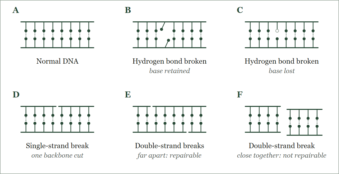
- Radiation produces lesions of increasing seriousness (Figure 1):
- Hydrogen bond broken, base retained (B).
- Hydrogen bond broken, base lost (C).
- Single-strand break: one backbone is cut (D). It is repaired easily, because the intact strand serves as the template.
- Double-strand breaks far apart on the two strands (E): the molecule holds together and repair is still possible.
- Double-strand breaks close together, within a few base pairs (F): the molecule separates into fragments and repair is unreliable. This lesion is the main cause of cell death, mutation and chromosome aberrations.
Chromosome aberrations
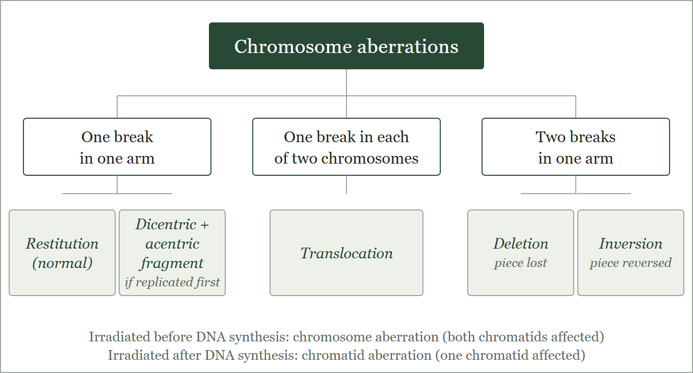
- Chromosomes are damaged in two ways: through mutations in their DNA, and by direct cleavage of an arm, producing single or double breaks.
- These structural changes are called aberrations (also anomalies or lesions).
- Chromatid aberration: irradiation after DNA synthesis, so only one of the two chromatids is affected.
- Chromosome aberration: irradiation before DNA synthesis, so the damaged chromatid is duplicated and both daughter cells carry damaged chromosomes (Figure 2).
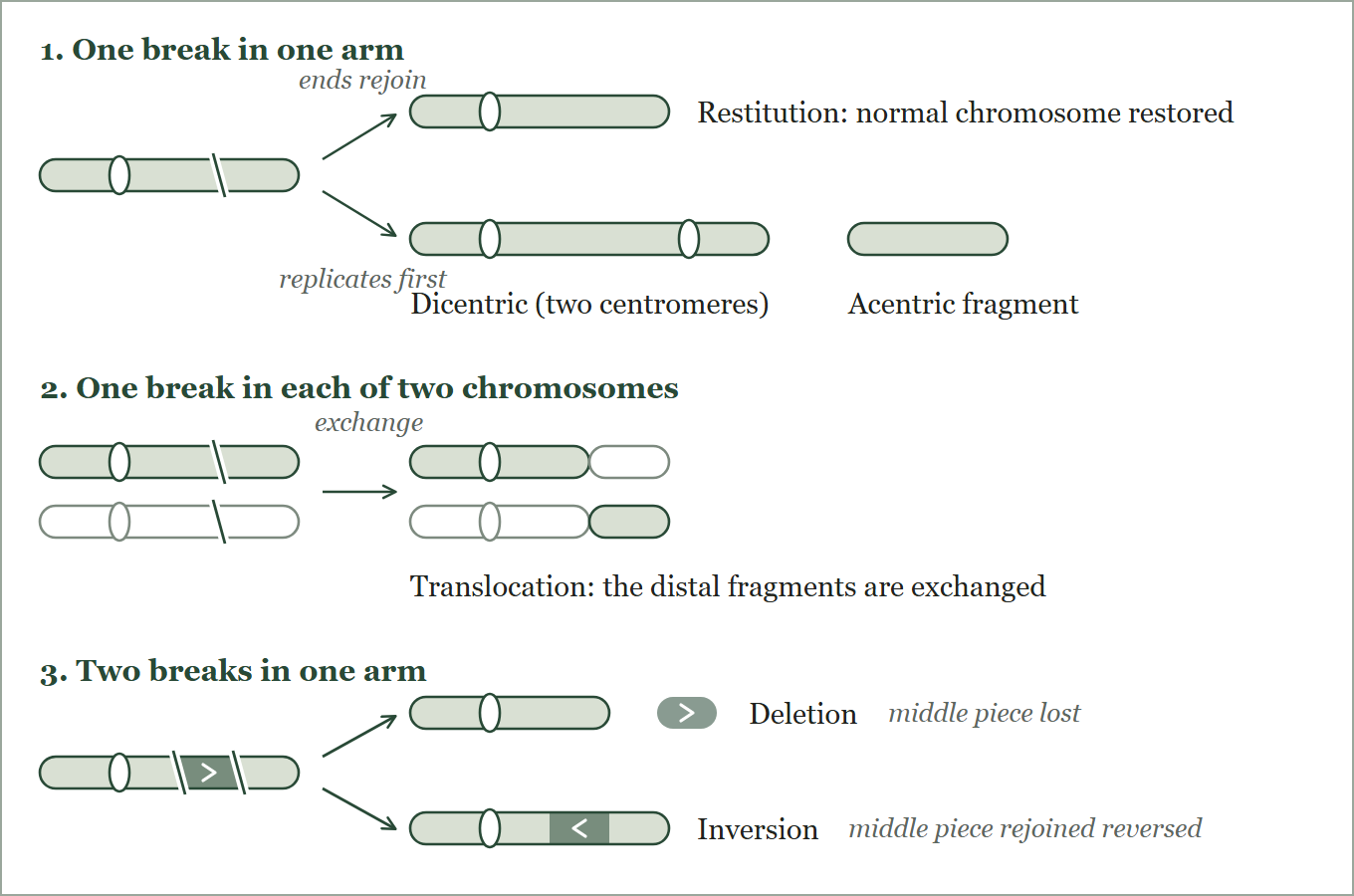
- One break in one arm (Figure 3, row 1): the two ends usually rejoin, a process called restitution. If the chromosome replicates before the ends rejoin, the broken fragments recombine at random, giving a dicentric chromosome (two centromeres) and an acentric fragment (no centromere).
- One break in each of two chromosomes (row 2): the four broken ends can rejoin in four ways. When a fragment carrying a centromere from one chromosome joins an acentric fragment from the other, a new chromosome is formed: a translocation.
- Two breaks in one arm (row 3): three fragments result. If the two outer fragments join and the middle piece is lost, it is a deletion. If all three rejoin with the middle piece reversed, it is an inversion.
- Dicentrics and acentric fragments are lost at mitosis, so these cells usually die. Balanced translocations and inversions can survive and be passed on, which links them to late effects.
Direct and indirect action
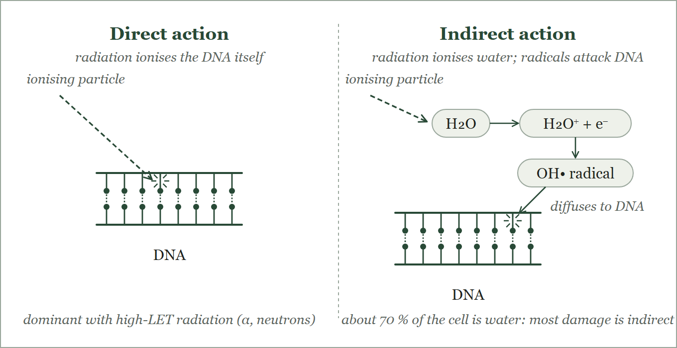
- Radiation damages DNA by two routes (Figure 4).
- Direct action: the ionising particle hits the DNA molecule itself and disrupts its structure. This route predominates with high-LET radiation such as α particles and neutrons, and at high doses.
- Indirect action: the particle ionises water, the main constituent of the cell, and the free radicals formed (mainly the hydroxyl radical, OH•) diffuse to DNA and damage it.
- The number of free radicals formed depends on the total dose, not on the dose rate.
- Water makes up about 70 % of the cell, so most radiation damage, about two thirds with x- and γ-rays, is by indirect action.
Radiosensitivity of cells
- Law of Bergonié and Tribondeau: cells that are undifferentiated and actively dividing are the most radiosensitive; fully differentiated, non-dividing cells are the least.
- Lymphocytes are an exception: although mature, they are highly radiosensitive, attributed to their large nucleus.
- Nerve and muscle cells are fully differentiated and highly radioresistant.
- After whole-body irradiation the blood counts fall in order of cell lifespan: lymphocytes the same day, granulocytes by about 3 days, platelets by about 6 days, red cells by about 10 days.
Cell cycle
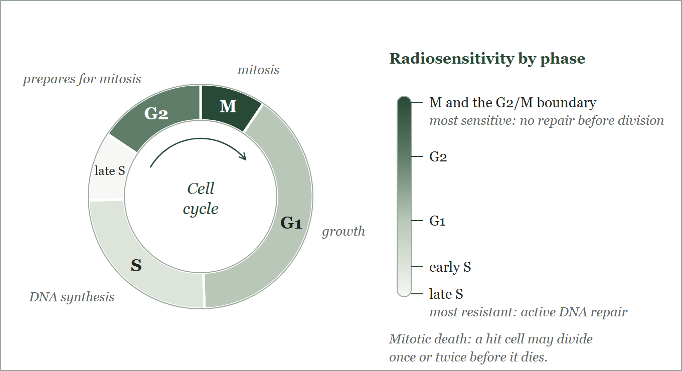
- Radiosensitivity varies through the cell cycle (Figure 5).
- Most sensitive: mitosis (M) and the G2/M boundary. The DNA is condensed and not being repaired, so damage is fixed.
- Least sensitive: the late S phase of DNA synthesis, when active repair mechanisms are at work.
- Mitotic death: an irradiated cell may still divide once or twice before it dies, so cell death is often delayed by one or two cycles.
Cell survival curves
- Radiation damage to a cell population depends on the total dose, the dose rate, the LET of the radiation, the phase of the cell cycle and the type of cell.
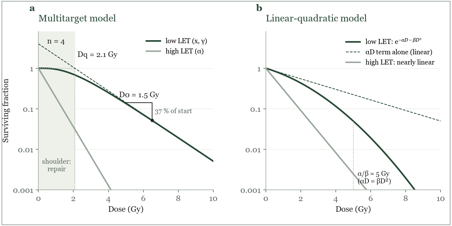
- The response is shown as a cell survival curve: dose on a linear x-axis, surviving fraction on a logarithmic y-axis (Figure 6).
- A surviving cell keeps its reproductive and functional capacity. Cell death means loss of function in differentiated cells and loss of reproductive ability in dividing cells.
Multitarget model
- Low-LET radiation (x- and γ-rays) gives a curve with an initial shoulder followed by a straight line. High-LET radiation (α particles, low-energy neutrons) gives a straight line from the lowest doses.
- The model has three parameters: D0, n and Dq.
- D0 is the dose that reduces survival to 37 % on the straight part of the curve, that is, it kills 63 % of the cells. It is found from the slope. A large D0 means a radioresistant cell type.
- n, the extrapolation number, is the intercept on the y-axis when the straight part is extrapolated back to zero dose. For mammalian cells it is between 1 and 10.
- Dq, the quasi-threshold dose, is the width of the shoulder. In this region almost all cells repair the small amount of damage, so there is little cell killing.
Equation
- ln n = Dq / D0
- S = 1 − (1 − e−D/D0)n
Linear-quadratic model
- The linear-quadratic model describes radiation cell killing more accurately.
Equation
- S = e−αD − βD^{2}
- S surviving fraction, D dose, α constant for single-hit (linear) killing, β constant for two-hit (quadratic) killing
- For low-LET radiation, βD2 is negligible at low doses, so survival falls linearly with dose; at higher doses the quadratic term dominates and the curve bends downward.
- For high-LET radiation the curve is linear throughout.
- The dose at which the linear and quadratic contributions are equal is α/β. It is high (about 10 Gy) for early-responding tissues and tumours, and low (about 3 Gy) for late-responding tissues.
Factors that modify radiation damage
Dose rate
- Dose rate is the dose delivered per unit time.
- At low dose rates mainly single-strand breaks occur and are repaired between ionising events. At high dose rates double-strand breaks occur, and the cell has too little time to repair them.
- In radiotherapy, when the total dose is given in fractions, the interval between fractions should be short enough to limit repair of the abnormal cells.
Linear energy transfer (LET)
- LET is the average energy deposited by a charged particle per unit length of its track in tissue, in keV/μm. It describes the density of ionisation along the track.
Equation
- LET = dE/dl (keV/μm)
- dE = energy deposited in a short length dl of the track
- LET ∝ z2/v2 (z charge, v speed of the particle)
- Factors that raise LET: a higher particle charge and a lower speed. So heavy charged particles (α, heavy ions) have high LET, and LET rises further towards the end of the track as the particle slows (the Bragg peak).
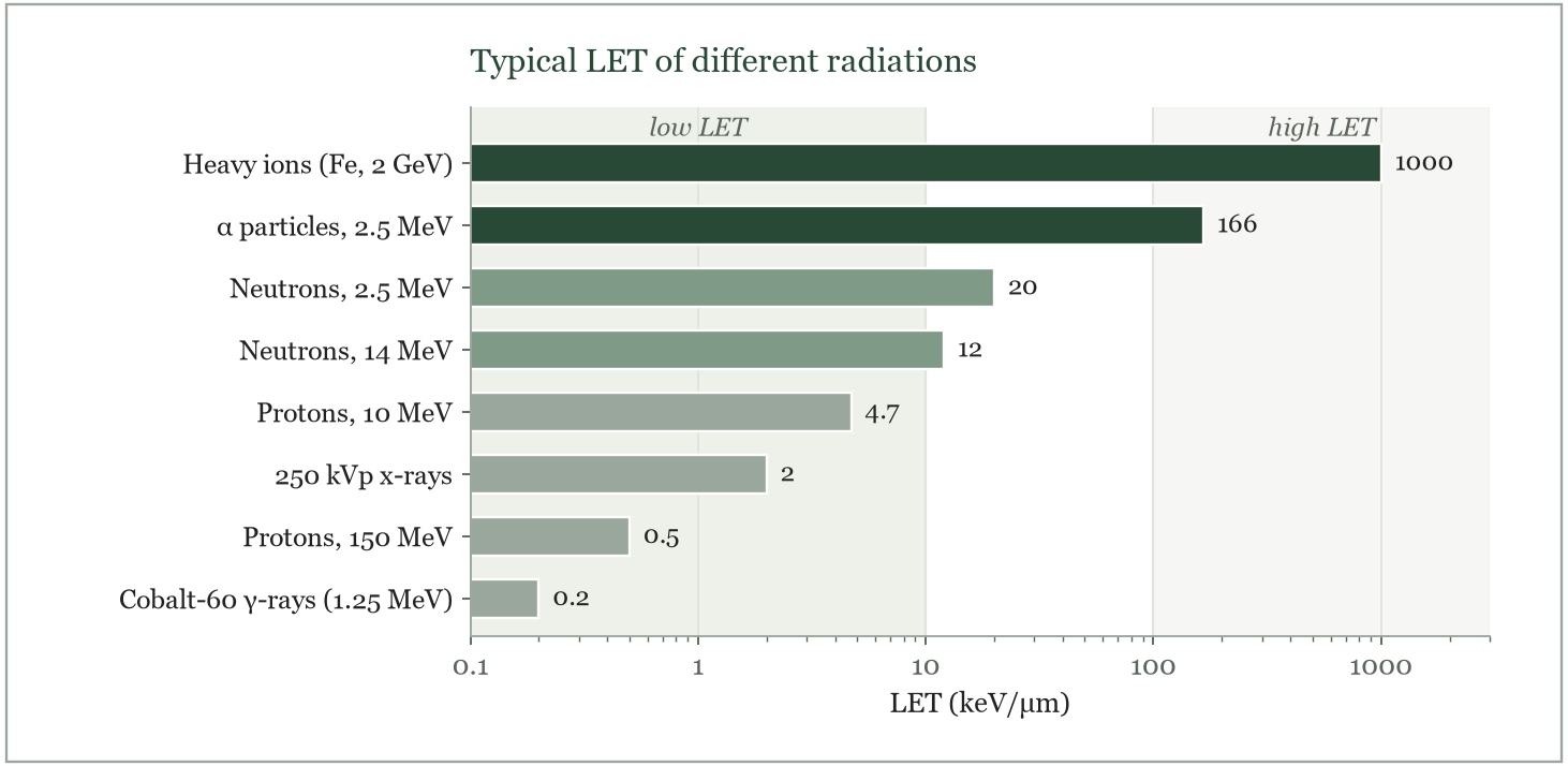
- Typical values (Figure 7): γ-rays and x-rays about 0.2 to 2 keV/μm; β particles about 0.3 keV/μm; protons 0.5 to 5; neutrons (through recoil protons) 10 to 20; α particles 100 to 200 keV/μm; heavy ions up to 1000 keV/μm. The table summarises the differences.
| Feature | High LET | Low LET |
|---|---|---|
| LET | > 20 keV/μm | < 3 keV/μm |
| Examples | α particles, neutrons, heavy ions | x-rays, γ-rays, β particles |
| Penetration in tissue | Low (μm to mm) | High (deep) |
| Ionisation along the track | Dense | Sparse |
| Relative biological effectiveness | High | Lower |
| Oxygen enhancement ratio | Low, about 1 | High, about 3 |
| Main type of damage | Direct | Indirect, through free radicals |
Relative biological effectiveness (RBE)
- RBE compares the biological effectiveness of a radiation with a reference radiation, usually 250 kVp x-rays.
Equation
- RBE = Dref / Dtest
- Dref = dose of the reference radiation (250 kVp x-rays) and Dtest = dose of the radiation being tested, each producing the same biological effect
- Example: if 2 Gy of neutrons kills the same fraction of cells as 6 Gy of x-rays, the RBE of the neutrons is 6/2 = 3.
- Factors affecting RBE:
| Factor | Effect on RBE |
|---|---|
| LET | Rises with LET to a maximum near 100 keV/μm, then falls: beyond this, energy is wasted on cells already killed (overkill) |
| Dose | Higher at low doses, because the low-LET reference curve has a shoulder and the high-LET curve does not |
| Fractionation and dose rate | Higher with fractionation or a low dose rate, because low-LET damage is repaired between fractions while high-LET damage is not |
| Biological endpoint and tissue | Differs with the effect measured (cell killing, mutation, cataract) and the tissue |
| Cell type and oxygen | Higher in hypoxic cells, because high-LET damage does not depend on oxygen |
- The radiation weighting factor (wR) is the regulatory simplification of RBE used in radiation protection. It converts the absorbed dose (D, in gray) to the equivalent dose (H, in sievert), which allows doses from different radiations to be added and compared.
Equation
- H = wR × D
- H = equivalent dose (Sv); wR = radiation weighting factor (no unit); D = absorbed dose (Gy)
| Radiation | wR (ICRP 103) |
|---|---|
| Photons (x-rays, γ-rays), all energies | 1 |
| Electrons, muons | 1 |
| Protons, charged pions | 2 |
| α particles, fission fragments, heavy ions | 20 |
| Neutrons | 2.5 to 20, a continuous function of energy, highest at about 1 MeV |
- Example: 1 mGy from α particles gives 20 mSv; 1 mGy from γ-rays gives 1 mSv.
Oxygen enhancement ratio (OER)
- Oxygen is the most important chemical modifier of radiation damage. The OER measures how much it sensitises cells.
Equation
- OER = Dhypoxic / Doxic
- Dhypoxic = dose needed in the absence of oxygen and Doxic = dose needed in the presence of oxygen, to produce the same biological effect
- Example: if hypoxic cells need 9 Gy of x-rays to reach the same survival as oxygenated cells at 3 Gy, the OER is 9/3 = 3. An OER of 3 means hypoxic cells are three times more resistant.
- Mechanism: oxygen reacts irreversibly with the free radicals formed on DNA by water radiolysis, fixing the damage before it can be repaired chemically. Oxygen must be present during irradiation or within milliseconds of it.
- Factors affecting OER: LET (about 2.5 to 3 for x- and γ-rays, falling to about 1 above 100 keV/μm); oxygen tension (most of the sensitisation is reached at a partial pressure of about 3 mmHg, and full effect by about 30 mmHg); dose (a little lower, about 2, at doses below 2 Gy).
- Clinical link: hypoxic tumour cells are radioresistant, and high-LET radiation or oxygen-mimic sensitisers (next section) are ways to overcome this.
Radiosensitisers and radioprotectors
Radiosensitisers
- Radiosensitisers increase the response of cells to radiation.
| Class | Examples | How they sensitise |
|---|---|---|
| Oxygen | Molecular oxygen | The strongest sensitiser: fixes free-radical damage on DNA so it cannot be repaired (see OER) |
| Halogenated pyrimidines | 5-chlorodeoxyuridine (ClUdR), 5-bromodeoxyuridine (BrUdR), 5-iododeoxyuridine (IUdR) | Thymidine analogues built into DNA during synthesis, making it more easily damaged; act only on dividing cells |
| Cytotoxic drugs | Actinomycin D, puromycin, methotrexate, 5-fluorouracil | Interfere with DNA synthesis and repair, with some direct cytotoxicity of their own |
| Oxygen mimics (nitroimidazoles) | Metronidazole, misonidazole, etanidazole | Electron-affinic drugs that imitate oxygen in hypoxic cells; etanidazole is less neurotoxic |
Radioprotectors
- Radioprotectors reduce radiation damage. They must be present at the time of irradiation.
- The classical agents contain a sulfhydryl (–SH) group: cysteine and cysteamine. They scavenge free radicals before the radicals reach DNA, and donate hydrogen to repair radical damage on DNA. They are therefore most effective against low-LET radiation, where indirect action dominates, and give little protection against high-LET radiation.
- The early compounds caused nausea and vomiting at effective doses. Phosphorothioate prodrugs, WR-638 and WR-2721 (amifostine), are better tolerated. Amifostine is given shortly before irradiation to protect normal rapidly dividing tissues such as bone marrow and salivary glands.
- The dose reduction factor (DRF) is the dose needed with the protector divided by the dose needed without it, for the same effect. For amifostine it is about 2 to 2.7 for bone marrow.
Deterministic and stochastic effects
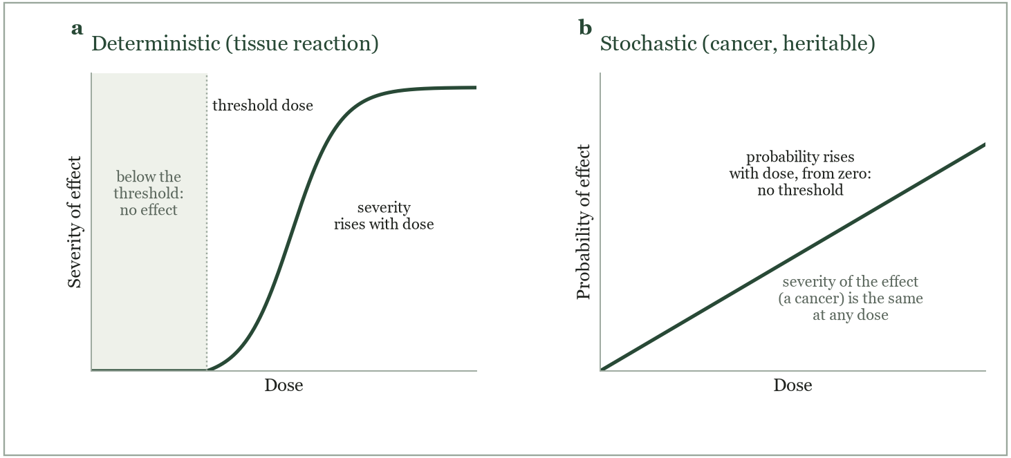
- Radiation effects on the body are classified by their dose–response relationship (Figure 8).
Deterministic effects (tissue reactions)
- Caused by the killing of a large number of cells, so that the tissue or organ can no longer function.
- There is a threshold dose, which differs between tissues. Below it no effect is seen.
- Above the threshold the severity increases with dose.
- Onset is usually hours to weeks after exposure.
- Examples: skin erythema and epilation, mucositis, cataract, sterility, the acute radiation syndromes, and bone marrow failure.
Stochastic effects
- Caused by damage to a single cell that escapes repair: a mutation that may lead to cancer in the exposed person or, in a germ cell, to a heritable effect in the offspring.
- The probability of the effect increases with dose, with no threshold: any dose, however small, carries some risk.
- The severity of the effect does not depend on the dose. A cancer induced by a small dose is no milder than one induced by a large dose.
- Onset is years to decades after exposure.
- Examples: leukaemia, thyroid, breast and other solid cancers; heritable disease.
- Radiation protection assumes a linear no-threshold (LNT) relationship for stochastic effects, so every dose is kept as low as reasonably achievable (ALARA).
Acute radiation syndromes
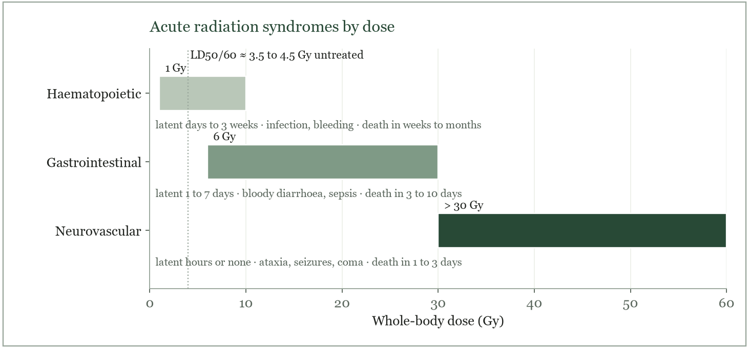
- Acute radiation sickness follows a large whole-body dose delivered over a short time. The syndromes are deterministic effects with threshold doses, and the organ affected depends on the dose (Figure 9).
- The lethal dose LD50/60 is the dose that kills 50 % of those exposed within 60 days. For humans it is about 3.5 to 4.5 Gy without treatment, and about 6 to 7 Gy with intensive supportive care (antibiotics, transfusions, growth factors).
- Each syndrome passes through four stages: a prodromal stage (nausea, vomiting, fatigue), a latent stage of apparent well-being, the stage of manifest illness, and then recovery or death. The higher the dose, the earlier and shorter each stage.
| Syndrome | Dose (Gy) | Target | Prodrome | Latent period | Manifest illness | Death, if untreated |
|---|---|---|---|---|---|---|
| Haematopoietic | 1 to 10 | Bone marrow stem cells | Hours to 1 day: nausea, vomiting, fatigue | Days to 3 weeks | Fever, infection, bleeding (petechiae), anaemia, from low white cells and platelets | Weeks to months, from infection or haemorrhage |
| Gastrointestinal | 6 to 30 | Small-bowel epithelium | Minutes to hours: severe vomiting, watery diarrhoea, abdominal pain | 1 to 7 days | Bloody diarrhoea, dehydration, electrolyte loss, sepsis, high fever | 3 to 10 days |
| Neurovascular | > 30 | Brain and blood vessels | Minutes: vomiting, confusion, loss of consciousness | Hours, or none | Ataxia, seizures, hypotension, circulatory collapse, coma | 1 to 3 days |
- The lymphocyte count in the first 48 hours is the most useful early guide to the dose received: the faster and deeper the fall, the higher the dose.
Long-term effects
- Late effects appear months to years after exposure, either after recovery from acute radiation sickness or after repeated low-dose exposure.
- Somatic effects occur in the exposed person: carcinogenesis, cataract, skin damage, damage to the reproductive organs, and life-shortening.
- Genetic (heritable) effects occur in the offspring, through mutations in germ cells.
Carcinogenesis
- Radiation is a complete carcinogen: it can both initiate and promote cancer.
- Initiation: DNA damage producing a mutation. Agents include chemicals, ultraviolet light, viruses and ionising radiation.
- Promotion: promoters cannot start a cancer, but encourage the growth of cells that already carry a mutation. Examples are oestrogen, phorbol esters, a high-fat diet and radiation itself.
- Progression: the mutated cells become increasingly abnormal, invade and metastasise.
Dose–response and risk estimates
- Human data come mainly from the atomic-bomb survivors, radiotherapy patients, occupationally exposed groups and the Chernobyl population.
- Below about 100 mGy the data are inconclusive, because of small numbers, lack of controls and uncertain dosimetry.
- The linear no-threshold (LNT) model assumes that risk is proportional to dose down to zero, with no safe dose. The BEIR VII report (2006) supports LNT for solid cancers; for leukaemia the curve is linear-quadratic, rising more steeply at higher doses.
Risk, BEIR VII
- 100 mSv (10 rem) to a population → about 1 extra cancer per 100 people over a lifetime, on top of about 42 that occur naturally
- Lifetime low-LET natural background (excluding radon) also gives roughly 1 in 100
- Leukaemia: about 20 % of radiation-induced cancer deaths; risk higher at young ages; shortest latent period, average 5 to 10 years; about 1 to 2.6 excess cases per 10,000 person-years per Gy.
- Breast cancer: risk highest for exposure at young ages; BEIR V excess risk per 10,000 person-years per Gy about 5 at age 25, 4 at 35, 2 at 45; BEIR VII about 10 per 10,000 person-years per Gy for exposure at age 50. Modern mammography carries a very low risk.
- Thyroid cancer: about 4.4 excess cases per 10,000 person-years per Gy; the risk is greatest for exposure in childhood (Chernobyl).
- Lung, bone and skin cancers are induced less often.
Skin
- The skin is sensitive because of its rapidly dividing basal layer, hair follicles and sebaceous glands.
- Skin effects are deterministic. Transient erythema appears within hours above about 2 Gy; the main erythema at about 6 Gy appears in the third or fourth week, with warm, red, oedematous, tender skin.
- Temporary epilation occurs above about 3 Gy, permanent epilation above about 7 Gy.
- Dry desquamation follows about 14 Gy, moist desquamation about 18 Gy, and necrosis above about 24 Gy. Normal skin takes 2 to 3 months to regrow.
- The effects depend on the dose, dose rate, LET and area exposed. Skin cancer is a rare late result.
Reproductive organs
- The gonads are highly radiosensitive. In the testis the spermatogonia are the most sensitive cells; spermatids and spermatozoa are resistant. In the ovary the oocytes in developing follicles are the most sensitive.
- Males: temporary sterility from about 0.15 Gy (15 rad); permanent sterility from an acute dose of about 3.5 to 6 Gy.
- Females: permanent sterility from about 2.5 to 6 Gy depending on age, lower in older women who have fewer oocytes left.
- Lymphocyte and sperm counts are both used as biological dosimeters after accidental exposure.
Life-shortening and hormesis
- In animal studies, whole-body irradiation shortened the life span, mainly through cancer.
- The evidence is inconsistent at low doses, where some studies found a longer life span. This led to the idea of radiation hormesis, that low doses are beneficial, possibly through stimulation of the immune system and DNA repair. It is not accepted as a basis for radiation protection.
Cataract
- The lens of the eye is radiosensitive, and radiation produces posterior subcapsular cataract.
- Cataract is a deterministic effect. ICRP 118 (2012) lowered the threshold to about 0.5 Gy, whether given in one or several exposures, and reduced the occupational lens limit to 20 mSv per year. Older texts give 2 Gy for a single exposure.
- High-LET radiation is more effective; neutrons roughly double the incidence.
- The latent period is at least 1 year and is shorter with higher doses.
Genetic effects
- Radiation damage to the DNA of germ cells causes mutations that are passed to future generations.
- Genes mutate spontaneously even without radiation. Radiation and chemical mutagens raise the frequency of mutation, not the kind: radiation-induced mutations are the same as spontaneous ones, only more frequent.
- Inherited disorders that could in principle increase include Down syndrome, achondroplasia, retinoblastoma, cystic fibrosis, sickle-cell anaemia and Tay–Sachs disease.
- No heritable effect of radiation has yet been demonstrated in humans, including the children of atomic-bomb survivors. The estimates come from mouse data.
- Doubling dose: the dose that doubles the spontaneous mutation rate in one generation. For humans it is estimated at about 1 Gy.
- Genetically significant dose (GSD): the average dose to the gonads of a population, weighted by the number of people exposed and their expected number of future children. It is a measure of the genetic burden of a practice such as diagnostic radiology.
Embryo and foetus
- The embryo is extremely radiosensitive because its cells are dividing and differentiating rapidly. The effect depends on the stage of development, the dose and the dose rate.
- Preimplantation (0 to 2 weeks after conception): the all-or-none period. The main effect is death of the embryo, with a threshold of about 100 mGy; an embryo that survives develops normally.
- Organogenesis (2 to 8 weeks): malformations, mainly of the central nervous system and skeleton, and growth retardation; threshold about 100 to 200 mGy.
- Early foetal period (8 to 15 weeks): the most sensitive time for the developing brain. Severe mental retardation has a threshold of about 300 mGy, and IQ falls by about 25 to 30 points per Gy above it. Between 16 and 25 weeks the sensitivity is lower.
- Late foetal period: foetal cells are more radioresistant and much higher doses are needed to cause death.
- Childhood cancer: in-utero exposure of about 10 mGy raises the risk of childhood leukaemia and other cancers by about 40 % (relative risk about 1.4), from a baseline of about 1 in 500.
- ICRP 84: a foetal dose below 100 mGy is not a reason to terminate a pregnancy; nearly all diagnostic nuclear medicine and radiology procedures give far less than this.
Test yourself
Revise this chapter with the Biological effects deck in Flashcards.
10 quick questions. Pick an answer to see the explanation.
1. Which lesion is the main cause of radiation-induced cell death?
Answer: C. Double-strand breaks within a few base pairs separate the molecule and are not reliably repaired; single-strand breaks and base damage are repaired using the intact strand.
2. A chromosome with two centromeres is seen after irradiation. It arose because
Answer: B. A single break that is replicated before restitution gives two centromere-bearing fragments that join (dicentric) and two acentric fragments that join.
3. Indirect action of radiation is mediated mainly by
Answer: B. Water is about 70 % of the cell; its radiolysis gives OH• radicals that diffuse to DNA.
4. The most radiosensitive phase of the cell cycle is
Answer: C. DNA is condensed and unrepaired at mitosis; late S, with active repair, is the most resistant.
5. In the multitarget model, D0 is the dose that
Answer: B. D0 is read from the slope of the exponential part; it kills 63 % of cells. Dq is the shoulder width and n the intercept.
6. In the linear-quadratic model, the curve bends downward at high doses because
Answer: B. βD2 grows faster than αD, so at high doses two-hit killing dominates and survival falls more steeply.
7. The oxygen enhancement ratio for α particles is about
Answer: C. High-LET damage is mostly direct and needs no oxygen to be fixed, so the OER approaches 1; for x- and γ-rays it is about 3.
8. Which statement describes a stochastic effect?
Answer: C. Stochastic effects (cancer, heritable disease) have no threshold; the dose changes the probability, not the severity.
9. A patient vomits within minutes of a whole-body exposure and dies in 2 days with seizures and coma. The dose was most likely
Answer: D. Vomiting within minutes, little or no latent period, and neurological death in 1 to 3 days is the neurovascular syndrome, above about 30 Gy.
10. According to ICRP 84, the foetal dose below which termination of pregnancy is not justified on radiation grounds is
Answer: C. ICRP 84 states that doses below 100 mGy should not be considered a reason for termination; diagnostic procedures give far less.
References
- Saha GB. Physics and Radiobiology of Nuclear Medicine. 4th ed. New York: Springer; 2013. Chapter 15.
- Hall EJ, Giaccia AJ. Radiobiology for the Radiologist. 8th ed. Philadelphia: Wolters Kluwer; 2019.
- ICRP. The 2007 Recommendations of the International Commission on Radiological Protection. ICRP Publication 103. Ann ICRP. 2007;37(2-4).
- ICRP. Pregnancy and medical radiation. ICRP Publication 84. Ann ICRP. 2000;30(1).
- ICRP. ICRP statement on tissue reactions and early and late effects of radiation in normal tissues and organs. ICRP Publication 118. Ann ICRP. 2012;41(1-2).
- National Research Council. Health Risks from Exposure to Low Levels of Ionizing Radiation: BEIR VII Phase 2. Washington, DC: National Academies Press; 2006.
Spotted an error or something out of date? Report an error