PET/CT in Lymphoma
- Lugano 2014 is still the standard. ¹⁸F-FDG PET/CT stages FDG-avid lymphomas and the Deauville five-point scale grades response; a 2025 Lugano workshop proposed refinements but kept the 2014 classification in force.
- Two references, one scan. Score the most intense residual site against the mediastinal blood pool and the liver: 1–3 is a complete metabolic response in routine practice; 4–5 is not.
- Know the trial threshold. De-escalation trials such as RAPID, HD16 and HD17 counted score 3 as positive; RATHL, AHL2011 and HD21 counted 1–3 as negative. Always state the score, not just 'positive' or 'negative'.
- Marrow biopsy is not routine in HL or DLBCL. Clearly focal marrow uptake is read as involvement (stage IV). Diffuse homogeneous marrow uptake, especially after G-CSF, is usually reactive.
- Interim PET guides therapy in Hodgkin lymphoma, not yet in DLBCL. PET-adapted strategies (RATHL, AHL2011, HD17, HD21) de-escalate or intensify safely; in DLBCL, escalation for a positive interim scan (PETAL) did not improve outcome.
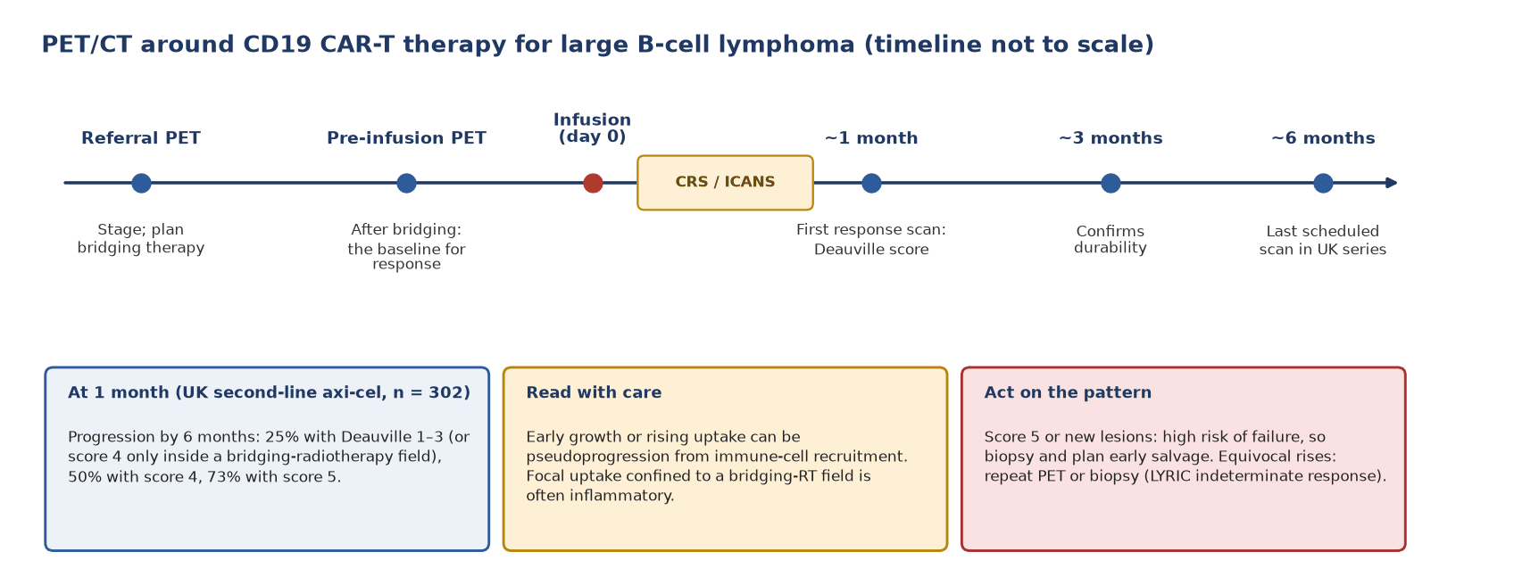
- Novel therapies need adapted reading. After CAR-T, the one-month Deauville score predicts failure; with checkpoint inhibitors or bispecific antibodies, flare and pseudoprogression are handled with LYRIC's indeterminate response.
- Avidity and transformation. Indolent subtypes vary in avidity; a site far hotter than the rest in CLL or follicular lymphoma is the biopsy target for transformation, but no SUV cut-off is diagnostic.
- No routine surveillance PET. Scans in patients in remission detect few relapses, create false positives and did not improve survival; image when relapse is suspected.
1. The clinical problem
Lymphomas are a family of more than 60 entities, but for imaging the questions are few: how far has the disease spread, is it responding, is there residual active disease at the end of treatment, and has an indolent lymphoma transformed? Two pathology classifications appeared in 2022, the World Health Organization 5th edition (WHO-HAEM5) and the International Consensus Classification (ICC). They largely agree on the entities that matter to the reporter; nomenclature differs in places, so copy the name on the pathology report into the PET report. PTLD is grouped by WHO-HAEM5 with other lymphoid proliferations associated with immune deficiency and dysregulation.
Staging: the Lugano classification (2014)
The Lugano classification (Cheson 2014, with the imaging consensus of Barrington 2014) made PET/CT the staging test for FDG-avid lymphomas and kept a modified Ann Arbor anatomical system. Tonsils, Waldeyer's ring and spleen count as nodal tissue. The A/B suffix for symptoms is kept only for Hodgkin lymphoma (HL), and the X suffix for bulk was dropped in favour of recording the longest diameter.
| Stage | Involvement | Extranodal (E) status | Treatment group |
|---|---|---|---|
| I | One node or a group of adjacent nodes | Single extranodal lesion without nodal involvement (IE) | Limited |
| II | Two or more nodal groups on the same side of the diaphragm | Stage I or II by nodal extent with limited contiguous extranodal involvement (IIE) | Limited |
| II bulky | Stage II with bulky disease | Not applicable | Limited or advanced, by histology and prognostic factors |
| III | Nodes on both sides of the diaphragm, or nodes above the diaphragm with spleen involvement | Not applicable | Advanced |
| IV | Additional non-contiguous extralymphatic involvement (for example marrow, liver, lung) | Not applicable | Advanced |
Lugano 2014 staging (after Cheson 2014).
- Bulk in HL: a single nodal mass of 10 cm or more, or greater than one third of the transthoracic diameter at any level of the thoracic vertebrae on CT.
- Bulk in non-Hodgkin lymphoma: no validated cut-off; Lugano noted suggested thresholds of 6–10 cm for DLBCL in the rituximab era. The 2025 EHA large B-cell lymphoma guideline notes that 7.5 cm is widely used and that total metabolic tumour volume (TMTV) above 220 mL has been proposed as a metabolic definition.
- Spleen: on PET, diffuse uptake, a solitary mass, miliary lesions or nodules indicate involvement; on CT, a vertical length above 13 cm defines splenomegaly.
Where nuclear medicine changes management
- Staging PET/CT upstages patients who look limited on CT and replaces staging marrow biopsy in HL and most DLBCL.
- Interim PET decides chemotherapy intensity and duration in HL (bleomycin omission, BEACOPP escalation, four versus six cycles of BrECADD).
- End-of-treatment PET decides consolidation radiotherapy: omitted after a complete metabolic response in early unfavourable HL (HD17) and in primary mediastinal B-cell lymphoma, and given to PET-positive residual disease.
- After CAR-T cell therapy the one-month scan identifies patients who need early salvage.
2. Tracers and why they work
¹⁸F-FDG is trapped as FDG-6-phosphate in cells with high glucose transport and hexokinase activity. Aggressive lymphomas are highly glycolytic. In classical HL the malignant Hodgkin and Reed–Sternberg cells are few, but the inflammatory microenvironment around them is intensely avid, so the whole node lights up. Uptake therefore tracks viable disease plus inflammation, which is why both lymphoma and its mimics are FDG-avid.
Avidity by subtype
| Histology | Avid at staging (Weiler-Sagie, n = 766) | Practical role of PET |
|---|---|---|
| Classical HL | 100% (233/233) | Staging, interim and end-of-treatment response |
| Burkitt lymphoma | 100% (18/18) | Staging and response; do not delay treatment for the scan |
| Mantle cell lymphoma | 100% (14/14) | Staging and response |
| DLBCL | 97% (216/222) | Staging, interim prognosis, end-of-treatment response |
| Follicular lymphoma | 95% (133/140) | Staging (especially before radiotherapy for limited disease) and end-of-treatment response; a much hotter site suggests transformation |
| T-cell lymphomas | 85% (34/40) | Staging and response in avid subtypes |
| Small lymphocytic lymphoma / CLL | 83% (24/29), usually low grade | CT staging; PET to find and target transformation |
| Nodal marginal zone lymphoma | 100% (8/8) | Variable intensity; PET with CT at staging, PET response if avid |
| Extranodal marginal zone (MALT) lymphoma | 55% (29/53) | Often non-avid; CT, endoscopy and MRI carry the load |
FDG avidity by histology (Weiler-Sagie 2010: avidity defined as at least one FDG-avid disease site).
Lugano recommends PET/CT staging for essentially all histologies except CLL/small lymphocytic lymphoma, lymphoplasmacytic lymphoma/Waldenström macroglobulinaemia, mycosis fungoides and marginal zone lymphomas, unless transformation is suspected. The 2025 workshop suggested PET/CT with contrast CT for marginal zone lymphoma staging, with response then assessed by PET only if the disease was avid at baseline. Avidity at staging is what matters: a non-avid baseline cannot be followed by Deauville scoring.
Transformation: where FDG helps most in indolent disease
- Richter transformation of CLL: in a 2025 meta-analysis of ten studies, an SUVmax threshold of about 5 gave pooled sensitivity 90% and specificity 54%; a threshold of about 10 gave 74% and 84%. Neither is diagnostic. Use PET to choose the hottest accessible site for biopsy, and treat SUVmax ≥10 as a strong warning, not a rule.
- Follicular lymphoma: the same meta-analysis found that transformation thresholds were higher and less discriminating than for Richter transformation. A site clearly hotter than the rest of the disease should be biopsied.
When FDG fails
False negatives arise in non-avid histologies (MALT, some CLL and T-cell lymphomas), in low-volume diffuse marrow infiltration (Lugano notes that PET can miss 10–20% diffuse involvement, often of discordant low-grade histology) and in organs with high background such as brain. Brain MRI, not PET, is the standard for parenchymal and leptomeningeal central nervous system (CNS) disease. Chemokine receptor CXCR4 PET is the most promising alternative for marginal zone lymphoma (section 8).
3. Indications by clinical scenario
| Scenario | Role of FDG PET/CT | Guideline position |
|---|---|---|
| Diagnosis | Not a diagnostic test; directs biopsy to the most accessible or most avid site | Excision biopsy preferred [I, B]; needle core acceptable (EHA LBCL 2025) |
| Staging, FDG-avid lymphoma | Standard staging test; contrast CT added only when needed (for example radiotherapy planning, vascular compression) | Lugano 2014; reaffirmed unanimously by the 2025 Lugano workshop; EHA HL 2026 and EHA LBCL 2025 |
| Marrow at staging | Replaces marrow biopsy in HL and in DLBCL (biopsy if PET is negative and discordant histology matters) | Lugano 2014; EHA HL 2026 (biopsy not recommended if PET done); EHA LBCL 2025 |
| Radiotherapy planning | Defines initially involved sites for involved-site radiotherapy; ideally scanned in the treatment position | Lugano 2014 (contrast CT preferred for planning) |
| Interim response, HL | Decides escalation or de-escalation; perform it when the chosen pathway uses the result | EHA HL 2026 |
| Interim response, DLBCL | Prognostic; high negative predictive value; ΔSUVmax above 70% at cycle 4 is favourable; score 5 (≥2 × liver) at cycle 4 prompts biopsy and consideration of salvage | EHA LBCL 2025 [III, A] |
| End of treatment | Defines remission; guides radiotherapy to PET-positive residual masses | Lugano 2014; EHA LBCL 2025 [III, A]; EHA HL 2026 |
| Residual score 4–5 at end of treatment (LBCL) | Positive predictive value only about 60% | Biopsy or repeat PET/CT at 8–12 weeks [III, A] (EHA LBCL 2025) |
| Suspected transformation | Chooses the biopsy site | Lugano 2014 (PET for indolent histologies when transformation is suspected) |
| Surveillance in remission | Not indicated | Lugano 2014 (discouraged); EHA HL 2026 and EHA LBCL 2025 (not recommended); 2025 workshop (abandon) |
| Suspected relapse, before transplant or CAR-T | Restaging and baseline for the next line; biopsy relapse before second-line therapy | EHA LBCL 2025 |
Recommendation strength is given where the guideline states it (EHA levels of evidence I–V and grades A–E). Lugano and the 2025 workshop are consensus statements without grades.
4. Evidence at a glance
| Study | Design, n | Key finding | What it changed |
|---|---|---|---|
| Weiler-Sagie 2010 | Retrospective, 766 | FDG avidity 100% in HL, Burkitt and mantle cell; 97% DLBCL; 95% FL; 55% extranodal marginal zone | Basis for which histologies are staged with PET |
| RAPID (Radford 2015) | Randomised, early-stage IA–IIA HL; 571 scanned | PET-negative (score 1–2) after 3 ABVD in 74.6%; 3-year PFS 94.6% with radiotherapy vs 90.8% without; non-inferiority not shown | Omitting radiotherapy trades a small loss of control for fewer late effects |
| H10 (André 2017) | Randomised, stage I–II HL, 1,925 with early PET | Early PET positive in 18.8%; switching to BEACOPPesc + INRT raised 5-year PFS from 77.4% to 90.6%; ABVD alone in PET-negative patients was not non-inferior | Escalate for a positive early PET; keep radiotherapy in PET-negative favourable disease |
| HD16 (Fuchs 2019) | Randomised, early favourable HL, 1,150 | PET-negative (score <3) after 2 ABVD: 5-year PFS 93.4% with 20 Gy vs 86.1% without; with a liver cut-off, PET-positive PFS was 80.9% | Radiotherapy cannot be dropped in favourable disease on PET alone; score 4 is the stronger risk marker |
| HD17 (Borchmann 2021) | Randomised, early unfavourable HL, 1,100 | PET-guided omission of radiotherapy after 2 + 2: 5-year PFS 95.1% vs 97.3% with standard radiotherapy (within the 8% margin) | Omit radiotherapy after a negative end-of-chemotherapy PET |
| RATHL (Johnson 2016) | Randomised, advanced HL, 1,214 | Interim PET negative (score 1–3) in 83.7%; 3-year PFS 85.7% (ABVD) vs 84.4% (AVD), just short of the non-inferiority margin, with less lung toxicity; PET-positive patients given BEACOPP had 3-year PFS 67.5% | Drop bleomycin after a negative interim PET |
| AHL2011 (Casasnovas 2019) | Randomised, advanced HL, 823 | 84% switched from BEACOPPesc to ABVD after a negative PET2; 5-year PFS 85.7% vs 86.2% (standard); treatment-related serious adverse events 28% vs 47% | PET-driven de-escalation from BEACOPP |
| HD21 (Borchmann 2024) | Randomised, advanced HL, 1,500 | PET2-guided BrECADD (4 cycles if score 1–3, 6 if ≥4) vs eBEACOPP: treatment-related morbidity 42% vs 59%; 4-year PFS 94.3% vs 90.9% | BrECADD becomes a standard for fit patients up to 60 (EHA 2026) |
| PETAL (Dührsen 2018) | Randomised, aggressive NHL, 862 | Interim PET positive (ΔSUVmax ≤66%) in 12.5%; intensive Burkitt protocol did not beat R-CHOP (2-year EFS 31.6% vs 42.0%) and was more toxic | Interim PET is prognostic in DLBCL but does not justify escalation |
| Danish–Swedish cohort (El-Galaly 2015) | Population-based, 1,221 DLBCL in first CR | Routine imaging follow-up (Denmark) gave no survival benefit over clinical follow-up (Sweden) | Evidence against surveillance scanning |
| Kuhnl 2026 | National cohort, 302 second-line axi-cel | Progression by 6 months after a one-month score 1–3: 25%; score 4: 50%; score 5: 73% | One-month PET stratifies post-CAR-T management |
5. Patient preparation and acquisition
Standard EANM tumour-imaging practice applies: fast for at least 4 hours, measure and record blood glucose, and keep the patient warm and relaxed in the uptake room. What differs in lymphoma is timing and history.
- Record treatment history on the request: date of the last chemotherapy cycle, granulocyte colony-stimulating factor (G-CSF), steroids, bridging therapy and radiotherapy fields. EANM notes marrow uptake after G-CSF and GM-CSF, chemotherapy, anaemia and infection.
- End-of-treatment timing: at least 3 weeks, preferably 6–8 weeks, after the last chemotherapy cycle; 2 weeks after G-CSF; 3 months after radiotherapy (Barrington 2014).
- Interim timing: follow the protocol of the treatment pathway, and keep scanner, reconstruction and uptake time the same as at baseline so that the Deauville score and ΔSUVmax are comparable.
- Brown fat: more common in young patients and in cold conditions; a warm waiting room reduces it. Uptake in supraclavicular fat must not be scored as nodal disease.
- Field of view: vertex (or skull base) to mid-thigh; include the whole of any limb with known disease.
- Quantification: use EANM Research Ltd (EARL) harmonised reconstructions when SUV-based measures such as ΔSUVmax or TMTV are reported.
- Suspected primary CNS lymphoma: brain MRI is the reference test; image and biopsy before corticosteroids whenever possible, because steroids shrink the lesions (21–68% volume reduction after about a week in one series).
6. Interpretation
The Deauville five-point scale
| Score | Definition (Lugano 2014) | Notes |
|---|---|---|
| 1 | No uptake above background | Usually a site no longer visible on CT |
| 2 | Uptake ≤ mediastinum | Reference: mediastinal blood pool |
| 3 | Uptake > mediastinum but ≤ liver | Complete metabolic response in routine practice; inadequate in some de-escalation trials |
| 4 | Uptake moderately > liver | Quantitatively above the SUVmax of a large region of normal liver (Barrington 2014) |
| 5 | Uptake markedly higher than liver and/or new lesions | Quantitatively 2–3 × the liver SUVmax (Barrington 2014); 2025 proposal: ≥2 × liver, split into 5a (existing lesions) and 5b (new lesions) |
| X | New areas of uptake unlikely to be related to lymphoma | For example infection or post-radiotherapy inflammation |
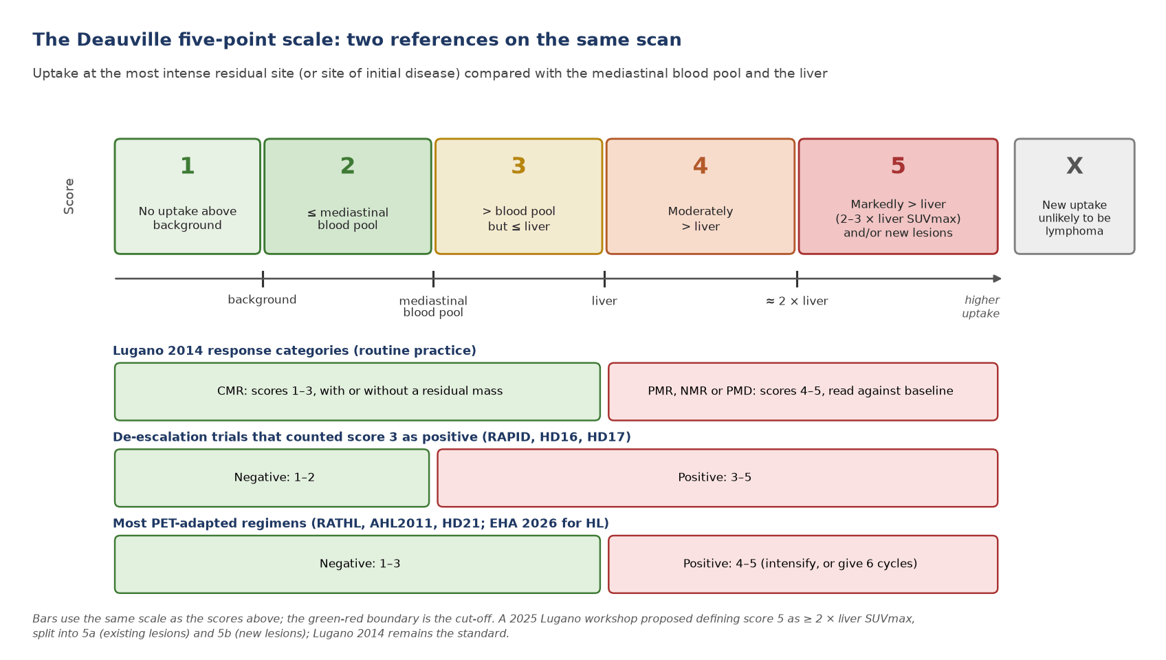
- Score the hottest residual site among sites of initial disease, on the same scan as the references. The score is visual; SUVmax supports it.
- High-background sites: in Waldeyer's ring, the bowel, or a spleen or marrow activated by chemotherapy or G-CSF, a complete metabolic response may be inferred when uptake at the initial site is no higher than the surrounding normal tissue (Lugano).
- Score 3 is the grey zone. Barrington 2014: score 3 probably represents a complete metabolic response at interim and a good prognosis at the end of standard treatment, but in de-escalation trials it may be preferable to count it as inadequate response. HD16 showed why: after 2 ABVD, PFS was lower with score 3 than with scores 1–2, and lower still with score 4.
- Quantitative extensions. qPET divides the lesion SUVpeak by the mean liver uptake; Deauville 3, 4 and 5 correspond to qPET values of about 0.95, 1.3 and 2.0 (Hasenclever 2014). It is used in paediatric HL trials and remains a research tool in adults.
Marrow and spleen at staging
Barrington 2014: focal uptake in bone or marrow, liver and spleen is highly sensitive for involvement in HL and aggressive non-Hodgkin lymphoma. In HL, diffuse marrow uptake without focal activity often represents reactive hyperplasia and should not be confused with involvement. Lugano: in HL a marrow biopsy is no longer required; in DLBCL, PET showing bone or marrow involvement is usually sufficient to designate advanced stage, and biopsy is indicated only when PET is negative and finding discordant histology would change management. In practice: clearly focal marrow FDG is read as involvement.
Special situations
- Primary mediastinal B-cell lymphoma and mediastinal grey zone lymphoma: residual anterior mediastinal masses are the rule. Score the residual uptake as usual; the 2025 EHA guideline recommends omitting mediastinal radiotherapy after a complete metabolic response. A score of 4 in a large residual mass is common and needs multidisciplinary review, with biopsy or repeat PET.
- Post-transplant lymphoproliferative disorder (PTLD): monomorphic and HL-type PTLD are staged and assessed like their counterparts; extranodal and graft involvement are common and infection or rejection can mimic disease (see the transplantation article).
- Primary CNS lymphoma: lesions are usually intensely avid (mean SUVmax 24.8, about three times normal grey matter, in one series), but MRI stages the brain. Report whole-body findings separately: systemic disease changes the diagnosis.
- Thymic rebound: a smooth, triangular, mildly avid anterior mediastinal soft-tissue in a young adult months after chemotherapy is usually thymic hyperplasia; compare shape with baseline and do not score it as residual lymphoma.
Pitfalls and mimics
| Pitfall | Typical pattern | How to resolve |
|---|---|---|
| Brown fat | Symmetrical supraclavicular, paravertebral and perirenal uptake in fat density on CT | Check CT density; keep patients warm; repeat if it obscures nodes |
| G-CSF or cytokine marrow activation | Diffuse homogeneous marrow uptake above liver, often splenic too, after chemotherapy | Score marrow as reactive; end-of-treatment scan at least 2 weeks after G-CSF |
| Thymic hyperplasia | Smooth anterior mediastinal soft tissue with mild uptake in young adults after treatment | Shape, symmetry and timing; follow-up if doubtful |
| Infection and inflammation | Consolidation, oesophagitis, catheter sites, abscess | Use score X; correlate clinically |
| Sarcoid-like reaction | Symmetrical mediastinal and hilar nodes, sometimes new after immunotherapy or chemotherapy | Biopsy before calling relapse (EHA LBCL 2025 lists it as a relapse mimic) |
| Injection-site and vaccination nodes | Ipsilateral axillary nodes after tracer extravasation or recent vaccination | Record injection arm and vaccination history |
| Post-radiotherapy change | Uptake conforming to the radiotherapy field, oesophagitis, pneumonitis | Wait 3 months; read shape against the field |
| Bowel and Waldeyer's ring | Physiological uptake that can exceed liver | Compare with surrounding normal tissue (Lugano rule) |
| Non-avid disease | Negative PET in MALT, some CLL or T-cell lymphomas | Check baseline avidity before calling a complete metabolic response |
Wording the conclusion
- Name the time point (staging, interim after n cycles, end of treatment), give the Deauville score of the most avid site and name that site, and state the Lugano category.
- When a pathway uses a different threshold, say so: 'Deauville 3; negative under the RATHL-type threshold (1–3), positive under a threshold of 1–2'.
- For new lesions, say whether they are consistent with lymphoma (score 5 or 5b) or unlikely to be lymphoma (score X), and recommend biopsy when management depends on it.
7. Response assessment and follow-up
Lugano metabolic response categories
| Category | PET definition (Lugano 2014) | Marrow |
|---|---|---|
| Complete metabolic response (CMR) | Score 1, 2 or 3 with or without a residual mass | No FDG-avid disease in marrow |
| Partial metabolic response (PMR) | Score 4 or 5 with reduced uptake compared with baseline and residual masses of any size; at interim this suggests responding disease, at end of treatment residual disease | Residual uptake above normal marrow but reduced compared with baseline |
| No metabolic response (NMR) | Score 4 or 5 with no significant change in uptake from baseline | No change from baseline |
| Progressive metabolic disease (PMD) | Score 4 or 5 with increased intensity of uptake, and/or new FDG-avid foci consistent with lymphoma | New or recurrent FDG-avid foci |
The 2025 workshop proposed clarifications: an increase in uptake in a single lesion constitutes progression even if others respond, and progression is split into 5a (existing lesions) and 5b (new lesions). These proposals are not yet a new classification.
Interim PET in Hodgkin lymphoma
- Advanced HL: RATHL (ABVD, drop bleomycin if interim score 1–3), AHL2011 (switch BEACOPPesc to ABVD if PET2 negative) and HD21 (four cycles of BrECADD if PET2 score 1–3, six if 4 or more) all used interim PET to spare treatment. EHA 2026 recommends BrECADD guided by PET2 for patients up to 60, or six cycles of nivolumab-AVD (N-AVD), followed in both cases by PET-guided 30 Gy radiotherapy to PET-positive residual disease.
- Early-stage HL: RAPID, H10 and HD16 showed that omitting radiotherapy after a negative interim scan costs some disease control; HD17 showed that after 2 eBEACOPP + 2 ABVD, radiotherapy can be omitted when the end-of-chemotherapy PET is negative.
- The trend: the 2025 workshop noted that recent regimens without interim adaptation also give outstanding outcomes; interim PET is essential only when the chosen pathway uses the result.
Interim and end-of-treatment PET in DLBCL
- Why escalation failed: in PETAL only 12.5% of patients had a positive interim scan (ΔSUVmax ≤66% after two cycles). Their prognosis was poor, but switching to a Burkitt-type protocol did not help (2-year event-free survival 31.6% vs 42.0% with continued R-CHOP) and added toxicity. A positive interim scan identifies biologically resistant disease that more of the same kind of chemotherapy does not overcome.
- ΔSUVmax: the fall in SUVmax of the hottest lesion from baseline; published thresholds are 66% after two cycles and 70% after four (Barrington 2014 records a range of 66–91%). EHA 2025 treats a ΔSUVmax above 70% at cycle 4 as a favourable response. It requires identical acquisition and harmonised reconstruction.
- End of treatment: EHA 2025 grades end-of-treatment PET [III, A]: scores 1–3 define CMR; the negative predictive value exceeds 90% but the positive predictive value is only about 60%, so scores 4–5 need biopsy or a repeat scan at 8–12 weeks before salvage.
Quantitative PET: prognostic, not yet standard
Baseline TMTV (the summed volume of all lesions), dissemination measures such as Dmax (the distance between the two lesions furthest apart) and qPET add prognostic information beyond clinical indices. The 2025 Lugano workshop endorsed the SUV 4.0 segmentation method for TMTV and encouraged reporting it, but kept the classification unchanged until the clinical benefit is proven. Report TMTV only with the method stated.
Immunotherapy, CAR-T and bispecific antibodies
- LYRIC (2016): adds an indeterminate response (IR) for immunomodulatory therapy. IR1: an increase of 50% or more in the sum of the product of diameters (SPD) of up to six lesions in the first 12 weeks without clinical deterioration; IR2: new lesions or growth of 50% or more in one or more lesions without overall progression; IR3: increased FDG uptake in one or more lesions without an increase in size or number. Progression is confirmed by biopsy or by repeat imaging within 12 weeks.
- RECIL (2017): for trials, uses the sum of longest diameters of up to three target lesions: partial response ≥30% decrease, a provisional minor response 10–29%, progression >20% increase; complete response needs Deauville 1–3 plus a decrease of more than 30%.
- CD19 CAR-T: scan after bridging and before infusion to set the baseline, then at about 1 and 3 months. In 302 patients treated with second-line axicabtagene ciloleucel, the risk of progression by 6 months was 25% after a one-month score of 1–3 (or of 4 confined to a bridging-radiotherapy field), 50% after a score of 4 and 73% after a score of 5. RECIL notes that CAR-T can cause pseudoprogression through immune-cell recruitment; the EHA guideline notes worse CAR-T outcomes with large tumour volume.
- Bispecific antibodies: glofitamab, a CD20×CD3 bispecific, caused tumour flare in 12% of patients, mostly in cycle 1 with a median onset of 2 days: pain and swelling at lymphoma sites or new pleural effusions. Avoid response scans in the flare window and read early rises cautiously.
Follow-up
Routine surveillance imaging is not recommended. In 1,221 patients with DLBCL in first complete remission, routine CT follow-up in Denmark gave no survival advantage over clinical follow-up in Sweden; the two-year relapse rate was 6% for an International Prognostic Index (IPI) of 2 or less and 21% above 2. Lugano cites a false-positive rate above 20% for surveillance scans. The 2025 workshop recommended abandoning routine surveillance. EHA 2025 allows one scan within 6–12 months of the end of treatment in high-risk LBCL patients who would be CAR-T candidates.
8. Theranostics and emerging tracers
| Agent | Target and use | Evidence level |
|---|---|---|
| ⁹⁰Y-ibritumomab tiuxetan | Anti-CD20 radioimmunotherapy for relapsed or refractory low-grade, follicular or transformed B-cell lymphoma; overall response 80% vs 56% with rituximab, complete response 30% vs 16% (phase III, 143 patients) | Approved, now rarely used |
| ⁶⁸Ga-pentixafor (CXCR4) | Staging of marginal zone lymphoma: in 22 patients it identified all with viable disease, changed stage in almost half and treatment in a third | Investigational |
| ¹⁸F-FDG quantitative metrics (TMTV, Dmax, qPET) | Risk stratification beyond Deauville and clinical indices | Prognostic, trial use; not standard |
No lymphoma theranostic pair is in routine use in 2026. Report CXCR4 PET only within studies or specialist pathways.
9. Structured report example
- Clinical question: interim response on a RATHL-type pathway (Deauville 1–3 negative).
- Technique: ¹⁸F-FDG 250 MBq, uptake 62 min (baseline 60 min), glucose 5.1 mmol/L, same scanner and EARL-harmonised reconstruction as baseline; no G-CSF in the previous 2 weeks.
- Reference regions: mediastinal blood pool SUVmax 1.9; liver SUVmax 2.8.
- Findings: anterior mediastinal mass 11.2 → 5.8 cm with residual uptake SUVmax 2.4, above blood pool and below liver (score 3). Cervical, axillary and para-aortic nodes, splenic lesions and the L1 marrow focus have resolved (score 1). Diffuse homogeneous marrow uptake, below liver, consistent with treatment effect. No new lesions.
- Conclusion: Deauville 3 (anterior mediastinum). Complete metabolic response (Lugano). Negative on a pathway that counts scores 1–3 as negative; it would be positive under a 1–2 threshold.
10. Take-home points
- Stage FDG-avid lymphomas with PET/CT using Lugano 2014; check baseline avidity before planning Deauville-based response assessment.
- Score the hottest residual site against the mediastinal blood pool and liver on the same scan; report the number, the site and the Lugano category.
- Score 3 is negative in routine practice and in RATHL, AHL2011 and HD21, but was positive in RAPID, HD16 and HD17: state the threshold in the report.
- Focal marrow uptake is involvement; diffuse homogeneous uptake after G-CSF or chemotherapy is reactive; marrow biopsy is not routine in HL or DLBCL.
- Interim PET adapts therapy in HL; in DLBCL it is prognostic only, and an end-of-treatment score of 4–5 needs biopsy or a repeat scan.
- Use LYRIC, RECIL and the one-month CAR-T score for immunotherapies; expect flare with bispecific antibodies.
- Do not scan patients in remission routinely.
Test yourself
5 quick questions. Pick an answer to see the explanation.
References
- Cheson BD, Fisher RI, Barrington SF, et al. Recommendations for initial evaluation, staging, and response assessment of Hodgkin and non-Hodgkin lymphoma: the Lugano classification. J Clin Oncol. 2014;32(27):3059-68.
- Barrington SF, Mikhaeel NG, Kostakoglu L, et al. Role of imaging in the staging and response assessment of lymphoma: consensus of the International Conference on Malignant Lymphomas Imaging Working Group. J Clin Oncol. 2014;32(27):3048-58.
- Organizing Committee of the Lugano Classification Workshop. Current status of revisions to the Lugano classification in lymphoma. Hematol Oncol. 2025;43(4):e70103.
- Alaggio R, Amador C, Anagnostopoulos I, et al. The 5th edition of the World Health Organization Classification of Haematolymphoid Tumours: lymphoid neoplasms. Leukemia. 2022;36(7):1720-48.
- Campo E, Jaffe ES, Cook JR, et al. The International Consensus Classification of mature lymphoid neoplasms: a report from the Clinical Advisory Committee. Blood. 2022;140(11):1229-53.
- Weiler-Sagie M, Bushelev O, Epelbaum R, et al. (18)F-FDG avidity in lymphoma readdressed: a study of 766 patients. J Nucl Med. 2010;51(1):25-30.
- Lee ONY, Kuruvilla J, Hodgson DC, et al. 18F-FDG PET or PET/CT in detecting high-grade transformation of chronic lymphocytic leukaemia and indolent lymphomas: a systematic review and meta-analysis. Br J Radiol. 2025;98(1169):669-78.
- Boellaard R, Delgado-Bolton R, Oyen WJG, et al. FDG PET/CT: EANM procedure guidelines for tumour imaging: version 2.0. Eur J Nucl Med Mol Imaging. 2015;42(2):328-54.
- Radford J, Illidge T, Counsell N, et al. Results of a trial of PET-directed therapy for early-stage Hodgkin's lymphoma. N Engl J Med. 2015;372(17):1598-607.
- André MPE, Girinsky T, Federico M, et al. Early positron emission tomography response-adapted treatment in stage I and II Hodgkin lymphoma: final results of the randomized EORTC/LYSA/FIL H10 trial. J Clin Oncol. 2017;35(16):1786-94.
- Fuchs M, Goergen H, Kobe C, et al. Positron emission tomography-guided treatment in early-stage favorable Hodgkin lymphoma: final results of the international, randomized phase III HD16 trial by the German Hodgkin Study Group. J Clin Oncol. 2019;37(31):2835-45.
- Borchmann P, Plütschow A, Kobe C, et al. PET-guided omission of radiotherapy in early-stage unfavourable Hodgkin lymphoma (GHSG HD17): a multicentre, open-label, randomised, phase 3 trial. Lancet Oncol. 2021;22(2):223-34.
- Johnson P, Federico M, Kirkwood A, et al. Adapted treatment guided by interim PET-CT scan in advanced Hodgkin's lymphoma. N Engl J Med. 2016;374(25):2419-29.
- Casasnovas RO, Bouabdallah R, Brice P, et al. PET-adapted treatment for newly diagnosed advanced Hodgkin lymphoma (AHL2011): a randomised, multicentre, non-inferiority, phase 3 study. Lancet Oncol. 2019;20(2):202-15.
- Borchmann P, Ferdinandus J, Schneider G, et al. Assessing the efficacy and tolerability of PET-guided BrECADD versus eBEACOPP in advanced-stage, classical Hodgkin lymphoma (HD21): a randomised, multicentre, parallel, open-label, phase 3 trial. Lancet. 2024;404(10450):341-52.
- Dührsen U, Müller S, Hertenstein B, et al. Positron emission tomography-guided therapy of aggressive non-Hodgkin lymphomas (PETAL): a multicenter, randomized phase III trial. J Clin Oncol. 2018;36(20):2024-34.
- Hasenclever D, Kurch L, Mauz-Körholz C, et al. qPET - a quantitative extension of the Deauville scale to assess response in interim FDG-PET scans in lymphoma. Eur J Nucl Med Mol Imaging. 2014;41(7):1301-8.
- Cheson BD, Ansell S, Schwartz L, et al. Refinement of the Lugano classification lymphoma response criteria in the era of immunomodulatory therapy. Blood. 2016;128(21):2489-96.
- Younes A, Hilden P, Coiffier B, et al. International Working Group consensus response evaluation criteria in lymphoma (RECIL 2017). Ann Oncol. 2017;28(7):1436-47.
- Kuhnl A, Kirkwood AA, Northend M, et al. Early PET response predicts the risk of relapse after 2L axi-cel in large B-cell lymphoma. Blood Adv. 2026;10(11):3777-86.
- Genentech. COLUMVI (glofitamab-gxbm) injection: US prescribing information. South San Francisco: Genentech; 2023.
- Eichenauer DA, André M, Borchmann P, et al. Hodgkin lymphoma: EHA Clinical Practice Guidelines for diagnosis, treatment, and follow-up. Hemasphere. 2026;10(6):e70422.
- Thieblemont C, Gomes Da Silva M, Leppä S, et al. Large B-cell lymphoma (LBCL): EHA Clinical Practice Guidelines for diagnosis, treatment, and follow-up. Hemasphere. 2025;9(9):e70207.
- El-Galaly TC, Jakobsen LH, Hutchings M, et al. Routine imaging for diffuse large B-cell lymphoma in first complete remission does not improve post-treatment survival: a Danish-Swedish population-based study. J Clin Oncol. 2015;33(34):3993-8.
- Yamaguchi S, Hirata K, Kaneko S, et al. Combined use of 18F-FDG PET and corticosteroid for diagnosis of deep-seated primary central nervous system lymphoma without histopathological confirmation. Acta Neurochir (Wien). 2015;157(2):187-94.
- Witzig TE, Gordon LI, Cabanillas F, et al. Randomized controlled trial of yttrium-90-labeled ibritumomab tiuxetan radioimmunotherapy versus rituximab immunotherapy for patients with relapsed or refractory low-grade, follicular, or transformed B-cell non-Hodgkin's lymphoma. J Clin Oncol. 2002;20(10):2453-63.
- Duell J, Krummenast F, Schirbel A, et al. Improved primary staging of marginal-zone lymphoma by addition of CXCR4-directed PET/CT. J Nucl Med. 2021;62(10):1415-21.
- Neck lumps and night sweats in a 26-year-old · Beginner