PET/CT in Osteosarcoma
1. What it is
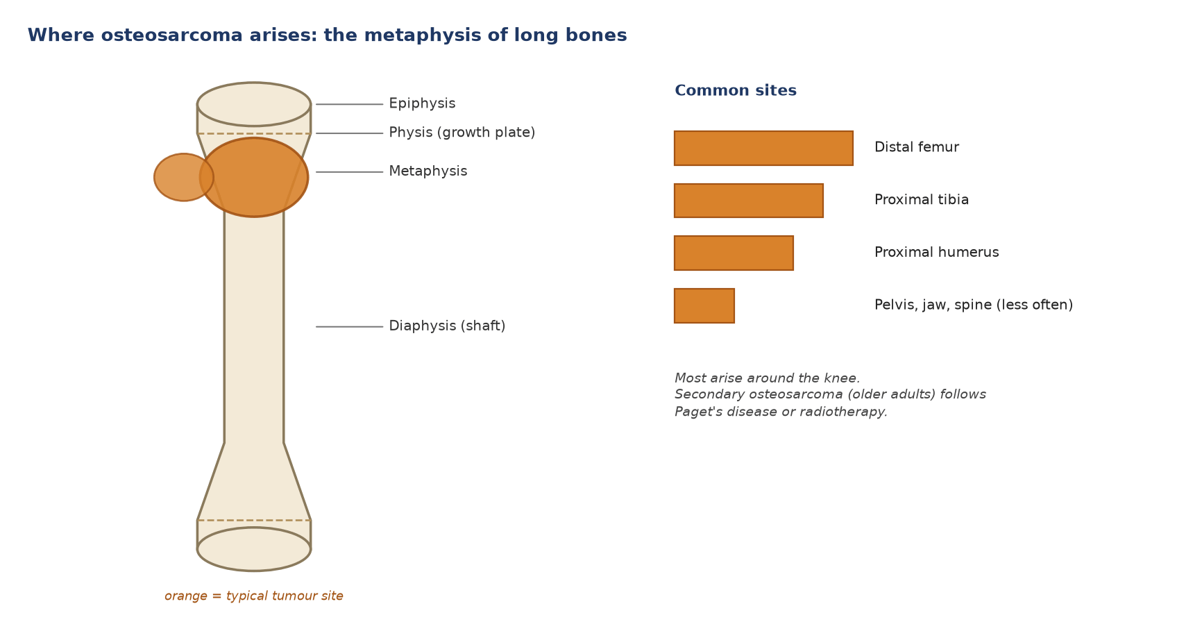
- A malignant bone tumour whose cells make osteoid (immature bone); the commonest primary malignant bone tumour.
- Two age peaks: adolescents during the growth spurt, and older adults, in whom secondary osteosarcoma follows Paget's disease or radiotherapy.
- Risk factors: Li–Fraumeni syndrome (TP53), hereditary retinoblastoma (RB1), Paget's disease, previous radiotherapy.
| Type | Key point |
|---|---|
| Conventional (high grade): osteoblastic, chondroblastic, fibroblastic | Most cases; intensely FDG avid |
| Telangiectatic | Blood-filled spaces with fluid levels; mimics an aneurysmal bone cyst |
| Surface: parosteal and periosteal | Parosteal: low grade, back of the distal femur, low uptake. Periosteal: intermediate grade, tibial or femoral shaft |
| Secondary | Older adults, after Paget's disease or radiotherapy; poor prognosis |
2. Where it arises
3. How the tumour looks on CT
| Feature | What CT shows |
|---|---|
| Site | Metaphysis of a long bone, often around the knee |
| Bone | Mixed lytic and sclerotic destruction with a wide zone of transition |
| Matrix | Cloud-like osteoid mineralisation in the bone and the soft-tissue mass: the key CT sign |
| Cortex and periosteum | Cortical breakthrough; sunburst (spiculated) periosteal reaction; Codman triangle |
| Soft tissue | Mass, often partly mineralised |
| Variants | Telangiectatic: lytic with fluid–fluid levels and little matrix. Parosteal: dense lobulated mass stuck to the back of the distal femur with a thin lucent cleft |
| Lungs | Metastases may calcify or ossify, and can cause pneumothorax |
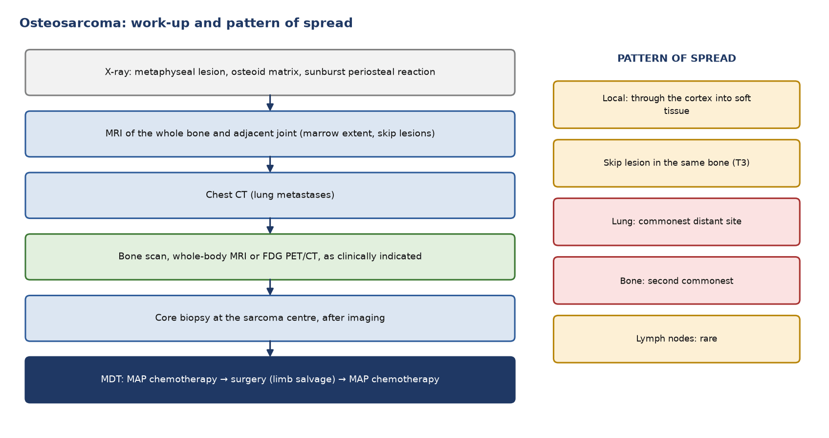
MRI shows the extent in the marrow, skip lesions in the same bone, and the relationship to the joint, vessels and nerves.
4. Work-up and pattern of spread
5. Staging (AJCC 8th edition, appendicular skeleton)
| Category | Definition |
|---|---|
| T1 / T2 | 8 cm or less / more than 8 cm |
| T3 | Discontinuous tumours (skip lesions) in the primary bone |
| N1 | Regional lymph nodes |
| M1a / M1b | Lung / bone or other distant sites |
- Stage groups: IA T1 low grade; IB T2–T3 low grade; IIA T1 high grade; IIB T2 high grade; III T3 high grade; IVA M1a; IVB N1 or M1b.
- Surgeons also use Enneking (MSTS): I low grade, II high grade, III metastatic; A within, B outside the compartment.
6. Indications for PET/CT
| Situation | Role |
|---|---|
| Staging | FDG PET/CT, bone scan or whole-body MRI for bone metastases and skip lesions, as clinically indicated. In a paediatric study PET detected nodal (95% vs 25%) and bone (90% vs 57%) metastases better than conventional imaging |
| Lung nodules | Chest CT decides: PET found only 25% of lung metastases that CT found |
| Biopsy target | The hottest solid part of the tumour |
| Response to neoadjuvant chemotherapy | Prognostic; does not replace histology |
| Suspected recurrence | When MRI is degraded by a prosthesis; whole-body restaging before salvage |
| Routine follow-up | Not indicated |
Other tracers: ⁹⁹ᵐTc-MDP bone scan for skeletal staging; ⁶⁸Ga-FAPI is investigational.
7. Response to chemotherapy
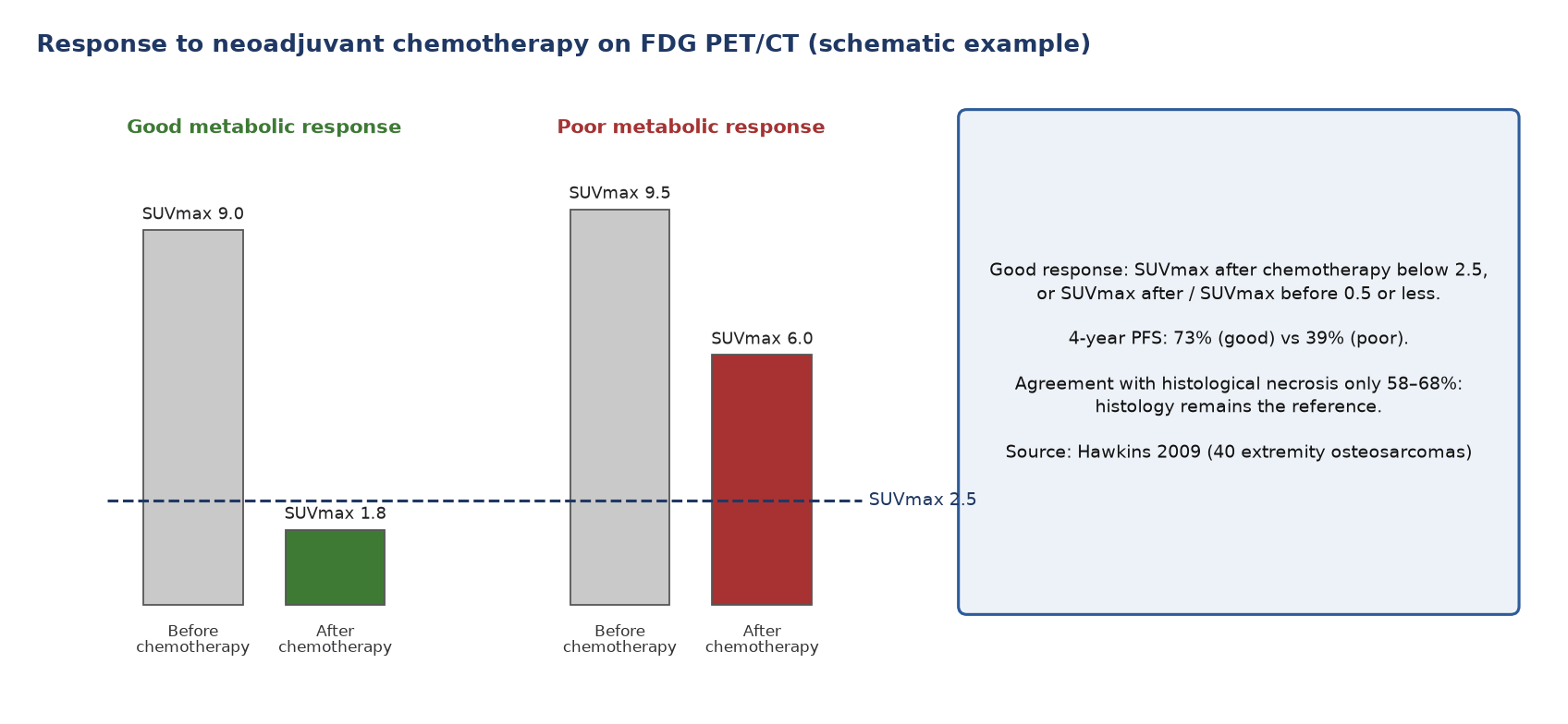
- Outcome: 4-year progression-free survival was 73% with SUVmax after chemotherapy below 2.5, against 39% above it (40 patients).
- Histology stays the reference: 90% or more tumour necrosis is a good response; PET agreed with histology in only 58–68%.
- No validated action yet: in EURAMOS-1, adding ifosfamide and etoposide for poor histological responders did not improve event-free survival.
8. Patient preparation and reporting
- Preparation: scan from vertex to toes including the whole affected limb; inject in the opposite arm; scan before biopsy if possible; keep the patient warm; use the same scanner and uptake time for response scans; record chemotherapy, biopsy, surgery, G-CSF and any prosthesis.
- Report: the hottest solid part and its SUVmax, the soft-tissue mass, skip lesions, lungs on CT, bone metastases, nodes (rare), the stage, and for response the SUVmax before and after chemotherapy and their ratio.
9. Pitfalls
| Pitfall | Why it misleads |
|---|---|
| Physes in adolescents | Symmetrical bands of uptake |
| Giant cell tumour, chondroblastoma, aneurysmal bone cyst, Langerhans cell histiocytosis | Benign but can be hot (giant cell tumours averaged SUV 4.6) |
| Healing fracture, biopsy track, postoperative change | Inflammation |
| G-CSF | Diffuse marrow uptake |
| Small lung nodules | FDG-negative but may be metastases |
10. When to do PET/CT: what the guidelines say
| Time point | Recommendation | Guideline |
|---|---|---|
| Staging | Chest CT plus bone scan and/or whole-body MRI and/or FDG PET/CT, as clinically indicated | ESMO–EURACAN–GENTURIS–ERN PaedCan 2021 |
| Response | No PET recommendation; histological necrosis is the reference | ESMO 2021 |
| Follow-up | Clinical review, imaging of the primary site and chest imaging; no routine PET | ESMO 2021 |
Test yourself
2 quick questions. Pick an answer to see the explanation.
1. A 17-year-old with distal femoral osteosarcoma has SUVmax 8.4 before and 2.1 after neoadjuvant MAP chemotherapy. The resected specimen shows 70% necrosis. How should these be reconciled?
2. Staging PET/CT for an osteosarcoma shows three 4-mm lung nodules without FDG uptake. What is the correct interpretation?
References
- Strauss SJ, Frezza AM, Abecassis N, et al. Bone sarcomas: ESMO-EURACAN-GENTURIS-ERN PaedCan Clinical Practice Guideline for diagnosis, treatment and follow-up. Ann Oncol. 2021;32(12):1520-36.
- Tanaka K, Ozaki T. New TNM classification (AJCC eighth edition) of bone and soft tissue sarcomas: JCOG Bone and Soft Tissue Tumor Study Group. Jpn J Clin Oncol. 2019;49(2):103-7.
- Völker T, Denecke T, Steffen I, et al. Positron emission tomography for staging of pediatric sarcoma patients: results of a prospective multicenter trial. J Clin Oncol. 2007;25(34):5435-41.
- Hawkins DS, Conrad EU, Butrynski JE, et al. [F-18]-fluorodeoxy-D-glucose-positron emission tomography response is associated with outcome for extremity osteosarcoma in children and young adults. Cancer. 2009;115(15):3519-25.
- Marina NM, Smeland S, Bielack SS, et al. Comparison of MAPIE versus MAP in patients with a poor response to preoperative chemotherapy for newly diagnosed high-grade osteosarcoma (EURAMOS-1): an open-label, international, randomised controlled trial. Lancet Oncol. 2016;17(10):1396-408.
- Aoki J, Watanabe H, Shinozaki T, et al. FDG PET of primary benign and malignant bone tumors: standardized uptake value in 52 lesions. Radiology. 2001;219(3):774-7.
- Boellaard R, Delgado-Bolton R, Oyen WJG, et al. FDG PET/CT: EANM procedure guidelines for tumour imaging: version 2.0. Eur J Nucl Med Mol Imaging. 2015;42(2):328-54.
Spotted an error or something out of date? Report an error