Nuclear Medicine in Bladder, Upper-Tract and Kidney Cancer
- Urine is the enemy. FDG is excreted in urine, so the bladder and upper-tract primary are usually unreadable on standard images. Forced diuresis with oral hydration and delayed pelvic images unmask bladder-wall and perivesical disease.
- Nodes: specific, not sensitive. In meta-analyses FDG PET/CT detects nodal metastases from bladder cancer with sensitivity of about 50–57% and specificity of about 91–92%. A negative scan does not replace lymph node dissection; a positive node is usually real.
- Distant staging is where FDG earns its place. In 711 patients with invasive bladder cancer, FDG PET/CT changed the stage in 26% and the treatment plan in 18%, and moved 9% from curative to palliative treatment. The EAU guideline rates this as level 2b evidence but still mandates CT for staging.
- Upper tract. The EAU upper-tract guideline states that FDG PET/CT may be used to rule out metastases in high-risk disease (strong rating).
- Kidney: FDG is the wrong first tracer. Clear-cell RCC is often only mildly FDG-avid (pooled sensitivity 62% for the renal primary). The EAU advises against FDG PET and bone scan for primary staging; high SUVmax in advanced disease carries a poor prognosis.
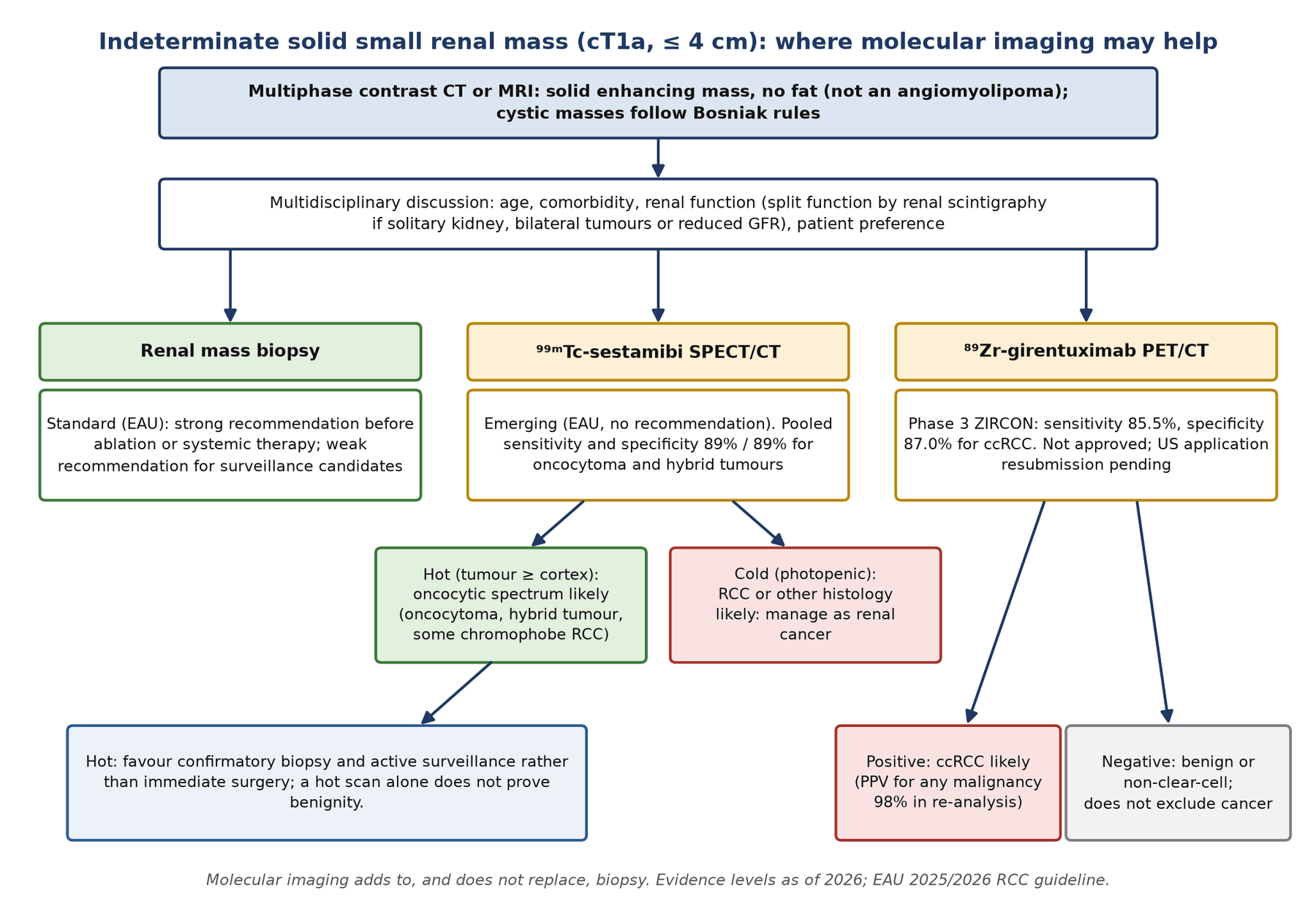
- Sestamibi for the small renal mass. Oncocytomas and hybrid oncocytic tumours take up ⁹⁹ᵐTc-sestamibi (mitochondria-rich); most RCCs are photopenic. Pooled sensitivity and specificity are both about 89%, but the evidence is low-grade and hot tumours still need biopsy.
- CAIX PET is close. ⁸⁹Zr-girentuximab PET/CT identified clear-cell RCC with sensitivity 85.5% and specificity 87.0% in the phase 3 ZIRCON trial. As of September 2026 it is not approved; the US application awaits resubmission after a complete response letter.
- Function matters too. Renal scintigraphy gives split function before nephrectomy when renal function is reduced or critical, and measured GFR can settle cisplatin eligibility near the 60 mL/min threshold.
1. The clinical problem
This article covers three related cancers that share one imaging obstacle, the urinary tract itself: urothelial carcinoma of the bladder, upper tract urothelial carcinoma (UTUC) of the renal pelvis and ureter, and renal cell carcinoma (RCC). Anatomical imaging (CT urography, multiphase CT and MRI) and cystoscopy or ureteroscopy remain the backbone of diagnosis. Nuclear medicine answers narrower questions: are there nodal or distant metastases before radical surgery, is a small renal mass likely benign, how much function will the patient lose with a nephrectomy, and is the kidney good enough for cisplatin?
What the multidisciplinary team needs
- Muscle-invasive bladder cancer (MIBC): local stage (if MRI is used it should be done before transurethral resection of bladder tumour, TURBT), nodes and distant metastases before radical cystectomy or trimodality therapy, and eligibility for cisplatin-based neoadjuvant chemotherapy. Cisplatin-ineligible patients now have an effective perioperative option: in KEYNOTE-905 (344 patients), enfortumab vedotin plus pembrolizumab around cystectomy raised 2-year event-free survival from 39.4% to 74.7% (hazard ratio 0.40) and pathological complete response from 8.6% to 57.1%.
- UTUC: risk stratification decides between kidney-sparing treatment and radical nephroureterectomy; high-risk disease needs systemic staging, and a chest CT is mandatory.
- RCC: most tumours are found incidentally as small renal masses. The central question is benign versus malignant and indolent versus aggressive, because a meaningful fraction of resected masses is benign: in a multicentre series of 542 resected small renal masses, benign pathology was found in 16% where biopsy was not used and 5% where it was.
- Advanced RCC: extent of disease, prognosis and response, where CT remains the standard and PET is adjunctive.
Staging systems in force
The EAU muscle-invasive bladder cancer guideline (2026 online edition) uses the TNM 9th edition (2025). The nodal categories are anatomical and matter for PET reporting. The EAU renal cell carcinoma guideline uses TNM 2017 (8th edition), in which T1a is a tumour of 4 cm or less and T1b more than 4 cm but no more than 7 cm, confined to the kidney.
| Category | Bladder (TNM 9th edition) | What PET must show |
|---|---|---|
| N1 | Single node in the true pelvis (hypogastric, obturator, external iliac or presacral) | One avid node below the common iliac bifurcation |
| N2 | Multiple nodes in the true pelvis | Count and name each station |
| N3 | Common iliac node(s) | Name the common iliac chain separately |
| M1a | Non-regional lymph nodes (for example para-aortic, inguinal) | Para-aortic nodes are M1a, not N3 |
| M1b | Other distant metastases (bone, lung, liver) | Biopsy a solitary PET-only lesion if it changes intent |
Kidney (TNM 2017): N1 is metastasis in any regional node; there is no N2.
Where nuclear medicine changes management
- Finding distant or non-regional nodal disease that turns cystectomy into systemic treatment.
- Characterising a small renal mass as probably oncocytic (sestamibi) or probably clear-cell (CAIX PET) so that surveillance, biopsy or surgery can be chosen with more confidence.
- Measuring split renal function and GFR, which decide nephron-sparing surgery and cisplatin eligibility.
2. Tracers and why they work
| Tracer | Target or mechanism | Where it helps | Main weakness |
|---|---|---|---|
| ¹⁸F-FDG | Glucose transport and hexokinase | Urothelial nodes and distant disease; aggressive or advanced RCC | Urinary excretion; low avidity of many clear-cell RCCs |
| ⁹⁹ᵐTc-sestamibi (SPECT/CT) | Lipophilic cation held by mitochondria; pumped out by multidrug-resistance proteins | Oncocytoma and hybrid oncocytic/chromophobe tumours (hot) | Chromophobe RCC can also be hot; low-grade evidence |
| ⁸⁹Zr-girentuximab (PET/CT) | Carbonic anhydrase IX (CAIX), driven by VHL loss in clear-cell RCC | Clear-cell RCC in an indeterminate mass | Not approved; imaging 5 days after injection; some non-clear-cell RCC express CAIX |
| PSMA ligands | PSMA on tumour neovasculature | Metastatic or recurrent clear-cell RCC | Investigational; heavy renal and urinary activity |
| ⁹⁹ᵐTc-DMSA, ⁹⁹ᵐTc-MAG3 | Tubular fixation (DMSA) or secretion (MAG3) | Split renal function before nephrectomy | Relative, not absolute, function |
| ⁵¹Cr-EDTA or ⁹⁹ᵐTc-DTPA plasma clearance | Glomerular filtration | Measured GFR before cisplatin | Needs timed blood samples |
VHL, von Hippel–Lindau tumour suppressor. PSMA, prostate-specific membrane antigen.
FDG in urothelial cancer
Urothelial carcinoma is usually FDG-avid, but unlike most tracers FDG is not reabsorbed after glomerular filtration, so it collects in the renal pelves, ureters and bladder. Tumour in the bladder wall is swamped by urine activity and blurred by the partial-volume effect, and a hot ureter lying next to an iliac vessel can mimic a node or hide one. Three strategies help: forced diuresis (intravenous furosemide with oral hydration) followed by delayed pelvic images, bladder priming or dual-time-point protocols, and reading with contrast-enhanced CT. A 2025 meta-analysis of 25 studies found that studies using bladder priming, dual-time-point imaging or contrast CT had better nodal sensitivity and specificity than standard protocols, and that PET/MRI (pooled sensitivity 90%, three studies) outperformed PET/CT (69%, five studies) for the primary tumour.
FDG in kidney cancer
Many clear-cell RCCs have only modest glucose metabolism and sit in a kidney that is itself excreting FDG, so the primary is often missed. A meta-analysis of 14 studies found pooled sensitivity and specificity of 62% and 88% for renal lesions but 79% and 90% for extrarenal lesions, rising to 91% and 88% with PET/CT. Uptake rises with grade, and in advanced RCC the highest SUVmax in a patient is prognostic: in 101 patients before systemic therapy, median overall survival was 41.9 months when the maximum SUVmax was below 7.0, 20.6 months between 7.0 and 12.0, and 4.2 months at 12.0 or above.
Sestamibi and the oncocytic tumours
Oncocytomas are packed with normal-appearing mitochondria that trap the cationic ⁹⁹ᵐTc-sestamibi. Most RCCs, particularly those from the proximal tubule, express multidrug-resistance pumps that export it, so they appear photopenic against the avid renal cortex. In the first SPECT/CT series oncocytomas had tumour-to-cortex ratios of 0.85–1.78 and RCCs 0.21–0.31. Hybrid oncocytic/chromophobe tumours (HOCT) and some chromophobe RCCs share the oncocytic phenotype and are also hot, which is the method's main limitation.
CAIX and PSMA
CAIX is a hypoxia-regulated surface enzyme that is constitutively expressed in clear-cell RCC because VHL loss stabilises hypoxia-inducible factor. Girentuximab is a chimeric antibody against CAIX; labelled with ⁸⁹Zr (half-life 78.4 h) it can be imaged after the several days an intact antibody needs to clear from blood. PSMA, despite its name, is expressed on the neovasculature of many solid tumours, including clear-cell RCC: a meta-analysis of nine studies (152 patients) found a pooled lesion detection rate of 0.83 overall, 0.74 for the primary and 0.87 for metastatic or recurrent disease.
3. Indications by clinical scenario
| Scenario | Test | Guideline position (strength) | Practical comment |
|---|---|---|---|
| MIBC, staging before cystectomy or trimodality therapy | FDG PET/CT | EAU MIBC: CT chest, abdomen and pelvis with CT urography is the standard (strong). FDG PET/CT 'can provide additional information to guide treatment' (level of evidence 2b); no formal recommendation. ACR: FDG PET/CT may be appropriate for nodal and metastatic disease | Most useful in cT3–4, cN+ or equivocal CT; use a diuretic protocol |
| Clinically node-negative MIBC | FDG PET/CT | Not routinely justified for nodal staging alone (EAU text, based on PURE-01) | Up to 20% of occult (micro)metastases are still missed |
| High-risk UTUC | FDG PET/CT | EAU UTUC: 'may be used to rule out metastases in high-risk disease' (strong) | Also an option when iodinated contrast is contraindicated |
| Response to neoadjuvant therapy (bladder) | FDG PET/CT | EAU: MRI to assess local response (weak); no PET recommendation | FDG did not beat contrast CT in a prospective study |
| Suspected recurrence after cystectomy | FDG PET/CT | No specific recommendation; CT is standard follow-up | Diversions (conduit, neobladder) are full of urine: read with CT |
| RCC, primary staging | FDG PET/CT, bone scan | EAU RCC: do not use bone scan and/or FDG PET/CT for primary staging (weak) | Routine bone imaging is not indicated; most bone metastases are symptomatic |
| Indeterminate small renal mass | ⁹⁹ᵐTc-sestamibi SPECT/CT; ⁸⁹Zr-girentuximab PET/CT | EAU RCC: 'emerging technologies' with a growing evidence base; no recommendation | Use within a biopsy and surveillance pathway (Figure 1) |
| Metastatic or recurrent RCC | FDG or PSMA PET/CT | No guideline recommendation | FDG SUVmax is prognostic; PSMA detects more ccRCC lesions in small series |
| Before nephrectomy or partial nephrectomy | Renal scintigraphy (DMSA or MAG3) | EAU RCC: estimate split function when renal function is compromised or clinically important, for example a solitary kidney or multiple or bilateral tumours (level 2b) | Predicts the function left after surgery |
| Before cisplatin | Measured GFR | Cisplatin-unfit definition includes creatinine clearance below 60 mL/min (Galsky consensus) | Measure when an estimate is close to the threshold |
EAU guidelines 2025 summaries and 2026 online editions. Strength ratings are those printed in the guideline.
- The EAU upper-tract guideline gives FDG PET/CT a strong rating to rule out metastases in high-risk UTUC, while the bladder guideline, based on a larger literature, gives no recommendation and requires CT. The difference reflects guideline wording rather than better evidence in UTUC, where the data are retrospective.
- The ACR rates FDG PET/CT as possibly appropriate for pretreatment staging of MIBC; the EAU considers its role still to be defined. In practice PET/CT is added when CT is equivocal or the tumour is locally advanced.
4. Evidence at a glance
| Study | Design and size | Key finding | What it changed |
|---|---|---|---|
| Ha et al., 2018 | Meta-analysis, 14 studies, 785 patients with new bladder cancer | Nodal sensitivity 57%, specificity 92%; LR+ 7.4, LR− 0.47 | Defined FDG as specific but insensitive for nodes; cited by the EAU |
| Abdlkadir et al., 2025 | Meta-analysis, 25 studies (18 on nodes) | Nodal sensitivity 50%, specificity 91%; priming, dual-time-point and contrast CT protocols performed better | Protocol matters; standardise diuresis |
| Voskuilen et al., 2022 | Retrospective, 711 invasive bladder cancers with CT and FDG PET/CT | Stage changed in 26%, treatment in 18%; curative to palliative in 9.1%; second primary confirmed in 3.9%; false-positive secondary findings in 8.1% | Main evidence for distant staging (EAU level 2b) |
| Anjos et al., 2007 | Prospective, 17 patients, delayed pelvic images after IV furosemide and oral hydration | 7 of 17 (41%) upstaged only on delayed images | Established the diuretic protocol |
| PURE-01 PET substudy (Marandino et al., 2021) | 108 cT2–4aN0 patients before and 105 after neoadjuvant pembrolizumab | Nodal sensitivity 27% before and 37.5% after; specificity 97–98% | No routine PET for cN0 nodal staging |
| Einerhand et al., 2023 | Prospective, 83 patients during neoadjuvant or induction chemotherapy | Accuracy for complete response 73% (PET) versus 78% (CT) | PET not better than CT for response |
| Voskuilen et al., 2020 (UTUC) | Multicentre retrospective, 117 patients, 62 with node dissection | Nodal sensitivity 82%, specificity 84%; PET-positive nodes predicted worse recurrence-free survival | Basis of the EAU UTUC statement |
| Wang et al., 2012 | Meta-analysis, 14 studies of RCC | Renal lesions: sensitivity 62%, specificity 88%; extrarenal with PET/CT: 91% and 88% | FDG not for the primary |
| Gorin et al., 2016 | Prospective, 50 cT1 renal masses, sestamibi SPECT/CT before surgery | Oncocytoma or HOCT: sensitivity 87.5%, specificity 95.2% | Proof of concept in a blinded prospective series |
| Basile et al., 2024 | Meta-analysis, 8 studies, 489 patients, 501 masses | Sensitivity 89%, specificity 89%; specificity 98% against clear-cell and papillary RCC | Low-certainty evidence; research use advised |
| ZIRCON (Shuch et al., 2024) | Phase 3, 36 centres, 300 dosed, 284 evaluable, cT1 masses up to 7 cm | Clear-cell RCC: mean sensitivity 85.5%, specificity 87.0% (three blinded readers) | Basis of the US licence application |
| KEYNOTE-905 (2026) | Phase 3, 344 cisplatin-ineligible or declining MIBC | Perioperative enfortumab vedotin–pembrolizumab: 2-year EFS 74.7% vs 39.4%, pCR 57.1% vs 8.6% | New standard; response imaging criteria not yet defined |
LR, likelihood ratio. EFS, event-free survival. pCR, pathological complete response.
5. Patient preparation and acquisition
Standard FDG preparation applies (fasting, glucose check, 60-minute uptake). What differs is the handling of urine and, for the renal tracers, the timing.
Diuretic FDG protocol for bladder and upper tract
- Whole-body acquisition at about 60 minutes as usual; the patient voids immediately before.
- Give intravenous furosemide and encourage oral hydration (in the original protocol images were acquired 1 hour later; a 2025 prospective series used 20 mg furosemide).
- Ask the patient to void again just before a delayed acquisition of the pelvis (and abdomen for UTUC).
- Read the delayed images for bladder-wall, perivesical, ureteric and pelvic nodal uptake; compare SUV between phases (malignant lesions usually retain or gain uptake).
- Check contraindications to furosemide (hypotension, dehydration, sulphonamide allergy, outlet obstruction or retention) and warn the patient about urgency. Patients with a catheter can have the bag emptied and the catheter flushed instead.
⁹⁹ᵐTc-sestamibi SPECT/CT of the kidney
- Johns Hopkins protocol: about 6 hours fasting; 925 MBq ⁹⁹ᵐTc-sestamibi intravenously; optional 30-minute dynamic planar images; SPECT/CT of the kidneys at about 75 minutes, when tumour-to-cortex contrast is better than immediately after injection.
- Correlate: use the diagnostic multiphase CT or MRI to place the region of interest on the mass; the low-dose CT may not show an isodense lesion.
⁸⁹Zr-girentuximab PET/CT
- ZIRCON protocol: a single intravenous dose of 37 MBq (±10%) with 10 mg antibody, and abdominal PET/CT 5 days (±2 days) later. Two visits are needed, and ⁸⁹Zr (78.4-hour half-life) requires attention to radiation protection for the days in between.
Renal function studies
- Split function: ⁹⁹ᵐTc-DMSA static imaging measures relative functioning cortical mass; ⁹⁹ᵐTc-MAG3 renography gives relative function plus drainage, which helps when a tumour or clot obstructs a collecting system. Either can predict function after nephrectomy (see the renal articles on this site).
- GFR: plasma clearance of ⁵¹Cr-EDTA or ⁹⁹ᵐTc-DTPA with timed blood samples is the reference; creatinine-based equations are least reliable in older, sarcopenic or post-nephrectomy patients, exactly those in whom cisplatin eligibility is borderline.
6. Interpretation
FDG in urothelial cancer
- No validated score: there is no Deauville-like scale for urothelial cancer. Nodes are called positive when uptake is clearly above blood pool and adjacent background and the structure is a node on CT, regardless of size; many metastatic nodes are under 10 mm.
- Name the station: external iliac, internal iliac (hypogastric), obturator and presacral nodes are N1–N2; common iliac nodes are N3; para-aortic, paracaval and inguinal nodes are M1a.
- Trace every ureter: follow the ureter on consecutive CT slices before calling a para-aortic or common iliac focus a node. Delayed post-diuretic images separate peristaltic urine from fixed nodal uptake.
- Distant disease: lung, bone, liver and peritoneum. The positive predictive value for distant lesions is high, but a solitary PET-only lesion that would change intent should be biopsied.
- Second primaries: in the 711-patient series, 15% had lesions suspicious for a second primary; 3.9% were confirmed. Report them, but expect false positives.
Sestamibi SPECT/CT
- Visual read: a mass with uptake equal to or greater than the adjacent normal renal cortex is sestamibi-positive; a photopenic mass is negative.
- Semi-quantitative: the tumour-to-normal-parenchyma uptake ratio; the studies pooled in the 2020 meta-analysis all used a ratio above 0.6 as positive.
- What positive means: an oncocytic tumour is likely (oncocytoma, HOCT, some chromophobe RCC). Specificity against chromophobe RCC alone was only 67% in that meta-analysis, so a hot mass is a reason for biopsy and surveillance, not proof of benignity.
- What negative means: RCC or another non-oncocytic histology is likely; some oncocytomas are cold.
⁸⁹Zr-girentuximab PET/CT
- ZIRCON used blinded visual reads by three independent readers; there is no validated SUV threshold. Intense uptake in the mass relative to normal kidney indicates CAIX expression and a high probability of clear-cell RCC.
- A re-analysis of ZIRCON found that some non-clear-cell RCCs, particularly papillary tumours with higher CAIX expression, were also positive; for any renal malignancy the positive predictive value was 98% with sensitivity 82% and specificity 87%.
- A negative scan argues against clear-cell RCC but does not exclude a non-clear-cell cancer.
Pitfalls and mimics
| Finding | Cause | How to tell |
|---|---|---|
| Bladder wall or diverticulum hidden or 'hot' | Urine activity, partial volume | Diuretic delayed images; contrast CT; MRI for local stage |
| Focal pelvic or para-aortic 'node' | Ureteric urine, ureteric kink or stasis, hydroureter | Trace the ureter on CT; delayed images; activity moves or clears |
| Bladder-wall uptake after TURBT | Post-resection inflammation | Know the resection date; stage the bladder with MRI before TURBT |
| Focal or multifocal prostate uptake after intravesical BCG | BCG granulomatous prostatitis | Seen in 35% after BCG versus 1% without; commoner within 1 year; median SUVmax 4.7; regressed without treatment in 14 of 16 |
| Avid nodes, lung or vascular lesions after BCG | Granulomatous inflammation or disseminated BCG infection (rare) | History of BCG and symptoms; biopsy with mycobacterial culture |
| Uptake in thyroid, stomach, lung or mediastinal nodes during checkpoint inhibitors | Immune-related inflammation | Pattern and timing; correlate with symptoms and laboratory tests |
| Ileal conduit or neobladder activity | Urine in the diversion | Read with CT; delayed images after drainage |
| Photopenic renal lesion on FDG or sestamibi | Simple or complex cyst, hydronephrotic calyx, angiomyolipoma | Correlate with CT or MRI; only solid enhancing masses should be characterised |
| FDG-negative renal mass | Low-grade clear-cell RCC | Does not exclude cancer |
| Sestamibi-positive renal mass | Oncocytoma, HOCT, but also chromophobe RCC | Biopsy before surveillance |
BCG, bacillus Calmette–Guérin. HOCT, hybrid oncocytic/chromophobe tumour.
Wording the conclusion
- Give the stage consequence: "FDG-avid left common iliac node (N3) and a left para-aortic node (non-regional, M1a)".
- State the limits: "a negative PET does not exclude micrometastatic nodal disease"; "the bladder primary cannot be assessed on the early images".
- For a small renal mass, give probability, not diagnosis: "sestamibi-positive mass; appearances favour an oncocytic tumour (oncocytoma or hybrid oncocytic tumour); chromophobe RCC is not excluded. Biopsy is recommended if surveillance is considered."
7. Response assessment and follow-up
- Neoadjuvant chemotherapy: in a prospective study of 83 patients imaged after two to three cycles, FDG PET/CT predicted complete response with accuracy 73% against 78% for contrast CT, and CT was more specific for downstaging; the authors concluded that routine FDG PET/CT adds little. MRI is the EAU choice for local response (weak).
- Checkpoint inhibitors: in PURE-01, PET after neoadjuvant pembrolizumab found nodal disease with sensitivity 37.5% and specificity 98%. Inflammatory uptake in the thyroid, stomach, lung and mediastinal nodes is common and must not be read as progression.
- Enfortumab vedotin–pembrolizumab: KEYNOTE-905 established perioperative use in cisplatin-ineligible disease with a pathological complete response rate of 57.1%. No PET response criterion has been validated for this regimen or for urothelial cancer generally; if FDG is used, report quantitative change against a baseline acquired with the same protocol and expect immune-related inflammatory uptake.
- Metastatic urothelial cancer: response is assessed with CT and RECIST 1.1 in trials; PET can clarify equivocal findings.
- RCC: CT and RECIST 1.1 remain standard. FDG SUVmax before systemic therapy is prognostic; PSMA PET may show more lesions than CT in metastatic clear-cell RCC but has no validated response criteria.
- Follow-up after cystectomy: CT-based. PET is a problem-solving tool for equivocal CT, remembering that the urinary diversion is full of FDG.
8. Theranostics and emerging tracers
| Agent | Target | Evidence level (September 2026) |
|---|---|---|
| ⁸⁹Zr-girentuximab PET/CT (TLX250-CDx) | CAIX | Phase 3 positive (ZIRCON). Not approved. A US licence application received a complete response letter in August 2025 over manufacturing (chemistry, manufacturing and controls) comparability; the company reports that resubmission is in preparation (company statements only) |
| ⁹⁹ᵐTc-sestamibi SPECT/CT | Mitochondria | Tracer approved for other uses; renal use is off-label, supported by prospective single-centre data and meta-analyses of low certainty |
| ¹⁷⁷Lu-girentuximab | CAIX | Phase 2 monotherapy: disease stabilisation in 9 of 14 patients, grade 3–4 myelotoxicity common. Trials with nivolumab (phase 2) or cabozantinib plus nivolumab (phase 1b/2) and a randomised phase 3 against investigator's choice (registered 2026) are ongoing |
| PSMA PET (⁶⁸Ga or ¹⁸F ligands) | Neovascular PSMA | Investigational in RCC; meta-analysed detection rate 0.83 |
| Nectin-4 PET (for example ⁶⁸Ga-N188) | Nectin-4, the enfortumab vedotin target | First-in-human and small series; shows heterogeneous expression between lesions. Investigational |
| FAPI PET | Fibroblast activation protein | 22 small retrospective reports (69 patients) across genitourinary cancers; urinary excretion limits the bladder and upper-tract primary. Investigational |
No radioligand therapy is approved for urothelial or renal cancer.
The logic of CAIX theranostics is attractive: the same antibody that identifies clear-cell RCC can carry ¹⁷⁷Lu. The limitation is also the antibody's: long blood residence irradiates the marrow, which is why myelotoxicity limited retreatment in the phase 2 trial. Nectin-4 imaging addresses a practical question raised by enfortumab vedotin, namely whether all lesions express the target, but there are no outcome data.
9. Structured report example
- Clinical question: 68-year-old man, high-grade urothelial carcinoma with muscle invasion at TURBT (3 weeks ago); CT urography shows a 4 cm left lateral wall tumour and a 9 mm left external iliac node. Staging before planned neoadjuvant chemotherapy and cystectomy.
- Technique: 3.5 MBq/kg ¹⁸F-FDG, glucose 5.9 mmol/L; skull base to mid-thigh at 62 min. Furosemide intravenously with oral hydration; the patient voided, and delayed pelvic images were acquired 60 min later. eGFR 58 mL/min.
- Bladder: early images non-diagnostic because of urine activity. On delayed images, focal uptake in the thickened left lateral wall (SUVmax 11.4) extending into perivesical fat, and mild diffuse uptake at the resection site.
- Nodes: left external iliac (9 mm, SUVmax 7.2) and left obturator (7 mm, SUVmax 5.9) nodes; left common iliac node (8 mm, SUVmax 6.4) separate from the ureter on CT and persisting on delayed images; left para-aortic node below the renal vein (10 mm, SUVmax 6.8).
- Distant: 8 mm FDG-avid nodule in the right lower lobe (SUVmax 5.1). No bone or liver lesions.
- Conclusion: Left lateral wall bladder primary with probable perivesical extension. Nodal disease in the pelvis including the common iliac chain (N3) and a para-aortic node, which is non-regional (M1a); a right lower lobe nodule is suspicious for M1b. Tissue confirmation of the para-aortic node or lung nodule is advised, as either would change treatment intent. eGFR is close to the cisplatin threshold: measured GFR is suggested if cisplatin remains an option.
10. Take-home points
- Use a diuretic protocol for any urothelial FDG study: furosemide, oral hydration, voiding and delayed pelvic images.
- FDG nodal staging in bladder cancer is specific (about 91–92%) but insensitive (about 50–57%): a negative scan does not replace node dissection.
- The biggest yield is distant disease: in 711 patients FDG changed treatment in 18%. EAU still requires CT; PET is level 2b add-on evidence.
- Para-aortic nodes are M1a, common iliac nodes N3: name the station.
- Do not use FDG or bone scan for primary staging of RCC (EAU, weak). High SUVmax in advanced RCC predicts poor survival.
- Sestamibi-hot small renal masses are probably oncocytic, but chromophobe RCC overlaps: biopsy before surveillance.
- ⁸⁹Zr-girentuximab PET identifies clear-cell RCC with about 86% sensitivity and 87% specificity; it is not yet approved.
- Measure split function before nephrectomy when renal function is reduced or critical, and measured GFR when cisplatin eligibility is borderline.
Test yourself
5 quick questions. Pick an answer to see the explanation.
References
- van der Heijden AG, Bruins HM, Carrion A, et al. European Association of Urology guidelines on muscle-invasive and metastatic bladder cancer: summary of the 2025 guidelines. Eur Urol. 2025;87(5):582-600.
- Masson-Lecomte A, Birtle A, Pradere B, et al. European Association of Urology guidelines on upper urinary tract urothelial carcinoma: summary of the 2025 update. Eur Urol. 2025;87(6):697-716.
- Bex A, Ghanem YA, Albiges L, et al. European Association of Urology guidelines on renal cell carcinoma: the 2025 update. Eur Urol. 2025;87(6):683-96.
- Ha HK, Koo PJ, Kim SJ. Diagnostic accuracy of F-18 FDG PET/CT for preoperative lymph node staging in newly diagnosed bladder cancer patients: a systematic review and meta-analysis. Oncology. 2018;95(1):31-8.
- Abdlkadir AS, Al-Adhami D, Allouzi S, et al. Diagnostic efficacy of [18F]FDG PET/CT and [18F]FDG PET/MRI in preoperative staging of locoregional urinary bladder cancer: a systematic review and meta-analysis. Discov Oncol. 2025;16(1):1241.
- Voskuilen CS, van Gennep EJ, Einerhand SMH, et al. Staging 18F-fluorodeoxyglucose positron emission tomography/computed tomography changes treatment recommendation in invasive bladder cancer. Eur Urol Oncol. 2022;5(3):366-9.
- Anjos DA, Etchebehere EC, Ramos CD, et al. 18F-FDG PET/CT delayed images after diuretic for restaging invasive bladder cancer. J Nucl Med. 2007;48(5):764-70.
- Marandino L, Capozza A, Bandini M, et al. [18F]Fluoro-deoxy-glucose positron emission tomography to evaluate lymph node involvement in patients with muscle-invasive bladder cancer receiving neoadjuvant pembrolizumab. Urol Oncol. 2021;39(4):235.e15-235.e21.
- Einerhand SMH, Voskuilen CS, van de Putte EEF, et al. Prospective evaluation of FDG-PET/CT for on-treatment assessment of response to neoadjuvant or induction chemotherapy in invasive bladder cancer. Bladder Cancer. 2023;9(1):49-57.
- Voskuilen CS, Schweitzer D, Jensen JB, et al. Diagnostic value of 18F-fluorodeoxyglucose positron emission tomography with computed tomography for lymph node staging in patients with upper tract urothelial carcinoma. Eur Urol Oncol. 2020;3(1):73-9.
- Kim CY, Lee SW, Choi SH, et al. Granulomatous prostatitis after intravesical bacillus Calmette-Guérin instillation therapy: a potential cause of incidental F-18 FDG uptake in the prostate gland on F-18 FDG PET/CT in patients with bladder cancer. Nucl Med Mol Imaging. 2016;50(1):31-7.
- Vulsteke C, Adra N, Danchaivijitr P, et al. Perioperative enfortumab vedotin and pembrolizumab in bladder cancer. N Engl J Med. 2026;394(13):1257-69.
- Galsky MD, Hahn NM, Rosenberg J, et al. Treatment of patients with metastatic urothelial cancer "unfit" for cisplatin-based chemotherapy. J Clin Oncol. 2011;29(17):2432-8.
- Wang HY, Ding HJ, Chen JH, et al. Meta-analysis of the diagnostic performance of [18F]FDG-PET and PET/CT in renal cell carcinoma. Cancer Imaging. 2012;12(3):464-74.
- Nakaigawa N, Kondo K, Tateishi U, et al. FDG PET/CT as a prognostic biomarker in the era of molecular-targeting therapies: max SUVmax predicts survival of patients with advanced renal cell carcinoma. BMC Cancer. 2016;16:67.
- Rowe SP, Gorin MA, Gordetsky J, et al. Initial experience using 99mTc-MIBI SPECT/CT for the differentiation of oncocytoma from renal cell carcinoma. Clin Nucl Med. 2015;40(4):309-13.
- Gorin MA, Rowe SP, Baras AS, et al. Prospective evaluation of (99m)Tc-sestamibi SPECT/CT for the diagnosis of renal oncocytomas and hybrid oncocytic/chromophobe tumors. Eur Urol. 2016;69(3):413-6.
- Wilson MP, Katlariwala P, Murad MH, et al. Diagnostic accuracy of 99mTc-sestamibi SPECT/CT for detecting renal oncocytomas and other benign renal lesions: a systematic review and meta-analysis. Abdom Radiol (NY). 2020;45(8):2532-41.
- Basile G, Fallara G, Verri P, et al. The role of 99mTc-sestamibi single-photon emission computed tomography/computed tomography in the diagnostic pathway for renal masses: a systematic review and meta-analysis. Eur Urol. 2024;85(1):63-71.
- Shuch B, Pantuck AJ, Bernhard JC, et al. [89Zr]Zr-girentuximab for PET-CT imaging of clear-cell renal cell carcinoma: a prospective, open-label, multicentre, phase 3 trial. Lancet Oncol. 2024;25(10):1277-87.
- Kaba A, Kanabur P, Calais J, et al. Performance of [89Zr]girentuximab PET/CT in predicting the presence of any renal malignancy: a reanalysis of the ZIRCON trial. Eur Urol. 2026 (published online 22 April 2026). doi:10.1016/j.eururo.2026.03.003.
- Sadaghiani MS, Baskaran S, Gorin MA, et al. Utility of PSMA PET/CT in staging and restaging of renal cell carcinoma: a systematic review and metaanalysis. J Nucl Med. 2024;65(7):1007-12.
- Muselaers CH, Boers-Sonderen MJ, van Oostenbrugge TJ, et al. Phase 2 study of lutetium 177-labeled anti-carbonic anhydrase IX monoclonal antibody girentuximab in patients with advanced renal cell carcinoma. Eur Urol. 2016;69(5):767-70.
- Duan X, Xia L, Zhang Z, et al. First-in-human study of the radioligand 68Ga-N188 targeting nectin-4 for PET/CT imaging of advanced urothelial carcinoma. Clin Cancer Res. 2023;29(17):3395-407.
- Hagens MJ, van Leeuwen PJ, Wondergem M, et al. A systematic review on the diagnostic value of fibroblast activation protein inhibitor PET/CT in genitourinary cancers. J Nucl Med. 2024;65(6):888-96.