PET/CT in Lung Cancer
- Stage everyone considered for cure with FDG PET/CT. Two randomised trials showed fewer futile thoracotomies: 41% to 21% in PLUS, and 38 to 21 futile operations in the Danish PET-CT trial.
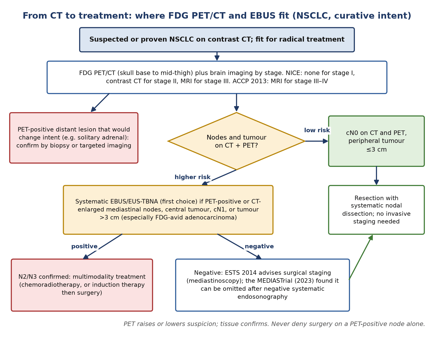
- PET selects the node; tissue decides. PET detects mediastinal metastases with a sensitivity of about 77% and specificity of about 86%, so a PET-positive node is sampled (EBUS/EUS-TBNA first) before anyone is denied surgery.
- A negative mediastinum is not always enough. ESTS 2014 advises invasive staging for central tumours, cN1 disease and tumours over 3 cm even when PET is negative.
- Ninth-edition TNM (2025). N2 is split into N2a (one ipsilateral mediastinal or subcarinal station) and N2b (several stations); M1c into M1c1 (one extrathoracic organ system) and M1c2 (several). Count stations and organ systems in the report.
- Nodules. For a solid nodule of 8 mm or more with a Brock risk of at least 10%, PET-CT feeds the Herder model. Pure ground-glass lesions, adenocarcinoma in situ, mucinous tumours and typical carcinoids are often FDG-negative; granulomas are the classic false positive.
- Brain needs MRI, not PET. Normal cortical glucose uptake hides small metastases.
- Radiotherapy. PET-defined target volumes without elective nodal irradiation were non-inferior in PET-Plan; dose painting to the FDG-avid subvolume (PET-Boost) remains experimental because of severe toxicity.
- Response and follow-up. Read with PERCIST; under immunotherapy confirm apparent progression 4–8 weeks later in a stable patient. After SABR, early uptake is expected; recurrence is suggested by high-risk CT features with SUVmax of 5 or more.
1. The clinical problem
Lung cancer is treated according to three things: histology and molecular profile, anatomical extent, and the patient's physiological reserve. Imaging contributes to all three. Non-small-cell lung cancer (NSCLC), mainly adenocarcinoma and squamous cell carcinoma, is staged with the TNM system and treated by surgery, radical radiotherapy, systemic therapy or combinations. Small-cell lung cancer (SCLC) and the other neuroendocrine tumours (typical and atypical carcinoid, large-cell neuroendocrine carcinoma, LCNEC) behave differently and need different tracers. Driver alterations (EGFR, ALK, ROS1, KRAS G12C and others) and programmed death-ligand 1 (PD-L1) expression are read from tissue or plasma, never from a PET scan.
At the tumour board the questions for the nuclear medicine physician are practical. Is this nodule likely to be cancer? Is the mediastinum involved, and which node should be sampled? Is there a distant metastasis that changes the intent from cure to control? Where exactly is the tumour for the radiotherapy plan? Has it responded, and is a new opacity after treatment scar or recurrence? And, for the surgeon, how much function will the patient lose?
Staging system in force: the ninth edition of TNM
The International Association for the Study of Lung Cancer (IASLC) published its ninth-edition proposals in 2024 from a database of 124,581 registered patients; the Union for International Cancer Control (UICC) and American Joint Committee on Cancer (AJCC) implemented them from January 2025. T categories are unchanged. The changes that matter to a PET reader are in N and M.
| Category | Ninth-edition definition | What the PET/CT report must give |
|---|---|---|
| N1 | Ipsilateral peribronchial, hilar or intrapulmonary nodes | Hilar (10) and interlobar (11) uptake; cN1 triggers invasive mediastinal staging |
| N2a | Single ipsilateral mediastinal or subcarinal nodal station | Name the station (for example 4R or 7) |
| N2b | Multiple ipsilateral mediastinal stations, with or without the subcarinal station | List every avid station; the count changes the stage |
| N3 | Contralateral mediastinal or hilar, or any scalene or supraclavicular node | Flag it first: it is the node to sample |
| M1a | Separate nodule(s) in a contralateral lobe; pleural or pericardial nodules or malignant effusion (unchanged) | Pleural uptake and effusion |
| M1b | Single extrathoracic metastasis in one organ (unchanged) | A solitary lesion needs confirmation if it removes curative options |
| M1c1 | Multiple extrathoracic metastases in one organ system | Count organ systems, not lesions |
| M1c2 | Multiple extrathoracic metastases in multiple organ systems | Both M1c subgroups remain stage IVB |
Stage groups were re-assigned: T1N1 moves to stage IIA, T1N2a to IIB, T3N2a to IIIA, and T2aN2b and T2bN2b to IIIB. The same TNM system is recommended for SCLC, although trials still use the older division into limited disease (confined to one hemithorax and its regional nodes) and extensive disease.
Where nuclear medicine changes management
- Selection for radical treatment: detection of unsuspected nodal or distant disease avoids non-curative surgery.
- Targeting tissue sampling: PET tells the bronchoscopist which station to needle first, starting with the one that would give the highest stage.
- Radiotherapy planning: separating tumour from collapsed lung and defining involved nodes.
- Response and recurrence: metabolic response, atypical responses to immunotherapy, and scar versus recurrence after radiotherapy.
- Fitness for surgery: quantitative perfusion scintigraphy predicts postoperative lung function.
2. Tracers and why they work
¹⁸F-fluorodeoxyglucose (FDG) enters tumour cells through glucose transporters, chiefly GLUT1, is phosphorylated by hexokinase and is trapped. Uptake rises with cell density, proliferation and hypoxia, so it tracks histological grade more than tumour type. Inflammatory cells use the same pathway, which is why infection, granulomas and treatment reactions are the recurring false positives.
| Tumour | Usual FDG avidity | Practical point |
|---|---|---|
| Squamous cell carcinoma | High | Often central with necrosis (photopenic centre) and post-obstructive collapse |
| Solid invasive adenocarcinoma | Moderate to high | Nodal and adrenal metastases are common |
| Adenocarcinoma in situ, minimally invasive and lepidic-predominant adenocarcinoma (ground glass) | Low or absent | PET does not characterise pure ground-glass nodules; CT growth and solid component do |
| Invasive mucinous adenocarcinoma | Often low | Can mimic consolidation; a negative PET is not reassuring |
| Typical carcinoid | Low | Somatostatin receptor (SSTR) PET is the tracer of choice |
| Atypical carcinoid | Variable | Consider both SSTR PET and FDG; FDG avidity suggests more aggressive biology |
| SCLC and LCNEC | High | SSTR expression is often low; FDG stages and plans radiotherapy |
The contrast between the two tracers in neuroendocrine tumours is striking. In a series of 18 pulmonary neuroendocrine tumours, all 11 typical carcinoids had high ⁶⁸Ga-DOTATATE uptake (SUVmax 8.2 or more) but four had negative or minimal FDG uptake (SUVmax 1.7–2.9). All higher-grade tumours were intensely FDG-avid (SUVmax 11.7 or more), while three of five showed only minimal DOTATATE uptake. DOTATATE also separated an endobronchial carcinoid from the collapsed lung beyond it better than FDG.
When FDG fails and what replaces it
- Small nodules: partial-volume loss and respiratory blurring make nodules under about 8 mm unreliable; CT surveillance is used instead.
- Ground glass and lepidic growth: CT morphology and follow-up.
- Carcinoid: SSTR PET with ⁶⁸Ga-DOTATATE or ⁶⁸Ga-DOTATOC.
- Brain: contrast-enhanced MRI.
- Lung function before resection: ⁹⁹ᵐTc-macroaggregated albumin (MAA) perfusion scintigraphy, planar or SPECT/CT.
3. Indications by clinical scenario
| Scenario | Role of nuclear medicine | Guideline position (strength where given) |
|---|---|---|
| Solid nodule of 8 mm or more | FDG PET/CT when the Brock risk is 10% or more and the nodule exceeds the local PET-CT detection threshold; re-estimate risk with the Herder model | BTS 2015. Fleischner 2017: for solid nodules over 8 mm consider CT at 3 months, PET/CT or tissue sampling |
| Subsolid or pure ground-glass nodule | Not a PET indication; CT surveillance of size and solid component | Fleischner 2017 and BTS 2015 |
| NSCLC, candidate for curative treatment | Whole-body FDG PET/CT for nodal and distant staging | ACCP 2013: recommended, with tissue confirmation of any finding suggesting metastasis (grade 1B); NICE NG122: offer PET-CT to everyone who could have curative treatment |
| Mediastinal staging | PET guides which node to sample | ESTS 2014: tissue confirmation of CT-enlarged or PET-positive nodes, EBUS/EUS-FNA first; invasive staging also for central tumours, cN1 and tumours over 3 cm |
| Brain | None: PET is insensitive | NICE: no routine brain imaging in stage I, contrast CT in stage II, MRI in stage III. ACCP 2013: MRI in stage III–IV (grade 2C) |
| Radical radiotherapy | PET/CT defines gross tumour and involved nodes | PET-Plan (randomised) supports PET-based volumes without elective nodal irradiation. SCLC: FDG PET recommended for volume delineation (ESMO 2021, III, A) |
| Limited-stage SCLC | Optional staging; findings that change treatment need confirmation | ESMO 2021: optional (II, C); bone scintigraphy if no PET-CT (V, B) |
| Lung carcinoid | SSTR PET for staging and receptor status | Based on comparative studies of SSTR and FDG PET |
| Response assessment | PERCIST in trials and problem-solving; not a routine test after every cycle | EANM/SNMMI/ANZSNM 2022 for immunotherapy: confirm suspected progression 4–8 weeks later |
| Suspected recurrence | PET/CT after high-risk CT change (for example after SABR); guides biopsy | Systematic review evidence (Huang 2012) |
| Planning resection in borderline function | Quantitative perfusion scintigraphy for predicted postoperative FEV₁ and DLCO | ERS/ESTS 2009; ACCP 2013 |
The solitary pulmonary nodule: Brock, then Herder
The British Thoracic Society (BTS) pathway starts with the Brock model, which uses age, sex, family history, emphysema, nodule size, type, upper-lobe location, number and spiculation. Solid nodules under 8 mm (or under 300 mm³) and nodules with a Brock risk below 10% go to CT surveillance. Above those thresholds PET-CT is offered, and the uptake is graded visually: absent, faint (at or below mediastinal blood pool), moderate (above blood pool) or intense. That grade enters the Herder model, which recalculates the probability of cancer. A Herder risk below 10% returns the patient to surveillance; 10–70% favours image-guided biopsy (or surveillance after discussion); above 70% favours excision or non-surgical treatment. Reading the grade honestly matters more than the SUV: the model was built on visual comparison with blood pool, not on a fixed SUV cut-off.
Two limits should be in every report. First, FDG-negative does not mean benign for ground-glass or part-solid lesions, mucinous tumours or carcinoids. Second, specificity depends on where the patient lives: in a meta-analysis of 70 studies and 8,511 nodules, pooled sensitivity was 89% and specificity 75%, but adjusted specificity fell to 61% in regions with endemic granulomatous infection, against 77% elsewhere.
Preoperative lung function: perfusion scintigraphy
Predicted postoperative FEV₁ (ppoFEV₁) is the preoperative FEV₁ multiplied by one minus the fraction of function that will be removed; the same calculation is made for the carbon monoxide transfer factor (DLCO). For a pneumonectomy the fraction comes from quantitative ⁹⁹ᵐTc-MAA perfusion scintigraphy (the geometric mean of anterior and posterior counts for each lung). For a lobectomy, segment counting is often used, but perfusion SPECT/CT with lobar segmentation is better when disease is uneven, for example central tumour, emphysema or obstruction.
- ACCP 2013: if ppoFEV₁ and ppoDLCO are both above 60% predicted, risk is low and no further tests are needed. If either lies between 30% and 60%, a low-technology exercise test follows (stair climb above 22 m or shuttle walk above 400 m is satisfactory). If either is below 30%, or the simple test is unsatisfactory, cardiopulmonary exercise testing is done: peak oxygen uptake below 10 mL/kg/min or 35% predicted means high risk; above 20 mL/kg/min or 75% predicted, low risk.
- ERS/ESTS 2009: after cardiological assessment, FEV₁ and DLCO above 80% predicted allow resection without further tests; otherwise exercise testing comes first, and split-function (perfusion-based) predicted values are used in the intermediate range, with values below 30% predicted marking high risk.
- On this site: the ppoFEV₁ calculator in Clinical Calculators applies the ACCP 2013 bands to the perfusion fraction you enter.
The two algorithms disagree on order: the European pathway puts formal exercise testing earlier, the American one starts from the predicted postoperative values. Either way, the perfusion fraction is only as good as the regions drawn, so state the method (planar lung, posterior oblique lobar, or SPECT/CT lobar) in the report.
4. Evidence at a glance
| Study | Design and size | Key finding | What it changed |
|---|---|---|---|
| PLUS (van Tinteren 2002) | Randomised, 188 patients, 9 hospitals | Futile thoracotomy 41% with conventional work-up versus 21% with added PET (relative reduction 51%, p=0.003) | PET entered routine preoperative staging |
| Fischer 2009 | Randomised, 189 patients, PET-CT versus conventional staging | Thoracotomies 60 versus 73; futile thoracotomies 21 versus 38 (p=0.05); survival similar | Confirmed the benefit with integrated PET-CT |
| ACCP 2013 evidence review (Silvestri) | Systematic review of staging tests | Mediastinal nodes: PET sensitivity about 77%, specificity 86%; CT 55% and 81%. EBUS-NA, EUS-NA and combined sensitivity about 89%, 89% and 91% | Needle techniques first; all abnormal scans confirmed by tissue |
| Deppen 2014 | Meta-analysis, 70 studies, 8,511 nodules | Sensitivity 89%, specificity 75%; specificity 61% in endemic infectious regions versus 77% elsewhere | Local calibration of PET for nodules |
| ASTER (Annema 2010) | Randomised, 241 patients needing mediastinal staging | Sensitivity for N2/N3: 94% with endosonography then surgical staging versus 79% with surgical staging alone; unnecessary thoracotomy 7% versus 18% | Endosonography became the first invasive test |
| MEDIASTrial (Bousema 2023) | Randomised non-inferiority, 360 patients after negative systematic endosonography | Unforeseen N2 8.8% with immediate resection versus 7.7% with mediastinoscopy first; mediastinoscopy found metastases in 8.0% | Confirmatory mediastinoscopy can be omitted (contrast ESTS 2014) |
| PET-Plan (Nestle 2020) | Randomised, 205 patients (172 per protocol), stage III chemoradiotherapy | 1-year locoregional progression 14% with PET-only volumes versus 29% with conventional volumes plus elective nodal irradiation (HR 0.57); toxicity similar | PET-based target volumes without elective nodal irradiation |
| PET-Boost (Cooke 2023) | Randomised phase II, 107 patients | 1-year freedom from local failure 97% (whole-tumour boost) and 91% (PET-subvolume boost), but 19 grade 5 events (18%) | FDG-guided dose escalation not adopted |
| Huang 2012 | Systematic review, 26 studies after SABR | Early SUV rise is common; SUVmax of 5 or more with high-risk CT change predicts recurrence | Follow-up algorithm after SABR |
| Kayani 2009 | 18 pulmonary neuroendocrine tumours, DOTATATE and FDG | Typical carcinoids SSTR-avid, 4 of 11 FDG-negative or minimal; high-grade tumours FDG-avid | SSTR PET for carcinoid |
5. Patient preparation and acquisition
Standard oncological FDG preparation applies: fasting for at least 4 hours (EANM; SNMMI 4–6 hours), blood glucose checked, uptake time about 60 minutes (55–75 minutes), and identical protocol, scanner and reconstruction for any scan that will be compared quantitatively. The points below are specific to the chest.
- Coverage: skull base to mid-thigh; include the skull base in patients on immunotherapy so hypophysitis is not missed. Extend to the vertex or feet only for a clinical question.
- Arms up when tolerated, to reduce beam-hardening and truncation across the chest and adrenals.
- Respiration: the CT used for attenuation correction should match the averaged PET position, so acquire it in shallow breathing or end-expiration rather than deep inspiration. Mismatch shifts lesions at the lung bases and liver dome. Respiratory-gated or 4D acquisition helps for small basal nodules and is used for radiotherapy planning.
- History to record: recent infection, bronchoscopy or EBUS (nodal reactivity), pleurodesis, prior radiotherapy with dates and fields, immunotherapy cycles, granulocyte colony-stimulating factor, and the site and date of any recent vaccination.
- Radiotherapy planning scans: a recent scan in the treatment position with the planning immobilisation, so that PET and planning CT register without deformation.
- SSTR PET for carcinoid: ⁶⁸Ga-DOTATATE or DOTATOC, acquired to the neuroendocrine tumour protocol, with somatostatin analogue treatment recorded.
6. Interpretation
The primary and the nodule
Describe uptake against the mediastinal blood pool using the four visual grades, give SUVmax as a supporting number, and say whether the lesion is solid, part-solid or ground glass. For a lesion under about 8–10 mm, state that absent uptake is not informative. Where FDG separates tumour from distal collapse or consolidation, say so; radiotherapy planners use it.
Mediastinal and hilar nodes
Report by IASLC station (for example 2R, 4R, 4L, 7, 10R). A node is positive when uptake clearly exceeds mediastinal blood pool, regardless of size; a normal-sized avid node matters more than an enlarged non-avid one. Symmetrical, low-to-moderate hilar and mediastinal uptake, especially in calcified or high-density nodes, suggests granulomatous disease or anthracosis rather than metastasis. List the stations in order of stage significance so the endoscopist samples the potential N3 node first, then N2.
Distant disease
The adrenals, bone, liver and distant nodes are the common sites. Adrenal uptake above liver is suspicious; a solitary adrenal or bone lesion that would change the intent of treatment must be confirmed, by biopsy or by targeted imaging, before cure is abandoned (ACCP 2013, grade 1B). For M1c, count organ systems: several liver lesions alone are M1c1; liver plus bone is M1c2.
Pitfalls and mimics
| Finding | Why it misleads | How to avoid the error |
|---|---|---|
| Granulomatous disease (tuberculosis, histoplasmosis, sarcoidosis), silicosis | Avid nodules and symmetrical hilar/mediastinal nodes | Look for calcification and symmetry; sample before upstaging; expect lower specificity in endemic areas |
| Reactive nodes after infection, bronchoscopy or EBUS | Inflammatory uptake in normal-sized nodes | Record recent procedures; scan before sampling where possible |
| Vaccine-related nodes | Axillary or supraclavicular uptake on the injected side | Record site and date; supraclavicular nodes on the vaccinated side need care before calling N3 |
| Talc pleurodesis | Intense nodular pleural uptake that persists long after the procedure | High-density talc on CT at the site; compare with pre-pleurodesis imaging |
| Rounded atelectasis | Pleural-based mass beside thickened pleura | Comet-tail of vessels and bronchi; usually low uptake |
| Radiation pneumonitis and fibrosis | Uptake for months after radiotherapy, especially SABR | Geometric shape matching the field; judge with CT features and time since treatment |
| Respiratory misregistration | A liver-dome lesion projects into the lung base, or a basal nodule into the liver | Check non-attenuation-corrected images and the CT; consider a gated acquisition |
| Brown fat | Symmetrical supraclavicular, paravertebral and mediastinal uptake | Fat density on CT; warm the patient and repeat if it hides nodes |
| Immune-related reactions | Sarcoid-like nodes, pneumonitis, thyroiditis, colitis, hypophysitis under checkpoint inhibitors | Know the treatment timeline; pattern-based reporting; biopsy a new node before calling progression |
| False negatives | Ground glass, mucinous or carcinoid tumours; small nodules; hyperglycaemia | Report as indeterminate, not benign; use SSTR PET for suspected carcinoid |
Wording the conclusion
- Give a clinical TNM using the ninth edition, stating which elements rest on imaging alone, for example: cT2a cN2b (4R, 7) cM0 on PET/CT; tissue confirmation of 4R and 7 recommended.
- Name the station or lesion that would upstage the patient and recommend how to sample it.
- For nodules: the visual grade, the Herder input and a plain statement of what a negative result does and does not exclude.
- Avoid ‘consistent with metastasis’ for a single lesion that would remove curative options; use ‘suspicious, confirmation recommended’.
7. Response assessment and follow-up
PERCIST 1.0
PERCIST uses the peak SUV normalised to lean body mass (SULpeak) in a 1 cm³ region over the hottest part of the most active lesion. A lesion is measurable if its SULpeak is at least 1.5 times the liver mean plus two standard deviations (3 cm region in the right lobe). Complete metabolic response is uptake no higher than liver and indistinguishable from surrounding blood pool; partial metabolic response is a fall in SULpeak of at least 30% and at least 0.8 SUL units; progressive metabolic disease is a rise of more than 30% and 0.8 units, a clear increase in extent, or a new lesion; anything else is stable metabolic disease. Scan at least 10 days after a chemotherapy cycle and about 8–12 weeks after radiotherapy.
Immunotherapy
Checkpoint inhibitors produce patterns that conventional criteria misread. Pseudoprogression, an initial increase in size or uptake followed by response, has been reported in up to 10% of patients; dissociated responses, where some sites shrink while others grow, in up to 10%; and hyperprogression, a sudden acceleration of growth, in 4–29% depending on definition. The joint EANM/SNMMI/ANZSNM guideline therefore advises that when progression and pseudoprogression cannot be separated, especially on the first scan after starting treatment, a confirmatory PET/CT is done 4–8 weeks later in a clinically stable patient who continues treatment. In iPERCIST terms, the first scan shows unconfirmed progression and the second either confirms it or reclassifies it; in metastatic lung cancer about one third of first-scan progressions are reclassified. The same scans show immune-related adverse events and immune activation (for example a rising spleen-to-liver ratio), which should be reported but not confused with tumour.
After radiotherapy and SABR
After conventional chemoradiotherapy, pneumonitis and fibrosis are FDG-avid for months and follow the treatment field. After stereotactic ablative radiotherapy (SABR) the early CT shows consolidation or ground glass and SUVmax may rise; late change is a modified fibrotic pattern that can keep evolving beyond 2 years. Recurrence is suggested by high-risk CT features: an enlarging opacity (especially after 12 months), sequential enlargement, a bulging margin, loss of a linear margin, loss of air bronchogram and craniocaudal growth. In a matched series, enlargement after 12 months was 100% sensitive and 83% specific, and three or more features exceeded 90% for both. PET is the next step: in the systematic review, SUVmax of 5 or more with high-risk CT change had a high predictive value for recurrence, and biopsy settles it where salvage is possible.
Targeted therapy and follow-up
Under tyrosine kinase inhibitors a new FDG-avid progression should prompt re-biopsy: resistance may be molecular or histological, and small-cell transformation occurs in 3–5% of EGFR-mutated NSCLC treated with EGFR inhibitors. Routine surveillance after curative treatment is CT-based; PET is used to characterise a new CT abnormality or to restage before salvage treatment. For SCLC, ESMO advises CT every 3–6 months for 2 years after curative treatment and brain MRI surveillance in patients who did not have prophylactic cranial irradiation.
8. Emerging tracers and theranostics
| Tracer | Target | Evidence level (2026) | What it may add |
|---|---|---|---|
| ⁶⁸Ga-DOTATATE / DOTATOC | Somatostatin receptor 2 | In clinical use for lung carcinoid | Staging, receptor status before somatostatin-based treatment |
| ⁶⁸Ga-FAPI | Fibroblast activation protein | Investigational | Both fibrotic interstitial lung disease and lung cancer take it up (density-corrected SUVmax at 60 minutes 11.1 and 16.7 in one study), so it does not by itself separate fibrosis from tumour |
| ⁸⁹Zr-DFO-SC16.56 (anti-DLL3) | Delta-like ligand 3 | Investigational (phase 1/2, 18 patients) | DLL3-avid tumours in 12 of 15; uptake matched immunohistochemistry in 15 of 16; possible selection for DLL3-directed therapy |
| ⁸⁹Zr-atezolizumab | PD-L1 | Investigational (22 patients, three tumour types) | Pre-treatment uptake correlated with response better than immunohistochemistry or RNA-based biomarkers |
DLL3 is expressed on the surface of most SCLCs with little expression in normal tissue. Tarlatamab, a bispecific DLL3-directed T-cell engager, received accelerated approval in 2024 and traditional US approval in November 2025 after the phase 3 DeLLphi-304 trial, in which median overall survival was 13.6 months versus 8.3 months with second-line chemotherapy (hazard ratio 0.60). DLL3 PET could in future show which patients and which lesions express the target, but it is a research tool; no DLL3 or PD-L1 imaging agent is approved, and there is no approved radioligand therapy for NSCLC or SCLC.
9. Structured report example
- Indication: 67-year-old; 32 mm spiculated right upper lobe mass, biopsy-proven adenocarcinoma; being considered for resection.
- Technique: ¹⁸F-FDG 260 MBq, uptake 62 min, glucose 5.8 mmol/L; skull base to mid-thigh, arms up; low-dose CT in shallow breathing.
- Primary: right upper lobe mass, SUVmax 14.2, well above blood pool; no pleural or chest-wall involvement on CT.
- Nodes: 4R 9 mm short axis, SUVmax 6.1; station 7, 11 mm, SUVmax 5.4; 4L 7 mm, SUVmax 3.8 (above blood pool 1.9); no hilar or supraclavicular uptake.
- Distant: left adrenal nodule 14 mm, SUVmax 7.9, above liver; no other lesions. Brain not assessed (MRI recommended).
- Conclusion: cT2a cN3 (4L) cM1b (left adrenal) on imaging, ninth-edition TNM stage IVA if confirmed. If 4L and the adrenal are negative, the ipsilateral nodes would make it cN2b.
- Recommendation: EBUS-TBNA of 4L first, then 4R and 7; tissue confirmation of the adrenal lesion before curative treatment is withdrawn; contrast brain MRI.
10. Take-home points
- FDG PET/CT belongs in the staging of every patient being considered for curative treatment of NSCLC; it reduces futile surgery.
- A PET-positive node or solitary distant lesion is a target for tissue, not a diagnosis. Sample the node that would give the highest stage first.
- Invasive mediastinal staging is still needed with a PET-negative mediastinum for central tumours, cN1 disease and tumours over 3 cm.
- Report nodal stations and extrathoracic organ systems so the ninth-edition N2a/N2b and M1c1/M1c2 categories can be assigned.
- In nodules, grade uptake visually against blood pool for the Herder model; a negative scan does not clear ground glass, mucinous tumours or carcinoid.
- Use MRI for the brain and SSTR PET for carcinoid.
- Under immunotherapy, confirm apparent progression after 4–8 weeks in a stable patient; after SABR, combine high-risk CT features with SUVmax before calling recurrence.
- Quantitative perfusion scintigraphy gives the fraction for ppoFEV₁ and ppoDLCO; state how the regions were drawn.
Test yourself
5 quick questions. Pick an answer to see the explanation.
References
- Rami-Porta R, Nishimura KK, Giroux DJ, et al. The International Association for the Study of Lung Cancer Lung Cancer Staging Project: proposals for revision of the TNM stage groups in the forthcoming (ninth) edition of the TNM classification for lung cancer. J Thorac Oncol. 2024;19(7):1007-27.
- Callister MEJ, Baldwin DR, Akram AR, et al. British Thoracic Society guidelines for the investigation and management of pulmonary nodules. Thorax. 2015;70(Suppl 2):ii1-ii54.
- MacMahon H, Naidich DP, Goo JM, et al. Guidelines for management of incidental pulmonary nodules detected on CT images: from the Fleischner Society 2017. Radiology. 2017;284(1):228-43.
- Deppen SA, Blume JD, Kensinger CD, et al. Accuracy of FDG-PET to diagnose lung cancer in areas with infectious lung disease: a meta-analysis. JAMA. 2014;312(12):1227-36.
- Silvestri GA, Gonzalez AV, Jantz MA, et al. Methods for staging non-small cell lung cancer: diagnosis and management of lung cancer, 3rd ed: American College of Chest Physicians evidence-based clinical practice guidelines. Chest. 2013;143(5 Suppl):e211S-e250S.
- De Leyn P, Dooms C, Kuzdzal J, et al. Revised ESTS guidelines for preoperative mediastinal lymph node staging for non-small-cell lung cancer. Eur J Cardiothorac Surg. 2014;45(5):787-98.
- Annema JT, van Meerbeeck JP, Rintoul RC, et al. Mediastinoscopy vs endosonography for mediastinal nodal staging of lung cancer: a randomized trial. JAMA. 2010;304(20):2245-52.
- Bousema JE, Dijkgraaf MGW, van der Heijden EHFM, et al. Endosonography with or without confirmatory mediastinoscopy for resectable lung cancer: a randomized clinical trial. J Clin Oncol. 2023;41(22):3805-15.
- van Tinteren H, Hoekstra OS, Smit EF, et al. Effectiveness of positron emission tomography in the preoperative assessment of patients with suspected non-small-cell lung cancer: the PLUS multicentre randomised trial. Lancet. 2002;359(9315):1388-93.
- Fischer B, Lassen U, Mortensen J, et al. Preoperative staging of lung cancer with combined PET-CT. N Engl J Med. 2009;361(1):32-9.
- National Institute for Health and Care Excellence. Lung cancer: diagnosis and management. NICE guideline NG122. London: NICE; 2019 (updated 2024).
- Nestle U, Schimek-Jasch T, Kremp S, et al. Imaging-based target volume reduction in chemoradiotherapy for locally advanced non-small-cell lung cancer (PET-Plan): a multicentre, open-label, randomised, controlled trial. Lancet Oncol. 2020;21(4):581-92.
- Cooke SA, de Ruysscher D, Reymen B, et al. 18F-FDG-PET guided vs whole tumour radiotherapy dose escalation in patients with locally advanced non-small cell lung cancer (PET-Boost): results from a randomised clinical trial. Radiother Oncol. 2023;181:109492.
- Huang K, Dahele M, Senan S, et al. Radiographic changes after lung stereotactic ablative radiotherapy (SABR): can we distinguish recurrence from fibrosis? A systematic review of the literature. Radiother Oncol. 2012;102(3):335-42.
- Huang K, Senthi S, Palma DA, et al. High-risk CT features for detection of local recurrence after stereotactic ablative radiotherapy for lung cancer. Radiother Oncol. 2013;109(1):51-7.
- Wahl RL, Jacene H, Kasamon Y, Lodge MA. From RECIST to PERCIST: evolving considerations for PET response criteria in solid tumors. J Nucl Med. 2009;50(Suppl 1):122S-50S.
- Lopci E, Hicks RJ, Dimitrakopoulou-Strauss A, et al. Joint EANM/SNMMI/ANZSNM practice guidelines/procedure standards on recommended use of [18F]FDG PET/CT imaging during immunomodulatory treatments in patients with solid tumors version 1.0. Eur J Nucl Med Mol Imaging. 2022;49(7):2323-41.
- Dingemans AMC, Früh M, Ardizzoni A, et al. Small-cell lung cancer: ESMO Clinical Practice Guidelines for diagnosis, treatment and follow-up. Ann Oncol. 2021;32(7):839-53.
- Kayani I, Conry BG, Groves AM, et al. A comparison of 68Ga-DOTATATE and 18F-FDG PET/CT in pulmonary neuroendocrine tumors. J Nucl Med. 2009;50(12):1927-32.
- Brunelli A, Charloux A, Bolliger CT, et al. ERS/ESTS clinical guidelines on fitness for radical therapy in lung cancer patients (surgery and chemo-radiotherapy). Eur Respir J. 2009;34(1):17-41.
- Brunelli A, Kim AW, Berger KI, Addrizzo-Harris DJ. Physiologic evaluation of the patient with lung cancer being considered for resectional surgery: diagnosis and management of lung cancer, 3rd ed: American College of Chest Physicians evidence-based clinical practice guidelines. Chest. 2013;143(5 Suppl):e166S-e190S.
- Tendler S, Dunphy MP, Agee M, et al. Imaging with [89Zr]Zr-DFO-SC16.56 anti-DLL3 antibody in patients with high-grade neuroendocrine tumours of the lung and prostate: a phase 1/2, first-in-human trial. Lancet Oncol. 2024;25(8):1015-24.
- Mountzios G, Sun L, Cho BC, et al. Tarlatamab in small-cell lung cancer after platinum-based chemotherapy. N Engl J Med. 2025;393(4):349-61.
- Röhrich M, Leitz D, Glatting FM, et al. Fibroblast activation protein-specific PET/CT imaging in fibrotic interstitial lung diseases and lung cancer: a translational exploratory study. J Nucl Med. 2022;63(1):127-33.
- Bensch F, van der Veen EL, Lub-de Hooge MN, et al. 89Zr-atezolizumab imaging as a non-invasive approach to assess clinical response to PD-L1 blockade in cancer. Nat Med. 2018;24(12):1852-8.