Parts of the stomach: cardia (at the oesophagogastric junction, OGJ), fundus, body, antrum and pylorus, with the lesser curvature on the right and the greater curvature on the left.
Junction rule: a tumour whose centre lies more than 2 cm below the OGJ is staged as gastric; within 2 cm it is staged as oesophageal.
Regional nodes: perigastric nodes along both curvatures, then nodes along the left gastric, common hepatic and splenic arteries and the coeliac axis. A D2 gastrectomy removes the perigastric nodes and those along these arteries.
Distant nodes (M1): para-aortic, retropancreatic and mesenteric nodes.
Named metastases: Virchow's node (left supraclavicular), Sister Mary Joseph nodule (umbilicus), Krukenberg tumour (ovary) and Blumer's shelf (pouch of Douglas).
2. Epidemiology and risk factors
Gastric cancer is the fifth most common cancer and the fifth leading cause of cancer death worldwide (about 968 000 new cases and 660 000 deaths in 2022).
Rates are highest in East Asia and are falling as Helicobacter pylori infection declines.
Sporadic
Hereditary
Helicobacter pylori infection (the main cause of non-cardia cancer)
Hereditary diffuse gastric cancer (CDH1)
Salted, smoked and preserved foods; low fruit and vegetable intake
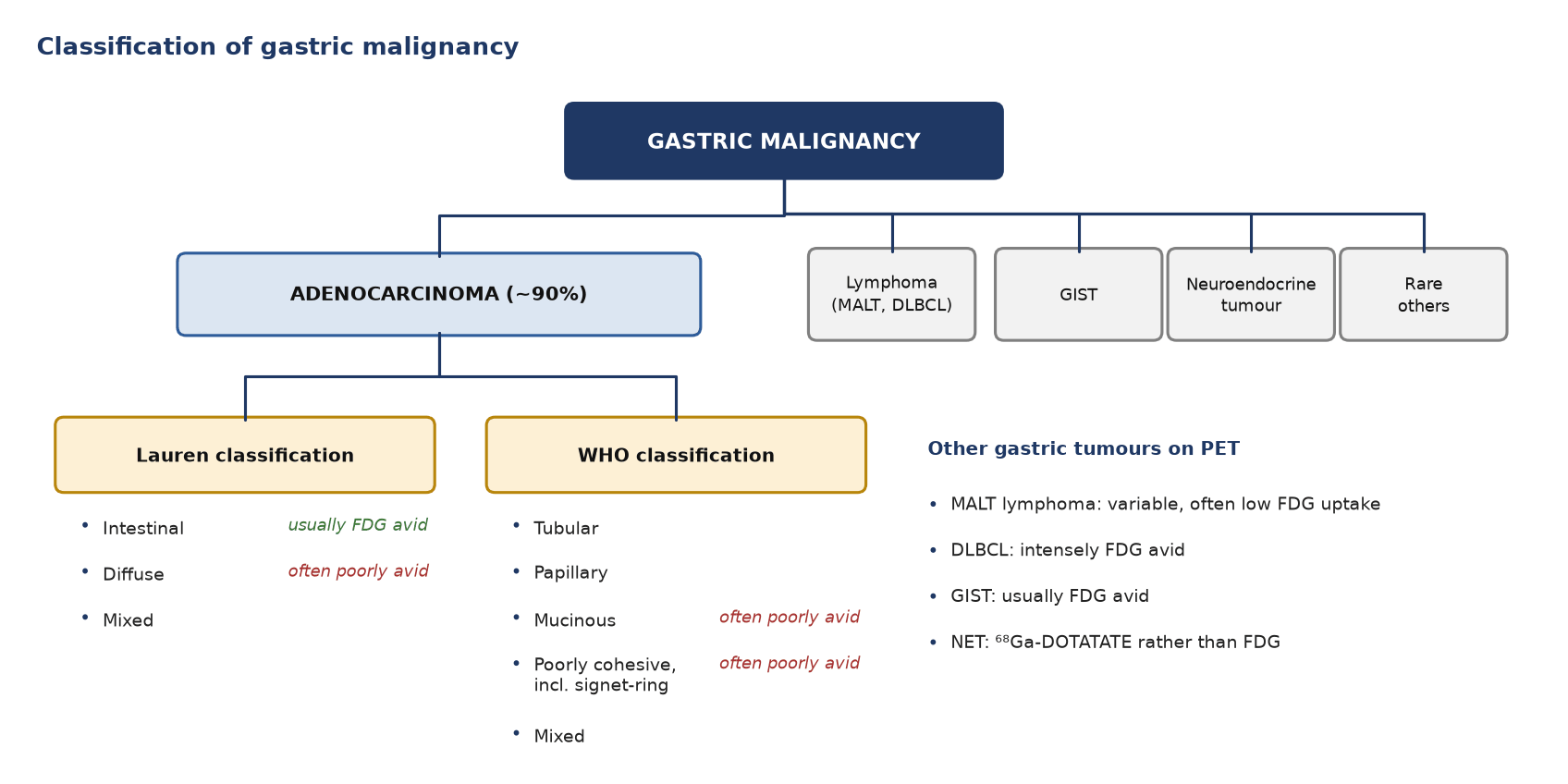
Figure 1. Classification of gastric malignancy. Adenocarcinoma is divided by the Lauren and WHO classifications; the histological type predicts FDG avidity.
Intestinal-type tumours are usually FDG avid. Diffuse, signet-ring and mucinous tumours often are not: they have fewer glucose transporters (GLUT1), more mucin and tumour cells spread thinly through the wall.
Type
FDG avidity
Intestinal (Lauren)
Usually high: 83% of primaries detected (Stahl 2003); 93% (Bosch 2020)
Diffuse, signet-ring, mucinous
Often low: 41% detected (Stahl 2003); 78% (Bosch 2020)
Gastric MALT lymphoma
Variable, often low (see PET/CT in Lymphoma)
Gastric DLBCL
High
GIST
Usually high (see the oesophagus and GIST article)
Gastric neuroendocrine tumour
Use ⁶⁸Ga-DOTATATE rather than FDG
4. Diagnosis and initial work-up
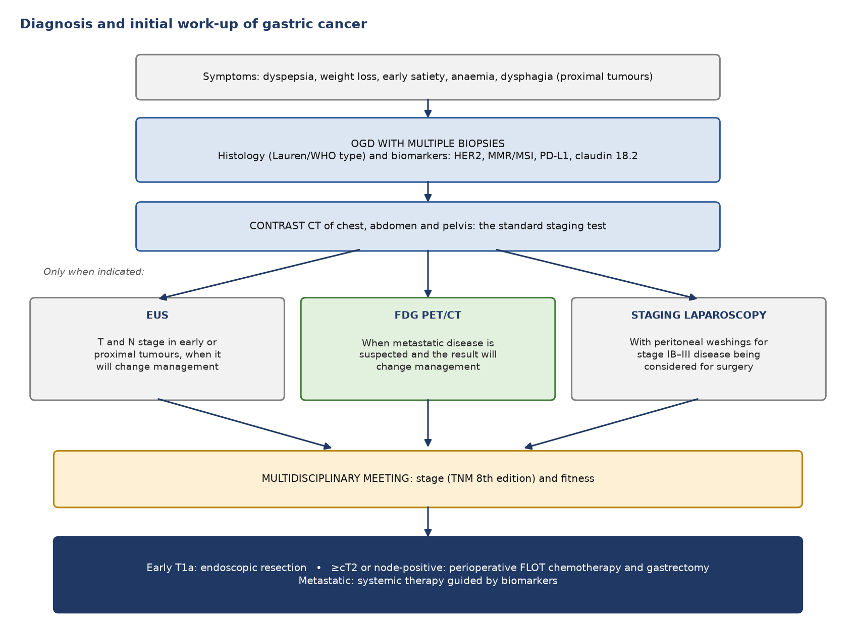
Figure 2. Diagnosis and initial work-up of gastric cancer. PET/CT is selective; staging laparoscopy is the test for the peritoneum.
Distant metastasis, including positive peritoneal cytology
Spread: direct (omentum, pancreas, transverse colon), lymphatic, peritoneal (commonest in the diffuse type) and haematogenous (liver first, then lung and bone).
What PET sees: liver, lung, bone and nodal metastases well; small peritoneal deposits poorly.
6. PET tracers
Tracer
Target
Main role
Main limitation
¹⁸F-FDG
Glucose metabolism (GLUT1)
Distant metastases; response; recurrence
Low avidity in signet-ring, mucinous and diffuse tumours; physiological gastric uptake; poor for the peritoneum
⁶⁸Ga/¹⁸F-FAPI
Fibroblast activation protein on cancer-associated fibroblasts
Staging, including diffuse-type tumours and the peritoneum
Investigational; limited availability
¹⁸F-FLT
Cell proliferation (thymidine kinase 1)
FDG-negative tumours; research response monitoring
Not in guidelines
⁶⁸Ga-DOTATATE/DOTATOC
Somatostatin receptors (mainly SSTR2)
Gastric neuroendocrine tumours; selection for PRRT
No role in adenocarcinoma
FDG: a glucose analogue trapped in glycolytic tumour cells (Warburg effect). Useful for occult distant metastases before surgery and for problem solving when CT is equivocal. Limited by low uptake in signet-ring, mucinous and diffuse tumours, by physiological uptake in normal gastric mucosa, and by poor sensitivity for the peritoneum.
FAPI: binds fibroblast activation protein on cancer-associated fibroblasts, a large part of gastric tumour stroma. Normal stomach has little FAP, so tumour-to-background ratios are high, including in signet-ring and diffuse tumours and in the peritoneum. Not yet in international guidelines.
FLT: a thymidine analogue that reflects proliferation, with less physiological gastric uptake than FDG. In 45 locally advanced gastric cancers it detected every primary, while 14 were FDG-negative. Research use only.
DOTATATE/DOTATOC: images and stages gastric neuroendocrine tumours and selects patients for PRRT. It has no role in adenocarcinoma.
7. Indications for FDG PET/CT
Clinical situation
Role of PET/CT
Why
Diagnosis
Not indicated
Endoscopy with biopsy makes the diagnosis
Staging, potentially curable, intestinal type
Selective, to find distant metastases missed on CT
Unsuspected metastases in 7% (Findlay 2019); stage changed in 19% (Bosch 2020)
Staging, diffuse or signet-ring type
Low value; laparoscopy is the key test
Primary and metastases are often not avid
Equivocal lesion on CT
Problem solving
Characterises liver, lung, bone or nodal lesions
Peritoneal disease
Not reliable
Staging laparoscopy with washings instead
Response to neoadjuvant chemotherapy
Investigational
Early metabolic response has been shown mainly for junctional tumours (MUNICON)
Standard FDG preparation: fasting, glucose check and a 60-minute uptake period.
Distend the stomach: many centres give water just before imaging so the stomach is not contracted; this helps separate tumour from normal wall.
Record: the date of the last endoscopy or biopsy, any stent, previous gastrectomy and the type of anastomosis, chemotherapy dates and G-CSF.
Timing: after neoadjuvant chemotherapy, scan at least 2–3 weeks after the last cycle; after surgery, allow time for postoperative inflammation to settle before scanning for recurrence.
10. What to look at when reporting
Primary: site (cardia, body, antrum), distance of its centre from the OGJ (gastric or oesophageal staging), SUVmax, and whether focal uptake matches wall thickening on CT.
Avidity: say whether the primary is avid; if it is not, say that its metastases may be missed too.
Regional nodes: perigastric, left gastric, common hepatic, splenic artery and coeliac; count them.
Distant nodes: para-aortic, retropancreatic, mesenteric and left supraclavicular (Virchow's) nodes are M1.
Organs: liver, lung, bone, adrenals, ovaries (Krukenberg tumour) and umbilicus.
Peritoneum: omental caking, nodules and ascites; always add that a negative PET does not exclude peritoneal disease.
After gastrectomy: anastomosis, gastric remnant, surgical bed and nodes.
Response scans: the change in SUVmax from baseline, on the same scanner and protocol.
11. Pitfalls
Pitfall
Why it happens
How to avoid it
Physiological gastric uptake
Normal mucosa, contracted stomach, hiatus hernia
Distend with water; call tumour only with focal uptake and wall thickening on CT
Report as non-diagnostic, not negative; laparoscopy
Peritoneal disease missed
Small, flat deposits below PET resolution
Staging laparoscopy with washings
Anastomosis after gastrectomy
Postoperative inflammation
Allow time; compare with baseline; endoscopy
Ovarian or endometrial uptake
Physiological in premenopausal women
Menstrual history; can mimic a Krukenberg tumour
Brown fat
Symmetrical supraclavicular uptake can mimic Virchow's node
Fat density on CT; keep the patient warm
Marrow and spleen after G-CSF
Diffuse reactive uptake
Record G-CSF; wait before scanning
12. When to do PET/CT: what the guidelines say
Time point
Recommendation
Guideline
Diagnosis
Endoscopy with multiple biopsies; PET/CT has no role
ESMO 2022
Staging, potentially curable
Consider FDG PET/CT only if metastatic disease is suspected and the result will guide management
NICE NG83
Not routinely recommended; contrast CT of the chest, abdomen and pelvis is the staging test
ESMO 2022
Consider only when the result may change management
RCR 2022 (UK)
Peritoneal staging
Staging laparoscopy with peritoneal washings for resectable disease; PET cannot replace it
ESMO 2022; NICE NG83
Response to chemotherapy
PET is not standard; used in trials
No guideline recommendation
Suspected recurrence
CT is standard; PET for equivocal findings
No guideline recommendation
FAPI PET
Research use
Not yet in guidelines
Test yourself
3 quick questions. Pick an answer to see the explanation.
1. A 58-year-old has a locally advanced signet-ring cell gastric adenocarcinoma. CT shows no metastases. On FDG PET/CT the primary is barely visible and there are no other avid lesions. What is the best next step before gastrectomy is planned?
2. Staging FDG PET/CT for a gastric antral cancer shows an avid node at the coeliac axis and an avid para-aortic node below the left renal vein. How should they be staged?
3. At staging laparoscopy for a gastric cancer, no peritoneal nodules are seen but the peritoneal washings contain malignant cells. What is the M category?
References
Lordick F, Carneiro F, Cascinu S, et al. Gastric cancer: ESMO Clinical Practice Guideline for diagnosis, treatment and follow-up. Ann Oncol. 2022;33(10):1005-20.
National Institute for Health and Care Excellence. Oesophago-gastric cancer: assessment and management in adults. NICE guideline NG83. London: NICE; 2018.
Royal College of Radiologists. Evidence-based indications for the use of PET-CT in the United Kingdom 2022. London: RCR; 2022.
Bray F, Laversanne M, Sung H, et al. Global cancer statistics 2022: GLOBOCAN estimates of incidence and mortality worldwide for 36 cancers in 185 countries. CA Cancer J Clin. 2024;74(3):229-63.
Gertsen EC, Brenkman HJF, van Hillegersberg R, et al. 18F-fludeoxyglucose-positron emission tomography/computed tomography and laparoscopy for staging of locally advanced gastric cancer: a multicenter prospective Dutch cohort study (PLASTIC). JAMA Surg. 2021;156(12):e215340.
Stahl A, Ott K, Weber WA, et al. FDG PET imaging of locally advanced gastric carcinomas: correlation with endoscopic and histopathological findings. Eur J Nucl Med Mol Imaging. 2003;30(2):288-95.
Bosch KD, Chicklore S, Cook GJ, et al. Staging FDG PET-CT changes management in patients with gastric adenocarcinoma who are eligible for radical treatment. Eur J Nucl Med Mol Imaging. 2020;47(4):759-67.
Findlay JM, Antonowicz S, Segaran A, et al. Routinely staging gastric cancer with 18F-FDG PET-CT detects additional metastases and predicts early recurrence and death after surgery. Eur Radiol. 2019;29:2490-8.
Foley KG, Coomer W, Coles B, Bradley KM. The impact of baseline 18F-FDG PET-CT on the management and outcome of patients with gastric cancer: a systematic review. Br J Radiol. 2022;95(1139):20220437.
Sim SH, Kim YJ, Oh DY, et al. The role of PET/CT in detection of gastric cancer recurrence. BMC Cancer. 2009;9:73.
Lordick F, Ott K, Krause BJ, et al. PET to assess early metabolic response and to guide treatment of adenocarcinoma of the oesophagogastric junction: the MUNICON phase II trial. Lancet Oncol. 2007;8(9):797-805.
Herrmann K, Ott K, Buck AK, et al. Imaging gastric cancer with PET and the radiotracers 18F-FLT and 18F-FDG: a comparative analysis. J Nucl Med. 2007;48(12):1945-50.
Ruan D, Zhao L, Cai J, et al. Evaluation of FAPI PET imaging in gastric cancer: a systematic review and meta-analysis. Theranostics. 2023;13(13):4694-710.
Zhao Y, Wang S, Du B, et al. Head-to-head comparison of [68Ga]Ga-FAPI-04 PET and [18F]FDG PET in the evaluation of primary gastrointestinal tumors and metastases: a systematic review and meta-analysis. Acad Radiol. 2025;32(11):6857-72.
Huang H, Altunay B, Schäfer L, et al. Diagnostic efficacy of FAPI-PET/CT versus [18F]FDG-PET/CT in upper-abdominal malignancies: a systematic review and meta-analysis. Diagnostics (Basel). 2026;16(4):520.