Home › PET/CT › Nasopharyngeal carcinoma › Full article
PET/CT in Nasopharyngeal Carcinoma
1. What it is
A carcinoma of the lining of the nasopharynx, most often starting in the fossa of Rosenmüller.
Where it is common: endemic in southern China and Southeast Asia; also more frequent in North Africa, the Arctic (Inuit) populations and North-East India. Rare elsewhere.
Causes: Epstein–Barr virus (EBV), salt-preserved fish eaten in childhood, smoking and genetic susceptibility.
EBV DNA: plasma EBV DNA is used for screening in endemic areas, for prognosis before treatment and for monitoring afterwards.
Type (WHO)
How common
Key point
FDG
Non-keratinising squamous cell carcinoma (differentiated or undifferentiated)
Over 95% in endemic areas; the majority worldwide
EBV-positive; very radiosensitive; better prognosis
Avid
Keratinising squamous cell carcinoma
Under 5% in endemic areas; about 20% worldwide, more in non-endemic countries
Less often EBV-related; linked with smoking; less radiosensitive
Avid
Basaloid squamous cell carcinoma
Rare
Aggressive
Avid
2. Anatomy
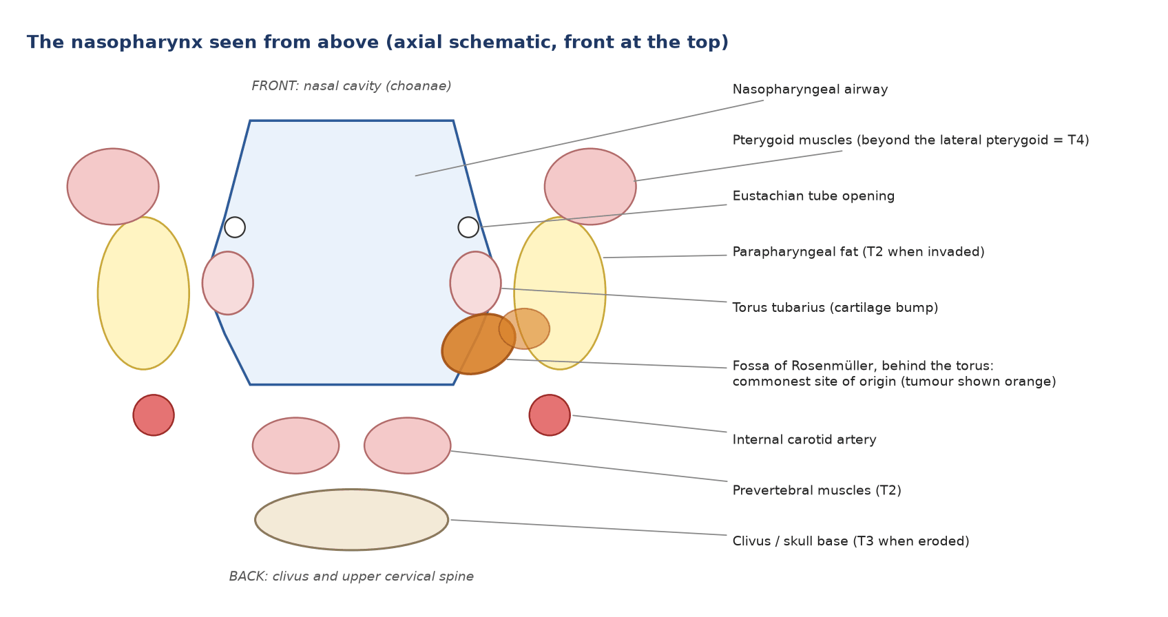
Figure 1. The nasopharynx seen from above. The fossa of Rosenmüller lies behind the torus tubarius; tumour there spreads sideways into the parapharyngeal fat and up to the skull base.
Boundaries: front, the choanae (back of the nasal cavity); roof, the sphenoid and clivus; back, the prevertebral muscles; below, the soft palate and oropharynx.
Side wall: the Eustachian tube opening, the torus tubarius behind it, and the fossa of Rosenmüller behind the torus.
Neighbours that decide T stage: parapharyngeal space, pterygoid muscles, skull-base foramina (lacerum, ovale), cavernous sinus and cranial nerves.
3. Pattern of spread
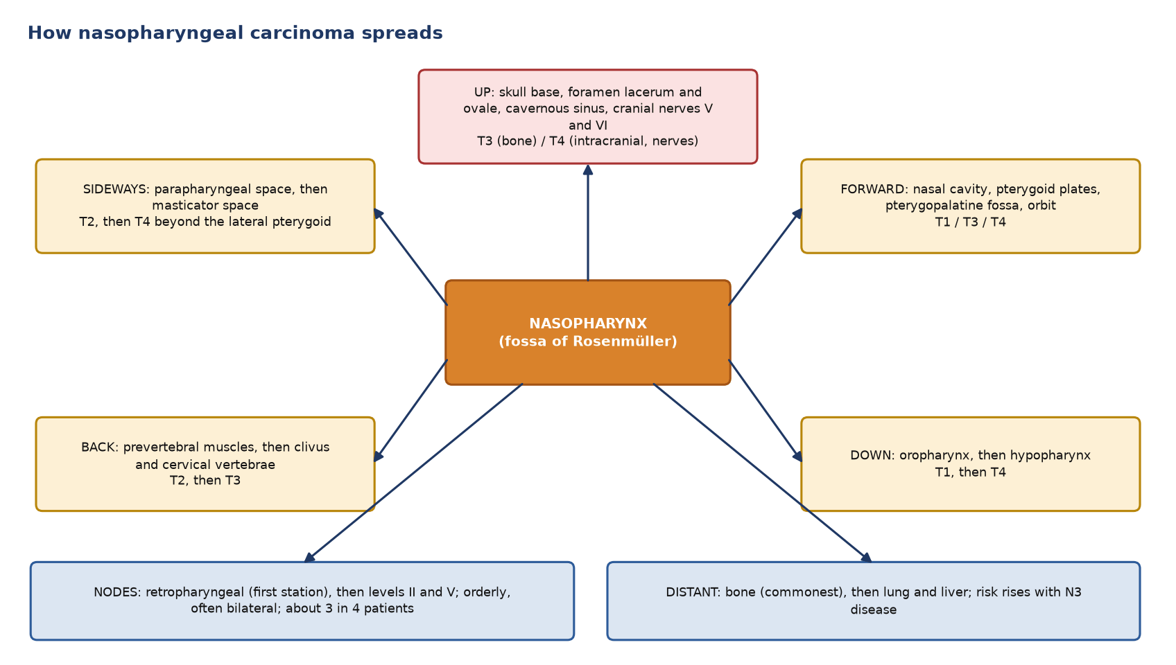
Figure 2. Directions of spread and the T stage each one implies, with the nodal and distant routes.
Route
How often
Nodes
About 3 in 4 patients have nodal disease (77% in a pooled analysis); retropharyngeal and level II nodes come first and skip spread is uncommon
Skull base
Common at diagnosis; MRI shows it best
Distant
About 4–5% at diagnosis; bone is the commonest site, then lung and liver. Risk is highest with N3 disease
4. How it looks on CT and MRI
Feature
CT
MRI
Primary
Asymmetric soft tissue filling one fossa of Rosenmüller
Best test for the primary and its extent
Skull base
Erosion or sclerosis of the clivus, pterygoid plates and foramina
Loss of normal fatty marrow; perineural spread along V2 and V3
Nodes
Retropharyngeal node 5 mm or more (short axis), or any node with necrosis
Better than PET for small retropharyngeal nodes next to the primary
Ear
Fluid in the middle ear and mastoid from a blocked Eustachian tube
5. Work-up and staging
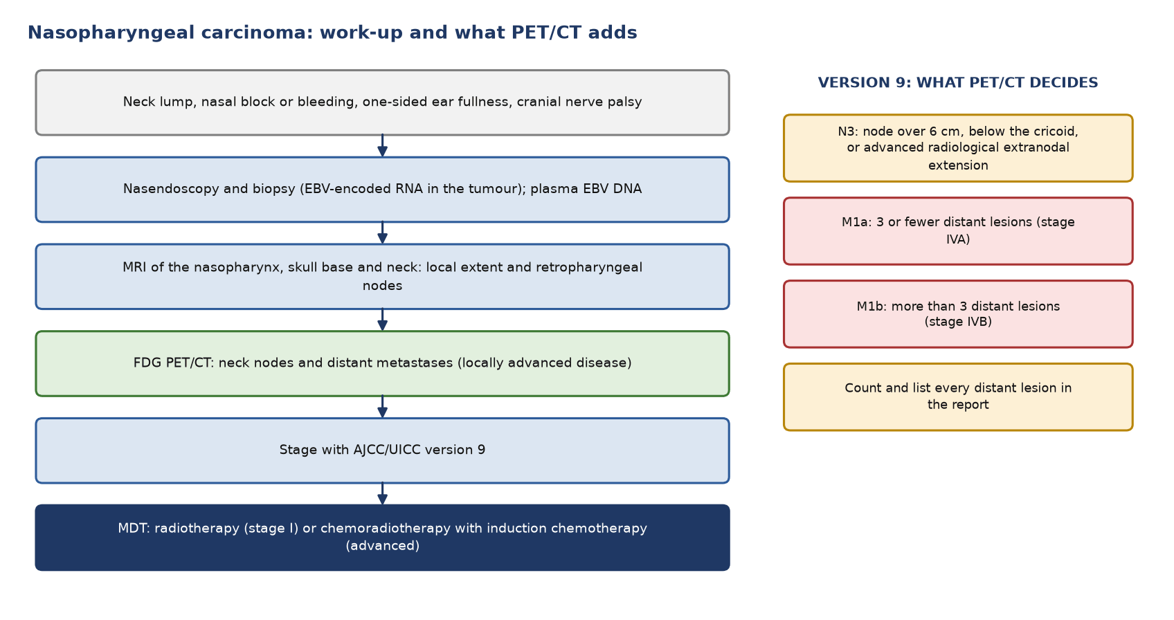
Figure 3. Work-up of nasopharyngeal carcinoma (left) and the version 9 categories that PET/CT helps decide (right).
AJCC/UICC version 9 (in use from January 2025)
Category
Definition
T1
Nasopharynx, or spread to the oropharynx or nasal cavity, without parapharyngeal involvement
T2
Parapharyngeal space, medial or lateral pterygoid, or prevertebral muscles
T3
Bone: skull base, cervical vertebrae, pterygoid structures, or paranasal sinuses
T4
Intracranial spread, cranial nerves, hypopharynx, orbit, parotid, or soft tissue beyond the lateral pterygoid
N1
One-sided neck nodes, or retropharyngeal nodes on one or both sides; 6 cm or less and above the cricoid
N2
Neck nodes on both sides; 6 cm or less and above the cricoid
N3
Over 6 cm, below the cricoid, or advanced radiological extranodal extension (into muscle, skin or neurovascular bundle)
M1a / M1b
3 or fewer distant lesions / more than 3
Stage groups: IA T1–2 N0; IB T1–2 N1; II T1–2 N2 or T3 N0–2; III T4 or N3; IVA M1a; IVB M1b.
What changed from the 8th edition: advanced extranodal extension now makes N3, stage IV is kept for metastatic disease only, and M1 is split by the number of lesions.
6. Indications for PET/CT
Situation
PET/CT recommended?
Details
Staging, locally advanced (T3–T4 or N2–N3)
Recommended
Best test for distant metastases; also stages the neck
Staging, early (T1–T2, N0–N1)
Optional
Chest CT or PET/CT, as available
Primary tumour and skull base
Not recommended
MRI decides local extent and perineural spread
Radiotherapy planning
Optional
Helps define involved nodes
Response, about 3 months after treatment
Selected cases
MRI first; PET/CT for an equivocal residual mass
Suspected recurrence (rising EBV DNA, symptoms or an MRI finding)
Recommended
Separates scar from tumour and restages the whole body
Routine follow-up
Not recommended
Clinical review, endoscopy, MRI and EBV DNA
7. Diagnostic accuracy
Question
FDG PET/CT
Comparison
Source
Nodal and distant staging
Sensitivity 89%, specificity 96%
Better than conventional work-up for distant metastases
Meta-analysis, 20 studies
Local recurrence or residual tumour after radiotherapy
Sensitivity 92%, specificity 89%
MRI 83% and 78%
Meta-analysis, 44 studies, 3,369 patients
Retropharyngeal nodes
Limited next to the primary
MRI is better
ESMO–EURACAN 2021
8. Patient preparation and reporting
Preparation: standard fasting; quiet uptake (no talking or chewing) to limit muscle uptake; keep warm; scan vertex to mid-thigh with arms down for the head and neck; record biopsy, radiotherapy and chemotherapy dates.
Report: the primary and its SUVmax with any skull-base or intracranial extension; retropharyngeal nodes named separately; neck nodes by level and side with the largest size and whether below the cricoid; signs of extranodal extension; the number and site of every distant lesion (bone, lung on CT, liver), because the count decides M1a or M1b.
9. Pitfalls
Pitfall
Why it misleads
Adenoid and Waldeyer's ring in young patients
Normal lymphoid tissue is avid and can be asymmetric
Small retropharyngeal nodes next to the primary
Merge with the primary; MRI is better
Skull base and intracranial spread
Hidden by normal brain uptake; MRI is better
Post-radiotherapy mucositis, osteoradionecrosis
Inflammation; scan 12 weeks or more after treatment
Muscles, brown fat, vocal cords
Physiological uptake, especially after talking or in the cold
Small lung nodules
Below PET resolution; judge them on CT
10. When to do PET/CT: what the guidelines say
Time point
Recommendation
Guideline
Staging, primary
MRI of the nasopharynx, skull base and neck
ESMO–EURACAN 2021
Staging, distant
FDG PET/CT is the best test for distant metastases; recommended at least in locally advanced disease
ESMO–EURACAN 2021
Staging, T4 or N3
Offer PET/CT
NICE NG36
Response
Clinical, endoscopic and MRI assessment; PET/CT for equivocal findings
ESMO–EURACAN 2021
Suspected recurrence
Imaging, including PET/CT, with endoscopy and biopsy
ESMO–EURACAN 2021
Follow-up
Clinical review and endoscopy; no routine PET
ESMO–EURACAN 2021
Test yourself
2 quick questions. Pick an answer to see the explanation.
1. Staging PET/CT for a T3 N2 nasopharyngeal carcinoma shows two FDG-avid rib lesions and one liver lesion. What is the stage under AJCC/UICC version 9?
2. A 6-mm right retropharyngeal node sits close to the primary tumour and shows no separate FDG focus. What is the best next step?
References
Bossi P, Chan AT, Licitra L, et al. Nasopharyngeal carcinoma: ESMO-EURACAN Clinical Practice Guidelines for diagnosis, treatment and follow-up. Ann Oncol. 2021;32(4):452-65.
Pan JJ, Mai HQ, Ng WT, et al. Ninth version of the AJCC and UICC nasopharyngeal cancer TNM staging classification. JAMA Oncol. 2024;10(12):1627-35.
Chen YP, Chan ATC, Le QT, et al. Nasopharyngeal carcinoma. Lancet. 2019;394(10192):64-80.
Ho FC, Tham IW, Earnest A, Lee KM, Lu JJ. Patterns of regional lymph node metastasis of nasopharyngeal carcinoma: a meta-analysis of clinical evidence. BMC Cancer. 2012;12:98.
Shen G, Zhang W, Jia Z, et al. Meta-analysis of diagnostic value of 18F-FDG PET or PET/CT for detecting lymph node and distant metastases in patients with nasopharyngeal carcinoma. Br J Radiol. 2014;87(1044):20140296.
Li Z, Li Y, Li N, Shen L. Positron emission tomography/computed tomography outperforms MRI in the diagnosis of local recurrence and residue of nasopharyngeal carcinoma: an update evidence from 44 studies. Cancer Med. 2019;8(1):67-79.
National Institute for Health and Care Excellence. Cancer of the upper aerodigestive tract: assessment and management in people aged 16 and over. NICE guideline NG36. London: NICE; 2016 (updated 2018).
Purohit BS, Ailianou A, Dulguerov N, et al. FDG-PET/CT pitfalls in oncological head and neck imaging. Insights Imaging. 2014;5(5):585-602.