Parts: fundus, body and neck, leading into the cystic duct, which joins the common hepatic duct to form the common bile duct.
Liver relationship: the gallbladder bed lies against segments IVB and V. There is no serosa on the hepatic side, so tumour reaches the liver early.
Wall: mucosa, lamina propria, a thin muscle layer and perimuscular connective tissue; serosa only on the peritoneal side. There is no muscularis mucosae and no submucosa.
2. Epidemiology and risk factors
The fifth most common gastrointestinal cancer and the commonest biliary tract cancer.
Women are affected 2–4 times as often as men; the average age at diagnosis is about 72 years.
Usually found late: early disease mimics gallstone symptoms; advanced disease brings right upper quadrant pain, weight loss, jaundice and a palpable mass.
Risk factor
Note
Gallstones
The commonest risk factor; present in 74–92% of patients
Porcelain gallbladder
Calcified wall
Gallbladder polyp of 10 mm or more
Removed because of malignant potential
Choledochal (congenital biliary) cysts
Anomalous pancreaticobiliary junction
Primary sclerosing cholangitis
Chronic Salmonella typhi carriage
Smoking, obesity
Genetic factors
3. Pathology
Type
Share or feature
Adenocarcinoma
About 85%; includes papillary (better prognosis) and mucinous (low FDG uptake) types
Liver (segments IVB/V), then colon, duodenum and pancreas
Lymphatic
Cystic and pericholedochal nodes, then periportal, hepatic artery and posterior pancreaticoduodenal nodes; coeliac, para-aortic and superior mesenteric nodes are M1
Haematogenous
Liver (the commonest distant site), then lung (the commonest site outside the liver)
Peritoneal
Seeding, especially after gallbladder perforation or laparoscopic surgery
After surgery
Port-site and wound metastases
5. Tumour markers
Marker
Role
CA 19-9
Raised in most patients; baseline and follow-up; also raised by biliary obstruction
CA-125
Supportive; raised with peritoneal disease
CEA
Supportive
6. Diagnosis and work-up
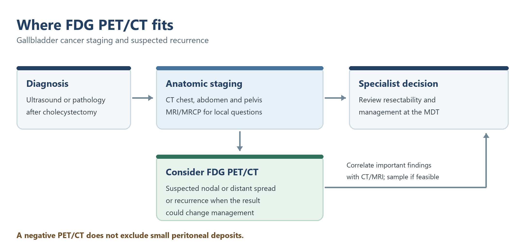
Figure 1. CT and MRI define anatomy; consider FDG PET/CT for suspected nodal or distant spread or recurrence when it may alter management. Correlate important PET findings before treatment decisions.
Incidental gallbladder cancer: found in the specimen after cholecystectomy. T1a tumours removed intact are observed; for T1b and above, restage and, if there are no metastases, re-resect the liver bed (segments IVB/V) with the hepatoduodenal nodes.
7. Staging (AJCC/UICC 8th edition)
T
Depth
T1a / T1b
Lamina propria / muscle layer
T2a / T2b
Perimuscular connective tissue on the peritoneal side / on the hepatic side
T3
Through the serosa, and/or into the liver and/or one other adjacent organ
T4
Main portal vein or hepatic artery, or two or more extrahepatic organs
N / M
Definition
N1 / N2
1–3 / 4 or more regional nodes (cystic, common bile duct, hepatic artery, portal vein)
M1
Distant metastases, including coeliac, para-aortic, pericaval and superior mesenteric nodes
8. CT appearance and its pattern on PET/CT
CT pattern
How often
PET/CT
Mass replacing the gallbladder
40–65%
Intense uptake; look for liver invasion
Focal or diffuse irregular wall thickening
20–30%
Uptake, but cholecystitis mimics it
Intraluminal polypoid mass
15–25%
Avid if 1 cm or more; small polyps may be missed
Contrast enhancement: hypodense on unenhanced CT; early, irregular peripheral enhancement in the late arterial phase; enhancement persists in the portal venous and delayed phases.
Associated findings: intrahepatic bile duct dilatation, invasion of the liver or nearby vessels, gallstones.
Uptake is variable; mobility or lack of enhancement supports sludge. Small lesions may be PET-negative.
Wall thickening
Carcinoma, acute or chronic cholecystitis, xanthogranulomatous cholecystitis, adenomyomatosis
FDG avidity overlaps. Intramural nodules, surrounding inflammation and MRI help; biopsy may be needed.
Mass in the gallbladder fossa
Gallbladder carcinoma or an adjacent biliary or gastrointestinal primary
Trace the anatomical origin on contrast CT/MRI; uptake alone cannot assign the primary site.
13. Patient preparation
Standard FDG preparation: fasting, glucose check and a 60-minute uptake period.
Record: the date of cholecystectomy, biliary stent, drain or ERCP, any recent cholecystitis, and the port-site locations.
Timing: allow a few weeks after surgery where possible, because postoperative inflammation is avid.
14. What to look at when reporting
Primary: pattern (mass, wall thickening or polyp) and SUVmax.
Local spread: invasion of the liver (segments IVB/V), bile ducts, duodenum and colon.
Nodes: regional nodes (N1/N2) versus coeliac and para-aortic nodes (M1).
Distant disease: liver metastases, peritoneum (omentum, gutters), port sites and wound after cholecystectomy, lung.
Conclusion: whether the disease looks resectable on PET/CT, and that a negative PET does not exclude small peritoneal deposits.
15. Pitfalls
False positives
False negatives
Xanthogranulomatous cholecystitis
Small tumours (under 1 cm)
Acute cholecystitis
Mucinous types
Chronic cholecystitis
Uptake masked by adjacent bowel
After surgery, stents or ERCP
Small peritoneal deposits
16. When to do PET/CT: what the guidelines say
Time point
Recommendation
Guideline
Diagnosis of the primary
FDG PET is not recommended for imaging the primary tumour; CT and MRI are standard
ESMO biliary tract cancer guideline
Initial staging
PET/CT may be considered when nodal or distant metastases are suspected, or to clarify equivocal CT or MRI
ESMO biliary tract cancer guideline
Incidental gallbladder cancer
Re-resection for T1b and above if there is no metastatic spread; restaging before it (PET/CT can be considered)
ESMO biliary tract cancer guideline
Response assessment
Supplementary to CT and MRI
No guideline recommendation
Suspected recurrence
PET/CT may be considered
ESMO biliary tract cancer guideline
Test yourself
3 quick questions. Pick an answer to see the explanation.
1. A 68-year-old woman has diffuse, irregular gallbladder wall thickening with intense FDG uptake (SUVmax 9) and intramural low-density nodules on CT. There is no liver invasion or adenopathy. What is the most important alternative to gallbladder cancer?
2. Gallbladder cancer is found in the specimen after laparoscopic cholecystectomy, invading the perimuscular connective tissue (T2). What is the best next step?
3. Staging PET/CT for gallbladder cancer shows avid pericholedochal and coeliac nodes. How are they staged?
References
Vogel A, Bridgewater J, Edeline J, et al. Biliary tract cancer: ESMO Clinical Practice Guideline for diagnosis, treatment and follow-up. Ann Oncol. 2023;34(2):127-40.
Amin MB, Edge SB, Greene FL, et al., eds. AJCC Cancer Staging Manual. 8th ed. New York: Springer; 2017.
Annunziata S, Pizzuto DA, Caldarella C, et al. Diagnostic accuracy of fluorine-18-fluorodeoxyglucose positron emission tomography in gallbladder cancer: a meta-analysis. World J Gastroenterol. 2015;21(40):11481-8.
Parida GK, Panda RA, Agrawal K. Impact of fluorine-18-fluorodeoxyglucose PET/computed tomography in staging of patients with gallbladder cancer: a systematic review and meta-analysis. Nucl Med Commun. 2021;42(8):846-54.
Hu JH, Tang JH, Lin CH, et al. Preoperative staging of cholangiocarcinoma and biliary carcinoma using 18F-fluorodeoxyglucose positron emission tomography: a meta-analysis. J Investig Med. 2018;66(1):52-61.
Batchu S, Veenstra MMK, de Jong DM, et al. Real-world evidence in a large cohort comparing FAPI and FDG PET/CT for hepatopancreatobiliary cancer diagnosis. Eur J Nucl Med Mol Imaging. 2026 (published online). doi:10.1007/s00259-026-08112-w.
Zhang Z, Dong H, Zhang L, et al. Xanthogranulomatous cholecystitis masquerading as gallbladder carcinoma on 68Ga-FAPI-04 PET. Clin Nucl Med. 2022;47(10):895-6.