PET/CT in Ewing Sarcoma
1. What it is
- A small round blue cell sarcoma of bone (or soft tissue) with an EWSR1–ETS gene fusion, most often EWSR1–FLI1, t(11;22)(q24;q12); CD99-positive.
- The second commonest bone sarcoma in children and adolescents; rare in people of African ancestry.
- WHO 2020: the look-alike undifferentiated round cell sarcomas (CIC-rearranged, BCOR-altered and EWSR1–non-ETS fused) are now separate from Ewing sarcoma.
2. Where it arises
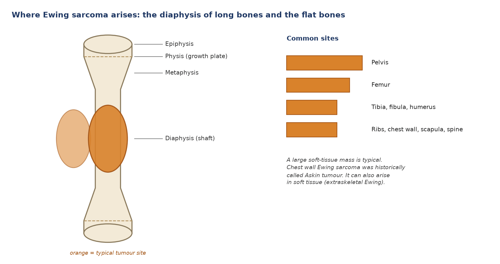
- Long bones: diaphysis or metadiaphysis of the femur, tibia, fibula and humerus.
- Flat bones: pelvis (the commonest single site), ribs, scapula and spine.
- Chest wall: Ewing sarcoma of the chest wall was historically called Askin tumour.
- Soft tissue: it can also arise outside bone (extraskeletal Ewing sarcoma).
3. How the tumour looks on CT
| Feature | What CT shows |
|---|---|
| Site | Diaphysis or metadiaphysis of a long bone; flat bones (pelvis, ribs, scapula, spine) |
| Bone | Permeative or moth-eaten lysis with a wide zone of transition; reactive sclerosis in some, especially in flat bones |
| Matrix | No tumour matrix mineralisation (unlike osteosarcoma) |
| Periosteum | Onion-skin (lamellated) periosteal reaction; Codman triangle; saucerisation of the outer cortex |
| Soft tissue | Large soft-tissue mass, out of proportion to the bone destruction |
| Lungs | Nodules (metastases), judged on chest CT |
4. Work-up and pattern of spread
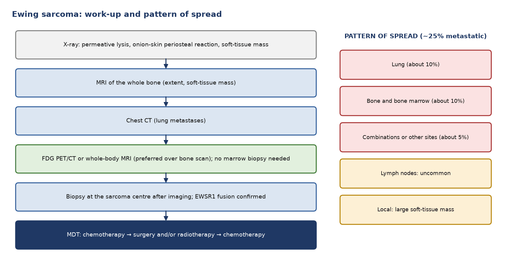
- Prognosis by site: multiple bone metastases carry a worse outlook than lung or pleural metastases (5-year survival under 20% versus 50–60%).
5. Staging
Ewing sarcoma of bone is staged with the AJCC 8th edition bone system (T1 8 cm or less, T2 more than 8 cm, T3 skip lesions; M1a lung, M1b bone or other sites). In practice the key split is localised versus metastatic, and lung-only versus bone or marrow metastases.
6. Indications for PET/CT
| Situation | Role |
|---|---|
| Initial staging | Recommended: FDG PET/CT or whole-body MRI is preferred over bone scan; bone marrow biopsy is not needed if PET/CT is done |
| Lung nodules | Chest CT decides |
| Response to induction chemotherapy | Prognostic; histology remains the reference |
| Radiotherapy planning | Shows active disease when the MDT asks |
| Suspected recurrence | High accuracy (sensitivity 90%, specificity 93%) |
| Routine follow-up | Not indicated |
7. Diagnostic accuracy
| Question | Sensitivity | Specificity |
|---|---|---|
| Bone metastases | 84% | 93% |
| Lung metastases | 76% | 92% |
| Recurrence | 90% | 93% |
Meta-analysis of 31 studies, 735 patients (Seth 2022). In a prospective paediatric study PET was better than conventional imaging for nodes and bone, but CT was far better for the lungs (Völker 2007).
8. Response to chemotherapy
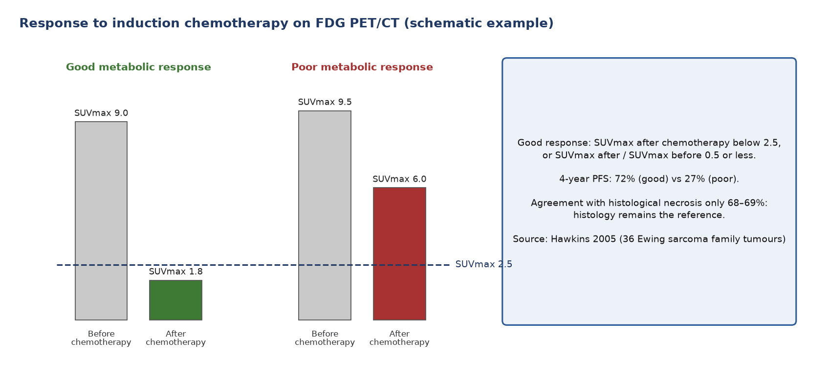
- Outcome: 4-year progression-free survival was 72% with SUVmax after chemotherapy below 2.5, against 27% above it (36 patients).
- Histology stays the reference: PET agreed with histological response in 68–69%.
9. Patient preparation and reporting
- Preparation: scan from vertex to toes; inject away from the tumour; keep young patients warm; record G-CSF, which makes marrow uptake diffuse.
- Report: the primary and soft-tissue mass with SUVmax; marrow (focal versus diffuse uptake); bone metastases; lungs on CT; nodes; a clear statement of localised, lung-only or bone/marrow metastatic disease; and for response the SUVmax before and after chemotherapy and their ratio.
10. Pitfalls
| Pitfall | Why it misleads |
|---|---|
| Physes | Symmetrical bands of uptake |
| G-CSF or reactive marrow | Diffuse uptake; true marrow metastases are focal |
| Osteomyelitis | Mimics Ewing sarcoma clinically and on imaging; FDG avid |
| Langerhans cell histiocytosis | Avid lytic lesion in a child |
| Small lung nodules | FDG-negative but may be metastases |
11. When to do PET/CT: what the guidelines say
| Time point | Recommendation | Guideline |
|---|---|---|
| Staging | FDG PET/CT or whole-body MRI preferred over bone scan; bone marrow biopsy and aspirate not mandated if FDG PET/CT is done | ESMO–EURACAN–GENTURIS–ERN PaedCan 2021 |
| Lungs | Chest CT | ESMO 2021 |
| Response | No PET recommendation; histology is the reference | ESMO 2021 |
| Follow-up | No routine PET | ESMO 2021 |
Test yourself
2 quick questions. Pick an answer to see the explanation.
1. A 15-year-old with pelvic Ewing sarcoma has staging FDG PET/CT with no bone or marrow lesions. The CT shows three 4-mm lung nodules without uptake. Which statement is correct?
2. After induction chemotherapy with G-CSF support, a Ewing sarcoma patient's PET/CT shows diffuse, homogeneous marrow uptake without focal lesions. What is the best interpretation?
References
- Strauss SJ, Frezza AM, Abecassis N, et al. Bone sarcomas: ESMO-EURACAN-GENTURIS-ERN PaedCan Clinical Practice Guideline for diagnosis, treatment and follow-up. Ann Oncol. 2021;32(12):1520-36.
- Sbaraglia M, Bellan E, Dei Tos AP. The 2020 WHO Classification of Soft Tissue Tumours: news and perspectives. Pathologica. 2021;113(2):70-84.
- Tanaka K, Ozaki T. New TNM classification (AJCC eighth edition) of bone and soft tissue sarcomas: JCOG Bone and Soft Tissue Tumor Study Group. Jpn J Clin Oncol. 2019;49(2):103-7.
- Seth N, Seth I, Bulloch G, et al. 18F-FDG PET and PET/CT as a diagnostic method for Ewing sarcoma: a systematic review and meta-analysis. Pediatr Blood Cancer. 2022;69(3):e29415.
- Völker T, Denecke T, Steffen I, et al. Positron emission tomography for staging of pediatric sarcoma patients: results of a prospective multicenter trial. J Clin Oncol. 2007;25(34):5435-41.
- Hawkins DS, Schuetze SM, Butrynski JE, et al. [18F]Fluorodeoxyglucose positron emission tomography predicts outcome for Ewing sarcoma family of tumors. J Clin Oncol. 2005;23(34):8828-34.
- Boellaard R, Delgado-Bolton R, Oyen WJG, et al. FDG PET/CT: EANM procedure guidelines for tumour imaging: version 2.0. Eur J Nucl Med Mol Imaging. 2015;42(2):328-54.
Spotted an error or something out of date? Report an error