PET/CT in Oesophageal Cancer
1. Anatomy and lymph node drainage
- Extent: the oesophagus runs from the upper oesophageal sphincter through the neck, chest and diaphragm to the oesophagogastric junction (OGJ).
- Describe the primary: record its epicentre, distance from the incisors, longitudinal extent and relation to the OGJ. These guide staging and radiotherapy planning.
- Junction rule: a tumour centred within 2 cm of the OGJ and extending into the oesophagus uses the oesophageal staging scheme; a more distal tumour uses the gastric scheme. [3]
- Longitudinal lymphatics: involved nodes may lie well above or below the primary.
- Regional nodes: the AJCC eighth-edition map extends from the upper oesophageal sphincter to the coeliac artery. Coeliac nodes are regional, even for a distal primary.
- Distant disease: non-regional nodes and organ deposits are M1. Inspect liver, lung, bone and adrenal glands. [3]
2. Epidemiology and risk factors
- Squamous cell carcinoma (SCC): the dominant histology worldwide; especially prominent in eastern Asia and parts of Africa. Tobacco and alcohol are important risk factors.
- Adenocarcinoma: relatively more common in some high-income countries; associated with reflux, Barrett’s oesophagus and obesity.
- Why histology matters: treatment pathways and FDG uptake patterns can differ. [4] [5]
3. Classification and the FDG signal
| Type | Usual site and context | PET/CT implication |
|---|---|---|
| SCC (~85% of oesophageal cancers worldwide) | Often upper or middle oesophagus; tobacco/alcohol risk | Usually FDG avid; inspect the upper aerodigestive tract for a possible second primary when clinically relevant. |
| Adenocarcinoma (~15%) | Usually distal oesophagus or OGJ; reflux/Barrett’s background | Often FDG avid, but uptake varies; record baseline avidity before any response scan. |
| Small, superficial or mucin-rich tumour | May have little visible metabolic volume | A faint or absent PET focus does not exclude the endoscopic tumour. |
- Tracer biology: FDG is a glucose analogue taken up by tumour and inflammatory cells; PET localises metabolism, not histology.
- Positive finding: focal uptake is more convincing with a matching lesion on diagnostic CT or endoscopy.
- Negative finding: tiny lesions can be missed because of limited spatial resolution. [2] [6]
4. Diagnosis and initial work-up
- Endoscopy and biopsy: establish histology and locate the lesion.
- Contrast-enhanced CT: map the primary, adjacent structures and visible metastases.
- FDG PET/CT: in a patient still eligible for radical treatment, search for additional disease that could change treatment intent or radiation fields.
- Selective EUS: resolve a local question, such as sampling a node outside the planned field. Endoscopic resection is more informative about depth in suspected early mucosal disease. [1] [2]
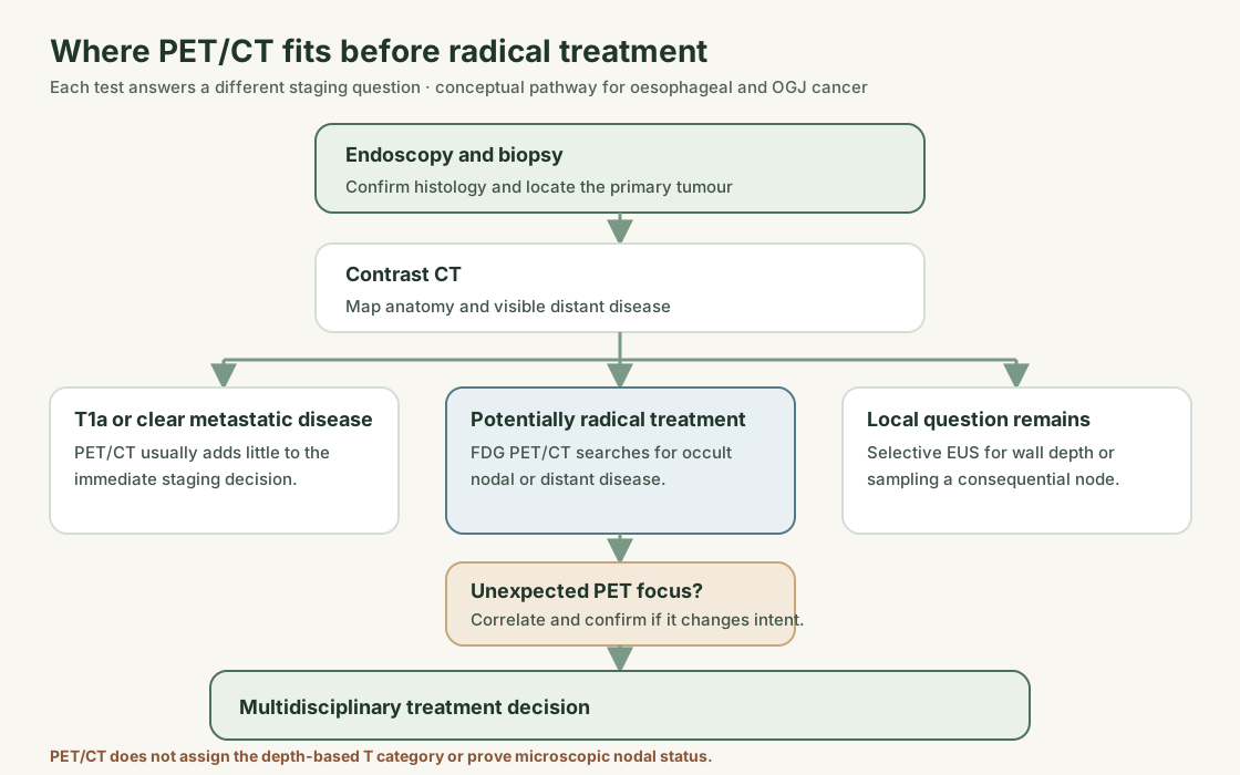
- NICE: offer FDG PET/CT after endoscopy and whole-body CT for oesophageal or OGJ tumours suitable for radical treatment, except T1a.
- ESMO: perform PET in candidates for oesophagectomy to seek occult metastases.
- EUS: use when its result guides management, not solely to distinguish T2 from T3.
- Laparoscopy: in locally advanced OGJ adenocarcinoma extending into the cardia, it can answer a peritoneal question PET may miss. [1] [2]
5. Staging: what PET can and cannot assign
| Category | Eighth-edition AJCC meaning | Main staging tool |
|---|---|---|
| T1a / T1b | Mucosa / submucosa | Endoscopic resection pathology; selective EUS for deeper or nodal questions |
| T2 / T3 | Muscularis propria / adventitia | EUS and anatomical imaging when they alter treatment |
| T4a / T4b | Invasion of potentially resectable adjacent structures / major unresectable structures such as airway or aorta | Contrast CT, EUS or bronchoscopy as appropriate |
| N1 / N2 / N3 | 1–2 / 3–6 / at least 7 involved regional nodes | Anatomical imaging and sampling; PET cannot count microscopic deposits reliably |
| M1 | Distant organ or non-regional nodal disease | CT and PET/CT; confirm an isolated consequential finding |
Table 1. Simplified AJCC eighth-edition categories; clinical and pathological stage groupings are separate and are not inferred from PET uptake alone. [3]
- PET strength: revealing disease outside the expected regional field.
- Nodal limits: a node beside an avid primary can merge with it; an involved small node can be PET-negative. Coeliac nodes remain regional N disease.
- Unexpected M1 focus: compare diagnostic CT, add targeted MRI or ultrasound when useful, and biopsy if feasible before abandoning radical treatment. [2] [3] [7]
6. PET tracers
- Established tracer: ¹⁸F-FDG for staging, selected response assessment and suspected recurrence.
- Strength and limit: it highlights metabolically active SCC and adenocarcinoma but is not cancer-specific and can miss microscopic disease.
- Other tracers: research tools in oesophageal carcinoma, without an established role in the NICE/ESMO staging pathway. They do not replace indicated contrast CT, EUS or tissue sampling. [1] [2] [6]
7. Indications for FDG PET/CT
| Clinical situation | Role | Decision it may change |
|---|---|---|
| New diagnosis, potentially radical treatment | Established after endoscopy and CT, except T1a under NICE | Finds unsuspected distant disease or a node outside planned treatment fields. |
| Early T1a disease | Usually low value | Endoscopic pathology establishes depth and local treatment options. |
| Equivocal lesion on CT | Problem solving | Guides targeted imaging or biopsy; PET uptake alone is not proof. |
| Radiotherapy planning | Adjunct | Helps identify avid tumour or nodes, alongside endoscopy and planning CT. |
| Early chemotherapy response | Trial strategy, not a universal rule | A protocol may use serial uptake to adapt treatment. |
| After chemoradiotherapy | Selective restaging | Looks for interval metastases; cannot independently establish complete local response. |
| New symptoms or equivocal follow-up imaging | Problem solving | Helps locate recurrence or choose a biopsy site. |
| Asymptomatic follow-up without residual disease | No routine scan solely to find recurrence under NICE | Scan when a clinical question arises. |
Table 2. Indications and limits, based chiefly on NICE, ESMO and the cited trials. [1] [2] [8] [9] [10]
8. Diagnostic performance and treatment response
- Distant metastases: an older staging meta-analysis reported pooled sensitivity of 71% for FDG PET versus 52% for CT. These pre-modern-scanner estimates are not a patient-specific prediction. [7]
- Regional nodes: EUS was more sensitive than PET in that analysis. PET adds a whole-body search; anatomical or endoscopic tests still answer wall and nearby-node questions. [7]
- Early response in a trial: CALGB 80803 studied oesophageal/OGJ adenocarcinoma. A fall in SUVmax of less than 35% after induction chemotherapy triggered a chemotherapy switch during chemoradiotherapy. Pathological complete response in the two switched groups was 18% and 20%. [8]
- Clinical meaning: that phase II result supports a studied response-adapted strategy, not a universal instruction to switch treatment. Serial scans require comparable scanner, reconstruction, uptake interval and glucose conditions. [6] [8]
- After chemoradiotherapy: radiation oesophagitis can remain FDG avid, while small residual cancer can be PET-negative.
- Complete-response question: preSANO used PET/CT for interval metastases and combined endoscopy, deep biopsies, EUS and suspicious-node sampling for local response. SANO studied surveillance only after a multimodal clinical complete response. A negative PET alone cannot justify omitting surgery or declaring pathological complete response. [9] [10]
9. Patient preparation and scan timing
- Standard preparation: follow the local validated FDG protocol, including fasting, a blood-glucose check and a documented uptake interval.
- For serial scans: record treatment dates; use the same scanner and acquisition method when possible.
- Before interpretation: ask about recent biopsy, dilatation, stent, surgery, radiation and systemic therapy. A low-dose non-contrast PET/CT component does not replace diagnostic contrast CT. [6]
- After chemoradiotherapy: early diffuse uptake along the treated segment is less specific than a new focal mass with a CT correlate. No single SUV threshold reliably separates inflammation from tumour; use endoscopic and clinical assessment when local treatment would change. [9]
10. What to look at when reporting
- Primary: histology, location, length, relationship to the OGJ, CT wall finding and baseline FDG avidity.
- Regional nodes: describe stations and side; recognise that the coeliac basin is regional in the oesophageal map.
- Distant sites: report each non-regional node or organ lesion separately, with its CT correlate and degree of certainty.
- Treatment context: state scan dates, recent procedures, radiation field, chemotherapy and comparability with baseline imaging.
- Clinical conclusion: identify the finding that could change intent and specify targeted imaging or biopsy when confirmation is needed. Avoid assigning T depth or complete pathological response from PET alone. [3] [6]
11. Pitfalls
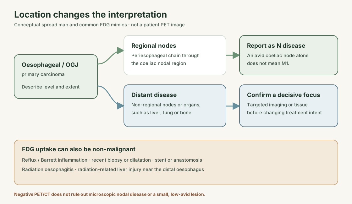
| Finding | Possible explanation | Useful check |
|---|---|---|
| Uptake at the distal oesophagus or OGJ | Reflux, Barrett-associated inflammation or tumour | Compare endoscopy, biopsy and CT wall morphology. |
| Uptake after biopsy, dilatation or stenting | Procedure-related inflammation | Record the dates and correlate with morphology. |
| Diffuse uptake in the treated oesophagus | Radiation oesophagitis | Correlate with timing; use endoscopy and biopsy for a consequential local question. |
| New focus in the adjacent left liver | Radiation-related liver injury can mimic metastasis | Compare the radiation field; confirm if it would alter treatment. |
| Avid focus near the gastric pull-up or anastomosis | Conduit mucosa, healing or recurrence | Trace anatomy on CT and use endoscopy where indicated. |
| PET-negative small CT lesion | Partial-volume loss or a poorly avid tumour | Do not dismiss a suspicious CT or endoscopic finding. |
Table 3. FDG is sensitive to inflammation and limited by lesion size. A focal PET abnormality needs anatomical context. [6] [9] [11]
12. What the guidelines say
| Time point | Recommendation |
|---|---|
| Baseline, suitable for radical treatment | NICE NG83: offer FDG PET/CT after endoscopy and CT except for T1a. |
| Candidate for oesophagectomy | ESMO 2022: carry out FDG PET to find occult distant metastases. |
| Local T/N question | NICE/ESMO: use EUS selectively when it changes management; PET does not replace it. |
| OGJ tumour extending into cardia | ESMO: laparoscopy for locally advanced adenocarcinoma crossing the diaphragm. |
| Asymptomatic after curative treatment | NICE: do not provide routine imaging solely to detect recurrence. |
Table 4. These are separate oesophageal/OGJ recommendations; the gastric-cancer PET pathway is more selective. [1] [2]
Test yourself
Two quick questions. Choose an answer to see the explanation.
1. A distal oesophageal adenocarcinoma has an avid coeliac node and no organ lesion. What is the correct staging interpretation?
2. After chemoradiotherapy, the primary is no longer visibly FDG avid. What can be concluded?
References
- NICE NG83: Oesophago-gastric cancer, recommendations 1.3 and 1.7.
- Obermannová R, et al. Oesophageal cancer: ESMO Clinical Practice Guideline. Ann Oncol. 2022;33:992–1004.
- Rice TW, et al. AJCC eighth-edition staging of oesophageal and OGJ cancer. CA Cancer J Clin. 2017.
- Morgan E, et al. Global landscape of oesophageal SCC and adenocarcinoma. Gastroenterology. 2022;163:649–658.e2.
- NCI Esophageal Cancer Treatment, health professional PDQ.
- EANM FDG PET/CT procedure guideline for tumour imaging, version 3.0. 2025.
- van Vliet EPM, et al. Staging investigations for oesophageal cancer: a meta-analysis. Br J Cancer. 2008;98:547–557.
- Goodman KA, et al. PET response-adapted therapy: CALGB 80803. J Clin Oncol. 2021;39:2803–2815.
- Noordman BJ, et al. Detection of residual disease after neoadjuvant chemoradiotherapy: preSANO. Lancet Oncol. 2018;19:965–974.
- van der Wilk BJ, et al. Active surveillance versus standard surgery after clinical complete response: SANO. Lancet Oncol. 2025.
- Grant MJ, et al. Radiation-induced liver disease as a PET/CT mimic of metastases. Abdom Imaging. 2014;39:963–968.
Further reading: Oesophageal cancer and GIST (extended article).
Spotted an error or something out of date? Report an error