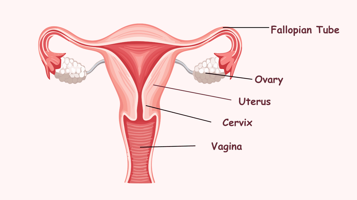
Figure 1. The uterus, fallopian tubes and ovaries.
The ovaries are paired intraperitoneal pelvic organs, 3–5 cm long, attached to the back of the broad ligament by the mesovarium.
Ligaments (routes of direct spread): utero-ovarian (to the uterus), suspensory or infundibulopelvic (to the pelvic side wall, carrying the ovarian vessels) and mesovarium (to the broad ligament).
Arteries: the ovarian artery (from the aorta) and the ovarian branch of the uterine artery.
Veins: the right ovarian vein drains to the IVC, the left to the left renal vein.
Route
Where it goes
How common
What to look for on PET/CT
Peritoneal seeding (commonest)
Cells follow peritoneal fluid: pouch of Douglas, paracolic gutters (especially the right), right subphrenic space, greater omentum
Most women already have stage III–IV disease at diagnosis, mostly peritoneal; the omentum, right subphrenic space and pouch of Douglas are the commonest sites
Omental cake; nodules on bowel serosa, liver and spleen capsule and right hemidiaphragm; ascites
Lymphatic
Along the ovarian vessels to para-aortic and paracaval nodes at the renal hilum; to pelvic nodes via the broad ligament; rarely to inguinal nodes via the round ligament
Clinical stage I–II epithelial cancer: 14% (para-aortic only 7%, pelvic only 3%, both 4%; serous 23%, mucinous 3%). Advanced disease: supradiaphragmatic nodes on PET/CT in 67% vs 33% on CT in one series
Para-aortic nodes up to the renal veins; cardiophrenic, internal mammary and other supradiaphragmatic nodes
Haematogenous (late)
Liver and spleen parenchyma, lung, pleura, bone, brain
Uncommon at diagnosis
Parenchymal (not capsular) liver lesions and a malignant pleural effusion are stage IV
Nodal figures from Kleppe 2011 (early stage) and Hynninen 2012 (supradiaphragmatic nodes).
2. Epidemiology and risk factors
About 325 000 new cases and 207 000 deaths worldwide in 2022: the most lethal gynaecological cancer.
Most women present with advanced (stage III–IV) disease because early disease causes few symptoms.
About 10% of cases are hereditary. The cumulative risk to age 80 is 44% for BRCA1 and 17% for BRCA2 carriers.
Increased risk
Reduced risk
Increasing age (peak 60–70 years for epithelial cancer)
Combined oral contraceptive use
BRCA1 or BRCA2 mutation
Pregnancy and breastfeeding
Lynch syndrome
Salpingectomy or salpingo-oophorectomy
Family history of ovarian or breast cancer
Tubal ligation
Endometriosis (clear cell and endometrioid types)
Nulliparity, early menarche, late menopause
3. Classification
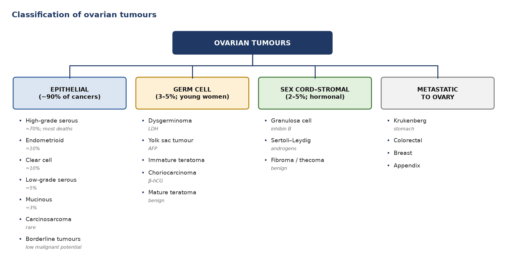
Figure 2. Classification of ovarian tumours by cell of origin, with the commoner types and their markers.
Category
Type
Share or key point
Epithelial (~90%; age 60–70)
High-grade serous carcinoma
≈70% of epithelial cancers; causes most deaths
Endometrioid carcinoma
≈10%; linked to endometriosis
Clear cell carcinoma
≈10%; linked to endometriosis
Low-grade serous carcinoma
≈5%
Mucinous carcinoma
≈3%; exclude a gastrointestinal primary
Carcinosarcoma
Rare, aggressive
Borderline (low malignant potential)
Serous borderline tumour
Younger women; more than 90% stage I
Mucinous borderline tumour
Younger women; more than 90% stage I
Germ cell (3–5%; young women)
Dysgerminoma
Commonest malignant germ cell tumour; LDH
Yolk sac tumour
AFP
Immature teratoma
Scattered fat and calcification
Choriocarcinoma
β-hCG
Mature teratoma (dermoid)
Benign; the commonest germ cell tumour
Sex cord–stromal (2–5%; hormonal)
Granulosa cell tumour
Oestrogen; inhibin B
Sertoli–Leydig cell tumour
Androgens; virilisation
Fibroma / thecoma
Benign; Meigs' syndrome
Metastatic to ovary (often bilateral)
Krukenberg tumour
Stomach (signet-ring cell)
Colorectal, breast, appendix
Look for a primary elsewhere
FDG uptake by type
Type
FDG uptake
High-grade serous, endometrioid, carcinosarcoma
Usually high
Dysgerminoma and other malignant germ cell tumours
Usually high
Clear cell
Variable
Low-grade serous, mucinous, borderline tumours
Often low: a negative scan does not exclude them
Calcified (psammomatous) serous deposits
May be low
Mature teratoma, fibroma, cystadenoma
Usually low
4. Diagnosis and initial work-up
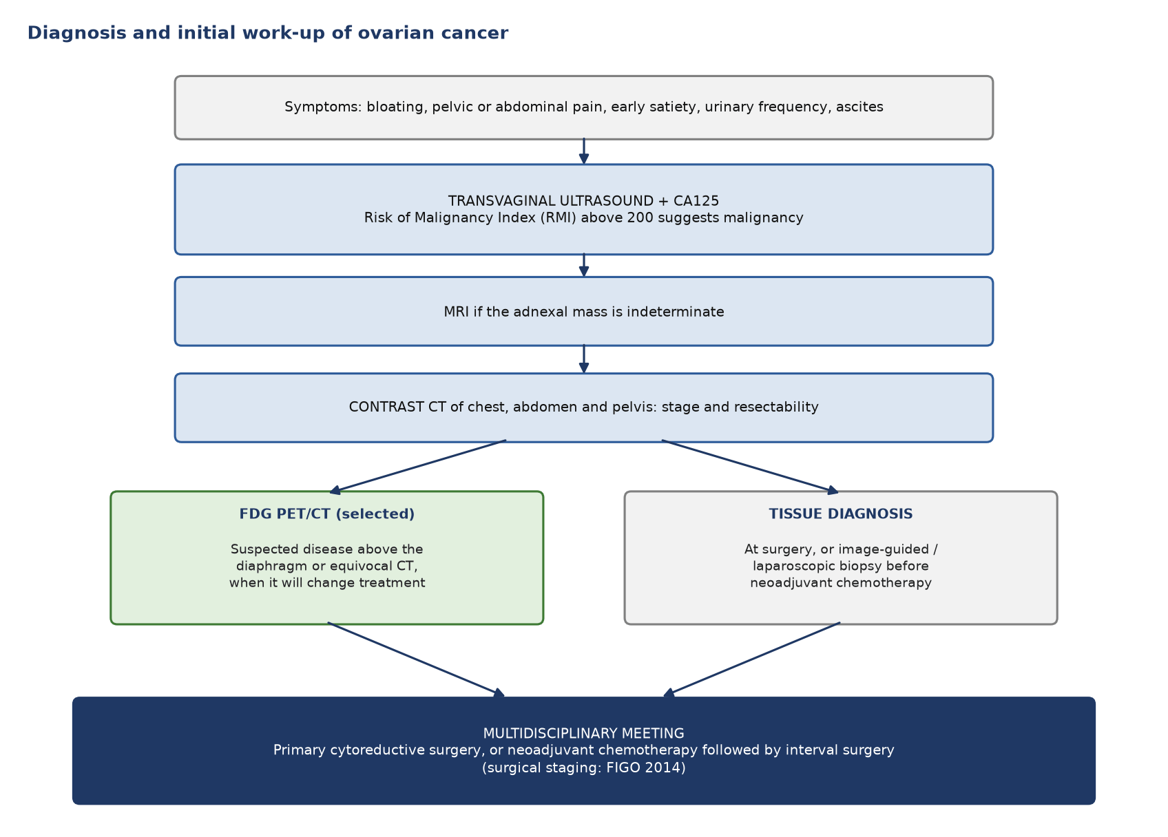
Figure 3. Diagnosis and initial work-up of ovarian cancer. Ultrasound and MRI characterise the mass; CT stages it; PET/CT is selective.
Tumour
Serum marker
Epithelial
CA125 (raised in about 80%)
Granulosa cell
Inhibin B (and oestradiol)
Yolk sac tumour
AFP
Choriocarcinoma
β-hCG
Dysgerminoma
LDH
Sertoli–Leydig
Testosterone
5. Staging (FIGO 2014)
Stage
Disease
I
Confined to the ovaries or tubes (IA one side; IB both sides; IC capsule rupture, surface tumour or malignant washings)
II
Extension to pelvic organs (IIA uterus or tubes; IIB other pelvic tissues)
III
Peritoneum outside the pelvis and/or retroperitoneal nodes: IIIA1 nodes only; IIIA2 microscopic peritoneal disease; IIIB deposits up to 2 cm; IIIC deposits over 2 cm, including the liver or spleen capsule
IV
Distant disease: IVA malignant pleural effusion; IVB liver or spleen parenchyma, nodes outside the abdomen (including inguinal), transmural bowel involvement
Staging is surgical: imaging maps the disease and helps decide whether it can be resected.
Two imaging calls change the stage: capsular liver or spleen implants (III) versus parenchymal metastases (IV), and supradiaphragmatic nodes (IV).
6. PET tracers
Tracer
Target
Role
¹⁸F-FDG
Glucose metabolism
Recurrence with a rising CA125; extra-abdominal staging
⁶⁸Ga-FAPI
Fibroblast activation protein in tumour stroma
Peritoneal and nodal disease (investigational)
¹⁸F-FES (fluoroestradiol)
Oestrogen receptor
Receptor status in low-grade serous cancer (investigational; approved only for breast cancer)
7. Indications for FDG PET/CT
Clinical situation
Role
Why
Characterising an adnexal mass
Not indicated
Ultrasound and MRI do this; physiological ovarian uptake in premenopausal women and low uptake in borderline tumours mislead
Screening
Not indicated
–
Initial staging of advanced disease
Selected cases, as an option alongside CT
Finds disease above the diaphragm that makes the stage IV
Before interval or secondary surgery
Useful
Excludes extra-abdominal disease that would make surgery pointless
Rising CA125 with negative or equivocal CT
Main indication
Highest pooled sensitivity for recurrence (91%)
Suspected recurrence on CT
Problem solving
Separates active disease from post-treatment change
Response to chemotherapy
Not established
CT with RECIST and GCIG CA125 criteria is standard
Radiotherapy planning (isolated nodal relapse)
Selected cases
Maps active nodes relative to the renal vessels
8. Diagnostic accuracy
Test
Question
Sensitivity
Specificity
FDG PET or PET/CT
Nodal metastases
73%
97%
CT
Nodal metastases
43%
95%
MRI
Nodal metastases
55%
88%
FDG PET/CT
Recurrence
91%
AUC 0.96
FDG PET/CT
Peritoneal metastases
70%
–
FAPI PET/CT
Peritoneal metastases
97%
–
Sources: Yuan 2012 (nodes); Gu 2009 (recurrence; CA125 alone had the highest specificity, 93%); Florit 2025 (peritoneum).
9. Patient preparation specific to ovarian cancer
Standard FDG preparation: fasting, glucose check and a 60-minute uptake period.
Menstrual history: record the first day of the last period, contraception and hormone therapy. For elective scans in premenopausal women, scan just before or a few days after menstruation to avoid physiological ovarian and endometrial uptake.
Exclude pregnancy.
Void just before imaging the pelvis. Oral contrast, if local practice uses it, helps separate bowel from peritoneal nodules.
Record: the date of surgery, chemotherapy cycles and dates, G-CSF, and the latest CA125 value and its trend.
10. What to look at when reporting
Adnexa: any mass, its uptake, and whether it is solid or cystic; ovarian uptake after the menopause is abnormal.
Peritoneum: omentum, paracolic gutters, right subphrenic space and diaphragm, liver and spleen surfaces, pouch of Douglas, bowel serosa and mesentery, ascites.
Nodes: pelvic, para-aortic up to the renal veins, cardiophrenic, internal mammary and other supradiaphragmatic nodes, and inguinal nodes (stage IVB).
Liver and spleen: capsular (stage III) or parenchymal (stage IV).
Thorax: pleural effusion or nodules, lung.
Resectability: small-bowel mesentery root, porta hepatis, lesser sac, para-aortic nodes above the renal vessels, pelvic side wall, hydronephrosis.
Conclusion: name the stage-changing findings, say that a negative PET does not exclude small peritoneal implants, and put the result in the context of the CA125 in recurrence.
11. Pitfalls
Pitfall
Why it happens
How to avoid it
Ovarian uptake in premenopausal women
Follicle or corpus luteum mid-cycle
Menstrual history; ultrasound; rim uptake on a thin-walled cyst is physiological
Endometrial uptake
Menstruation and ovulation
Thin, central uptake that matches the cycle day
Bowel uptake
Physiological activity
Follow the loop on CT; delayed images
Urine in ureters and bladder
Excreted FDG
Trace the ureter; void and re-image the pelvis
Small peritoneal implants
Below PET resolution and next to bowel
Report them as not excluded; surgery remains the reference
Ultrasound first and MRI if indeterminate; PET/CT is not a characterisation test
Standard practice
Initial staging, advanced disease
Contrast CT of the chest, abdomen and pelvis, MRI or FDG PET/CT, with a structured report, are options
ESGO-ESMO-ESP 2024 [III, A]
Suspected recurrence or rising CA125
PET/CT when CT is negative or equivocal
Gu 2009 meta-analysis
Follow-up
Clinical review and CA125; image when relapse is suspected. Treating a rising CA125 alone did not improve survival
MRC OV05/EORTC 55955 (Rustin 2010)
FAPI / FES PET
Research use
Not yet in guidelines
Test yourself
3 quick questions. Pick an answer to see the explanation.
1. Two years after treatment for high-grade serous ovarian cancer, CA125 has risen from 18 to 95 U/mL. Contrast CT is normal. What is the most useful next test?
2. A 34-year-old on day 14 of her cycle has staging PET/CT for lymphoma. There is rim-like uptake (SUVmax 4.0) around a 2 cm thin-walled cyst in the left ovary. What is the most likely explanation?
3. Staging imaging in advanced ovarian cancer shows a 3 cm deposit on the surface of the liver and a separate lesion inside the right lobe. How do they affect the FIGO stage?
References
Prat J; FIGO Committee on Gynecologic Oncology. Staging classification for cancer of the ovary, fallopian tube, and peritoneum. Int J Gynaecol Obstet. 2014;124(1):1-5.
Ledermann JA, Matias-Guiu X, Amant F, et al. ESGO-ESMO-ESP consensus conference recommendations on ovarian cancer: pathology and molecular biology and early, advanced and recurrent disease. Ann Oncol. 2024;35(3):248-66.
Bray F, Laversanne M, Sung H, et al. Global cancer statistics 2022: GLOBOCAN estimates of incidence and mortality worldwide for 36 cancers in 185 countries. CA Cancer J Clin. 2024;74(3):229-63.
Kuchenbaecker KB, Hopper JL, Barnes DR, et al. Risks of breast, ovarian, and contralateral breast cancer for BRCA1 and BRCA2 mutation carriers. JAMA. 2017;317(23):2402-16.
Kleppe M, Wang T, Van Gorp T, et al. Lymph node metastasis in stages I and II ovarian cancer: a review. Gynecol Oncol. 2011;123(3):610-4.
Hynninen J, Auranen A, Carpén O, et al. FDG PET/CT in staging of advanced epithelial ovarian cancer: frequency of supradiaphragmatic lymph node metastasis challenges the traditional pattern of disease spread. Gynecol Oncol. 2012;126(1):64-8.
Lee IO, Lee JY, Kim HJ, et al. Prognostic significance of supradiaphragmatic lymph node metastasis detected by 18F-FDG PET/CT in advanced epithelial ovarian cancer. BMC Cancer. 2018;18(1):1165.
Yuan Y, Gu ZX, Tao XF, Liu SY. Computer tomography, magnetic resonance imaging, and positron emission tomography or positron emission tomography/computer tomography for detection of metastatic lymph nodes in patients with ovarian cancer: a meta-analysis. Eur J Radiol. 2012;81(5):1002-6.
Gu P, Pan LL, Wu SQ, et al. CA 125, PET alone, PET-CT, CT and MRI in diagnosing recurrent ovarian carcinoma: a systematic review and meta-analysis. Eur J Radiol. 2009;71(1):164-74.
Florit A, de Koster EJ, Sassano S, et al. Head-to-head comparison of fibroblast activation protein inhibitors (FAPI) radiopharmaceuticals and [18F]FDG in gynaecological malignancies: systematic literature review and meta-analysis. Eur J Nucl Med Mol Imaging. 2025;52(11):3975-89.
Rustin GJ, van der Burg ME, Griffin CL, et al. Early versus delayed treatment of relapsed ovarian cancer (MRC OV05/EORTC 55955): a randomised trial. Lancet. 2010;376(9747):1155-63.
Lerman H, Metser U, Grisaru D, et al. Normal and abnormal 18F-FDG endometrial and ovarian uptake in pre- and postmenopausal patients: assessment by PET/CT. J Nucl Med. 2004;45(2):266-71.
Nishizawa S, Inubushi M, Okada H. Physiological 18F-FDG uptake in the ovaries and uterus of healthy female volunteers. Eur J Nucl Med Mol Imaging. 2005;32(5):549-56.
van Kruchten M, de Vries EF, Arts HJ, et al. Assessment of estrogen receptor expression in epithelial ovarian cancer patients using 16α-18F-fluoro-17β-estradiol PET/CT. J Nucl Med. 2015;56(1):50-5.