Radioactive decay
Types of radioactive decay
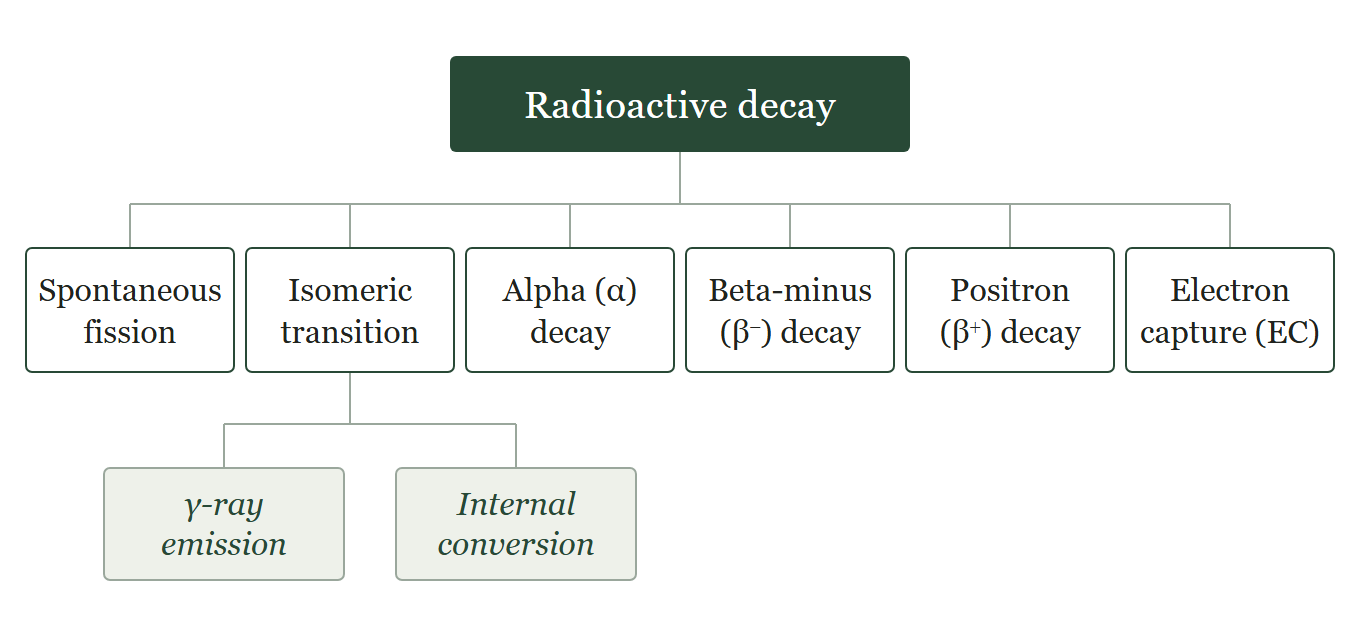
Spontaneous fission
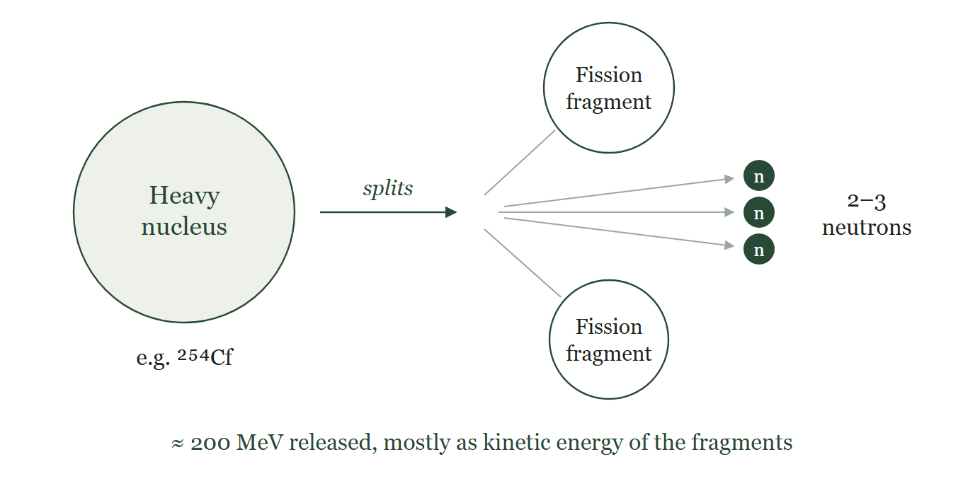
- Spontaneous fission is a process in which a heavy nucleus splits on its own into two smaller nuclei accompanied by emission of 2–3 neutrons.
- The neutrons carry a mean energy of about 2 MeV and about 200 MeV of energy is released as heat in this process.
- It occurs only in very heavy nuclei; the probability is low and increases with mass number of nuclei.
Isomeric transition
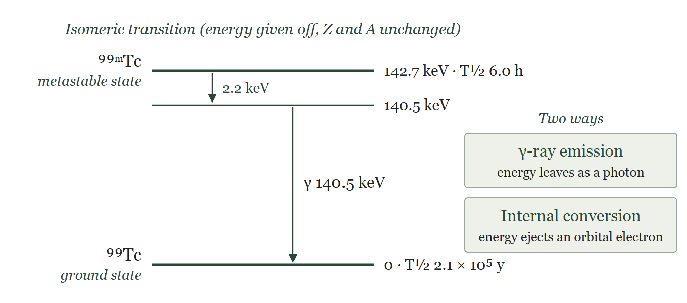
- A nucleus can exist in different energy states above its ground state; these excited states are called isomeric states.
- An isomeric state is called metastable when its half-life is long enough to be measured, conventionally longer than about 10⁻⁹ s (1 ns). Most excited states decay within about 10⁻¹² s. Metastable states are denoted by "m", as in ⁹⁹ᵐTc (T½ 6.0 h).
- An excited nucleus decays to a lower energy state by giving off its energy; such transitions are called isomeric transitions (IT).
- Isomeric transitions occur in two ways: γ-ray emission and internal conversion.
γ-ray emission
- The common mode of isomeric transition is the emission of electromagnetic radiation called the γ-ray.
- The energy of the γ-ray is the difference between the energies of the two isomeric states.
Internal conversion
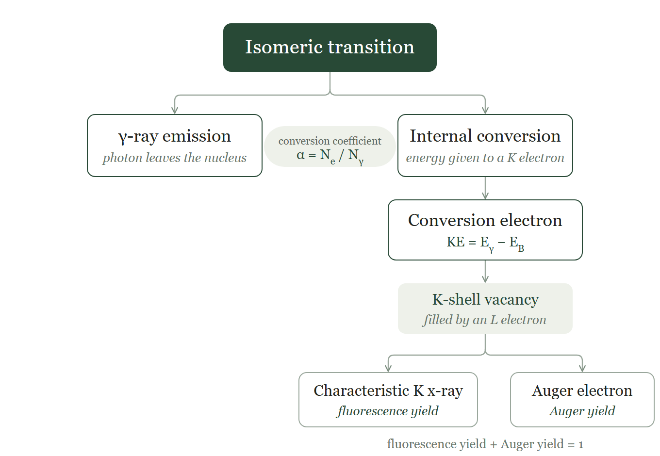
- The excited nucleus transfers its excitation energy to an orbital electron of its own atom, preferably a K-shell electron, which is then ejected from the shell.
- This occurs only if the excitation energy is greater than the binding energy of the electron.
- The ejected electron is called the conversion electron and carries kinetic energy equal to Eγ − EB, where Eγ is the excitation energy and EB is the binding energy of the electron.
- K-shell electrons are the most likely to be ejected because of their proximity to the nucleus.
- The conversion coefficient (α) is the ratio of the number of conversion electrons (Ne) to the number of observed γ-rays (Nγ): α = Ne/Nγ.
- Internal conversion leaves a vacancy in the shell, which is filled by an electron from a higher shell.
- When an L-shell electron fills a K-shell vacancy, the energy difference between the K and L shells is emitted as a characteristic K x-ray.
- Alternatively, this energy may be transferred to another orbital electron, which is ejected; this electron is called an Auger electron, and the process is called the Auger process.
- The Auger electron carries kinetic energy equal to the characteristic x-ray energy minus its own binding energy; Auger electrons are therefore monoenergetic.
- The fraction of vacancies filled with emission of characteristic x-rays is the fluorescence yield; the fraction filled by the Auger process is the Auger yield.
Alpha (α) decay
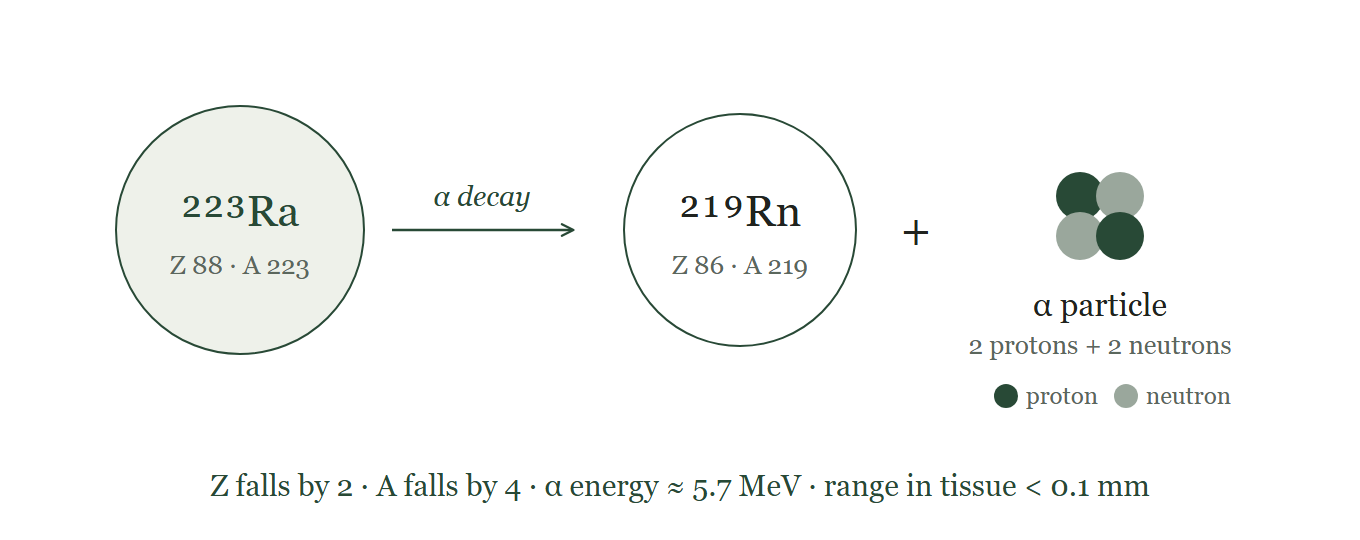
- α decay occurs mostly in heavy nuclides such as uranium, radon and plutonium.
- α particles are helium ions: a nucleus of two protons and two neutrons, with the two orbital electrons of the helium atom removed.
- After α decay, the atomic number of the nucleus falls by 2 and the mass number by 4.
- The energy of the α particle equals the energy difference between the two nuclear levels and ranges from 1 to 10 MeV.
- The range of α particles in matter is very short: less than 0.1 mm (about 40–100 µm) in body tissue.
- α particles can be stopped by a sheet of paper, a few centimetres of air, or gloves.
- Example: ²²³Ra → ²¹⁹Rn + α (Z 88 → 86, A 223 → 219; α energy about 5.7 MeV; T½ 11.4 days). ²²³Ra dichloride is used to treat bone metastases from prostate cancer.
Beta-minus (β⁻) decay
- β⁻ decay occurs in neutron-rich nuclides, whose N/Z ratio is higher than that of the nearest stable nuclide.
- The nucleus emits a β⁻ particle (an electron created in the nucleus) together with an antineutrino (ν̄): n → p + β⁻ + ν̄.
- The daughter's atomic number rises by 1, and its mass number stays the same.
- The energy released is called the transition (decay) energy, Eₘₐₓ.
- Eₘₐₓ is shared between the β⁻ particle and the antineutrino, so β⁻ particles have a spread of energies; their average is about one-third of Eₘₐₓ.
- The antineutrino has no charge and almost no mass. It carries away the rest of Eₘₐₓ in each decay, which is why it was proposed: to keep energy conserved.
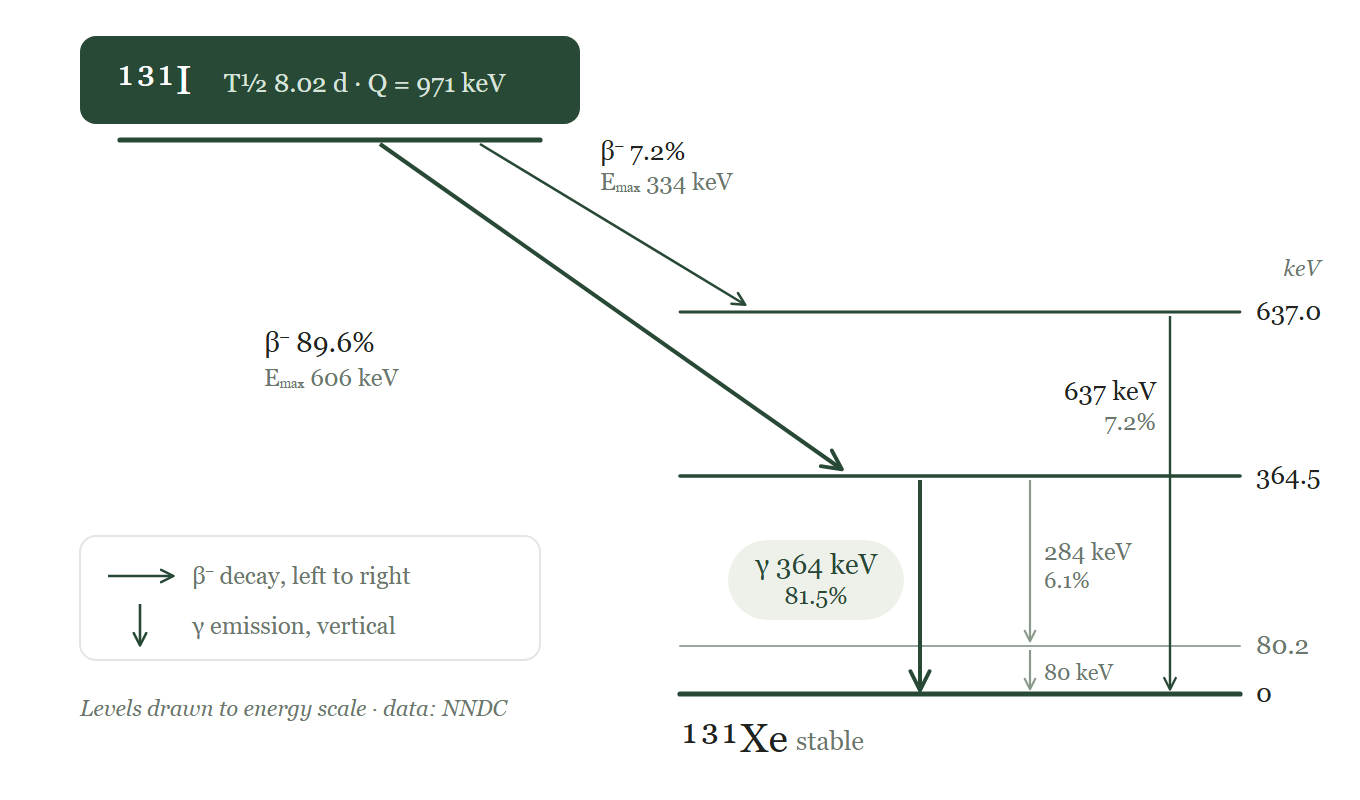
- A decay scheme shows how a radionuclide decays. β⁻ decay is drawn as an arrow running left to right from parent to daughter, and isomeric transition as a vertical arrow between two energy levels (Figure 6).
- Example: ¹³¹I → ¹³¹Xe + β⁻ + ν̄ (T½ 8.0 days). Most decays reach an excited level of ¹³¹Xe, which then emits the 364 keV γ-ray used for imaging and dosimetry.
Positron (β⁺) decay
- β⁺ decay occurs in proton-rich nuclides, whose N/Z ratio is lower than that of the nearest stable nuclide.
- A proton in the nucleus changes into a neutron, emitting a positron (β⁺) and a neutrino (ν), the counterpart of the antineutrino: p → n + β⁺ + ν.
- The daughter's atomic number falls by 1, and its mass number stays the same.
- β⁺ decay is only possible if the parent has at least 1.022 MeV more mass–energy than the daughter. This is the mass of two electrons (2 × 0.511 MeV).
- Any energy above 1.022 MeV is shared as kinetic energy between the positron and the neutrino, so positrons also have a spread of energies up to Eₘₐₓ.
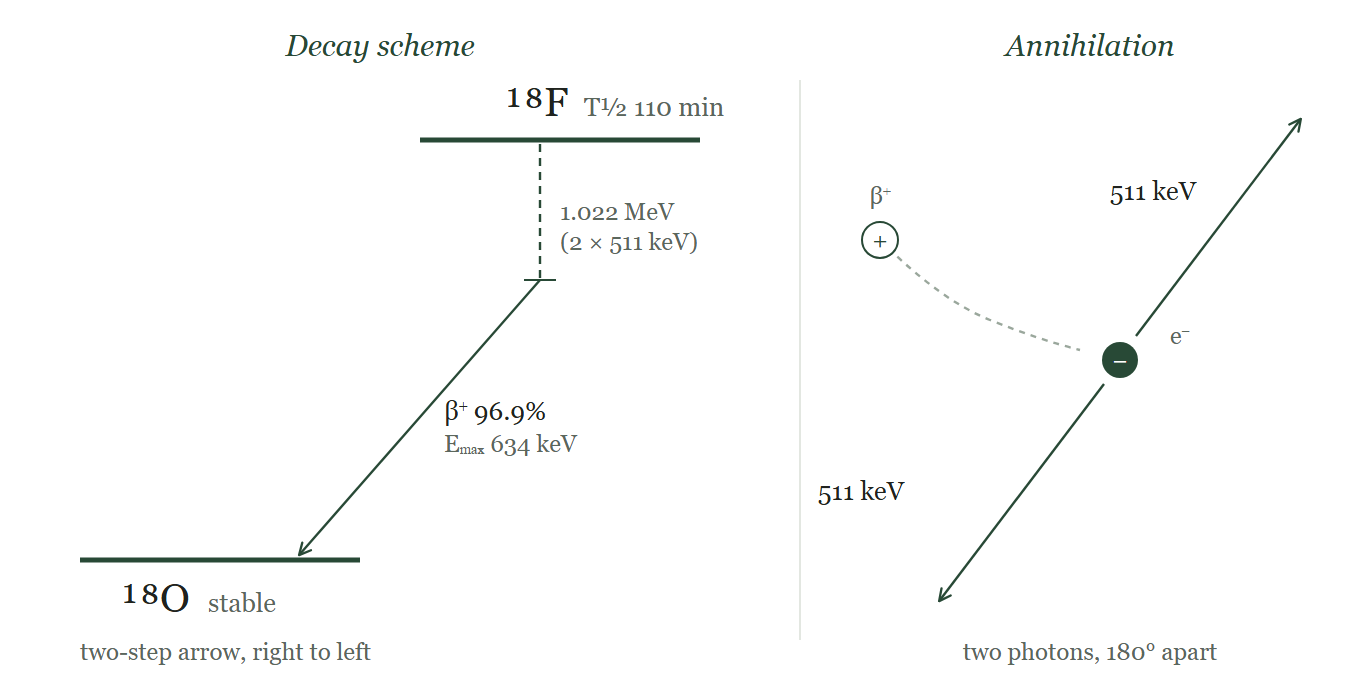
- Examples: ¹⁸F → ¹⁸O + β⁺ + ν and ⁶⁸Ga → ⁶⁸Zn + β⁺ + ν.
- Once the positron has lost almost all its energy, it combines with an electron and both are annihilated. Their mass becomes two 511 keV photons emitted in opposite directions. This annihilation radiation is what PET detects.
- In a decay scheme, β⁺ decay is drawn as a two-step arrow running right to left: a vertical drop of 1.022 MeV, then a slanted arrow to the daughter (Figure 7).
Electron capture (EC)
- Electron capture is the alternative to β⁺ decay for proton-rich nuclides, whose N/Z ratio is lower than that of the stable nuclide.
- An orbital electron, usually from the K shell because it lies closest to the nucleus, is captured by a proton, which becomes a neutron; a neutrino is emitted to conserve energy: p + e⁻ → n + ν.
- The daughter's atomic number falls by 1, and its mass number stays the same.
- EC is the usual route when the parent has less than 1.022 MeV more mass–energy than the daughter, too little for β⁺ decay.
- When the energy difference is above 1.022 MeV, both EC and β⁺ decay can occur; the higher the energy, the more likely β⁺ decay becomes.
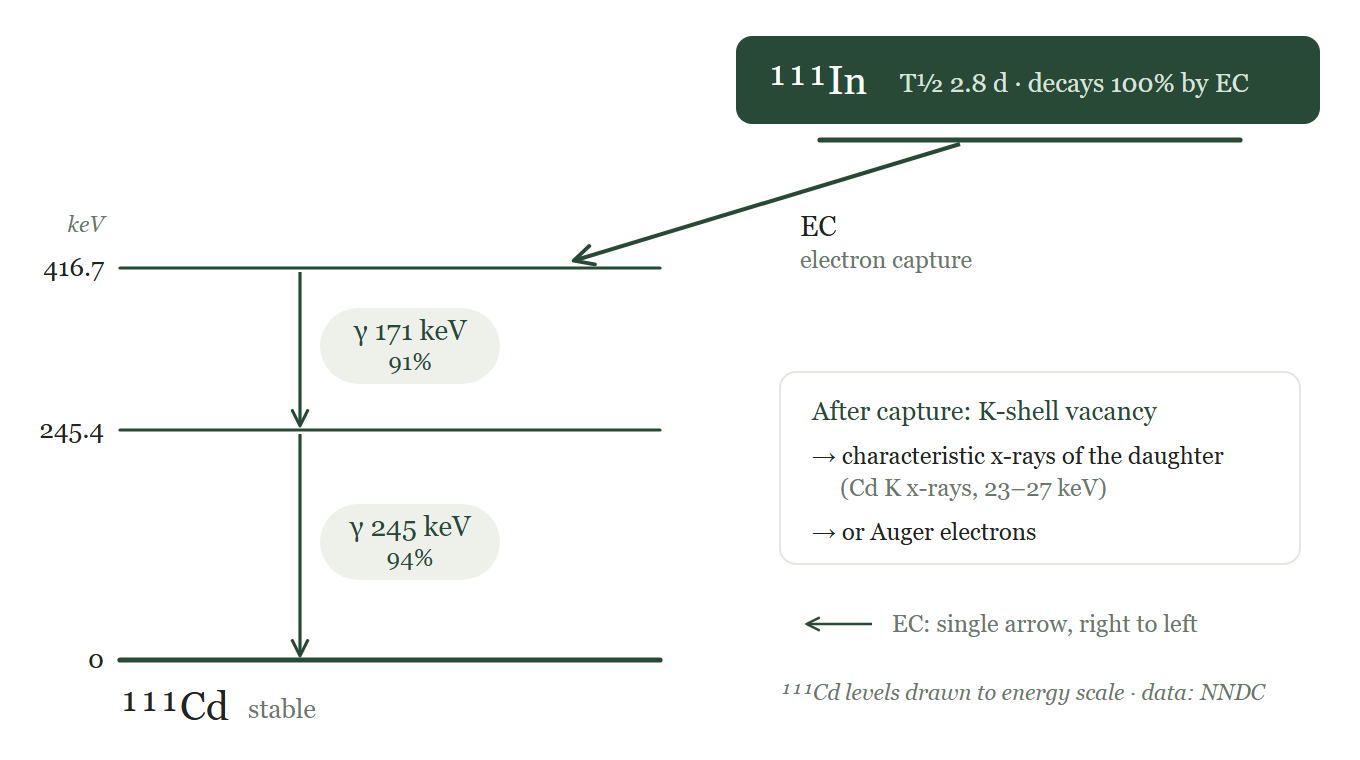
- Example: ¹¹¹In + e⁻ → ¹¹¹Cd + ν (T½ 2.8 days). The daughter emits γ-rays of 171 and 245 keV, which are used for imaging (Figure 8).
- Capture leaves a vacancy in the inner shell. It is filled by an electron from a higher shell, giving off a characteristic x-ray of the daughter or, alternatively, an Auger electron (the Auger process).
Test yourself
Revise this chapter with the Radioactive decay deck in Flashcards.
10 quick questions. Pick an answer to see the explanation.
1. Which mode of decay changes neither the atomic number nor the mass number?
Answer: C. Isomeric transition only releases excess energy; the numbers of protons and neutrons stay the same.
2. In ⁹⁹ᵐTc, a 140.5 keV transition is internally converted and ejects a K-shell electron (binding energy 21.0 keV). What is the kinetic energy of the conversion electron?
Answer: B. Kinetic energy = excitation energy − binding energy = 140.5 − 21.0 = 119.5 keV.
3. A transition has a conversion coefficient (α) of 0.25. What fraction of these transitions gives a γ-ray?
Answer: D. α = Nₑ/N_γ = 0.25, so for every 4 γ-rays there is 1 conversion electron: 4 of 5 transitions (80%) give a γ-ray.
4. Which statement about β⁻ particles is correct?
Answer: B. The decay energy is shared with the antineutrino, so β⁻ energies form a spread with a mean of about Eₘₐₓ/3.
5. What is the minimum energy difference between parent and daughter needed for β⁺ decay?
Answer: B. β⁺ decay needs at least two electron masses: 2 × 0.511 = 1.022 MeV.
6. A proton-rich nuclide has 0.8 MeV more mass–energy than its daughter. How will it decay?
Answer: B. Below 1.022 MeV there is not enough energy for β⁺ decay, so only electron capture is possible.
7. ²²³Ra (Z = 88) decays by α emission. What is the daughter?
Answer: A. α decay lowers Z by 2 and A by 4: ²²³Ra (88) → ²¹⁹Rn (86).
8. A patient is imaged after ¹³¹I therapy. Which nucleus emits the 364 keV γ-ray that is detected?
Answer: C. The γ-ray comes from an excited level of the daughter, ¹³¹Xe, after β⁻ decay of ¹³¹I.
9. What does the fluorescence yield describe?
Answer: B. The fluorescence yield is the fraction of vacancies filled by x-ray emission; the rest are filled by the Auger process.
10. In a decay scheme, a two-step arrow running from right to left shows which decay?
Answer: C. β⁺ decay is drawn as a vertical drop of 1.022 MeV followed by a slanted arrow from right to left.
References
- Saha GB. Physics and Radiobiology of Nuclear Medicine. 4th ed. New York: Springer; 2013. Chapter 2.
- Cherry SR, Sorenson JA, Phelps ME. Physics in Nuclear Medicine. 4th ed. Philadelphia: Elsevier Saunders; 2012.
- National Nuclear Data Center, Brookhaven National Laboratory. NuDat 3: evaluated nuclear structure and decay data.
Spotted an error or something out of date? Report an error