PET in Adult Diffuse Glioma
1. Classification
- Scope: Adult-type diffuse glioma. Brain metastasis and primary CNS lymphoma enter the differential, but have separate clinical pathways.
- Integrated diagnosis: WHO CNS5 combines histology with molecular testing. Imaging can suggest a pattern and select a biopsy site; it cannot establish IDH status, 1p/19q codeletion or CNS WHO grade. [1]
| Adult-type diffuse glioma | Defining molecular finding | CNS WHO grade | Imaging clue, not a diagnosis |
|---|---|---|---|
| Astrocytoma, IDH-mutant | IDH1/2 mutation; no whole-arm 1p/19q codeletion | 2, 3 or 4 | Often infiltrative; enhancement varies. |
| Oligodendroglioma, IDH-mutant and 1p/19q-codeleted | Both IDH mutation and whole-arm 1p/19q codeletion | 2 or 3 | Cortical–subcortical lesion; calcification is frequent, but not specific. |
| Glioblastoma, IDH-wildtype | IDH-wildtype with defining histological or molecular features | 4 | Necrosis and irregular enhancement are common; a less dramatic scan does not exclude it. |
Table 1. WHO CNS5 adult-type diffuse gliomas. Tissue and molecular tests remain decisive. [1] [2]
2. Workup: presentation to tissue
- Presentation: A first seizure, progressive focal deficit, cognitive change or raised intracranial pressure often starts the workup. Stabilise an acute neurological problem before refining tumour type. [2]
- Urgent CT: Non-contrast CT is quick for haemorrhage, hydrocephalus, marked mass effect and calcification. Contrast CT can show enhancement when MRI cannot be obtained, but is less useful for mapping infiltrative tumour. [2]
- Diagnostic MRI: T1 before and after contrast, T2/FLAIR, diffusion and susceptibility sequences define anatomy and the enhancing and non-enhancing components. Perfusion or spectroscopy can answer a further question. MRI remains the main study for surgical planning and follow-up. [2] [3]
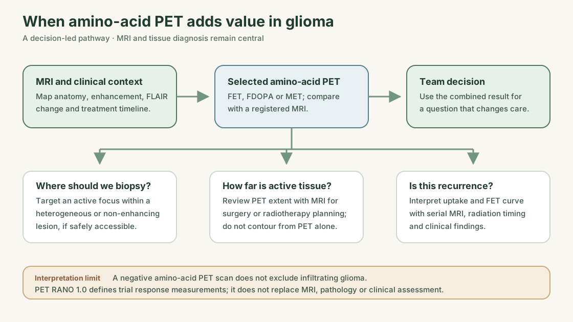
- Tissue: Maximal safe resection or stereotactic biopsy supplies histology and molecular markers. If the lesion is heterogeneous, MRI and selected amino-acid PET can identify a viable, safely accessible target. [1] [3]
- PET/CT detail: The CT acquired for attenuation correction is usually low-dose and non-contrast. Do not treat it as a substitute for diagnostic CT or MRI. Review recent MRI registered to PET. [4]
3. CT appearance
- Infiltration: A diffuse glioma may be an ill-defined low-attenuation area with expansion of the affected brain. CT often underestimates its extent; FLAIR MRI is more useful. [2]
- Enhancement: Little or no enhancement does not guarantee a low grade. Irregular or ring enhancement suggests blood–brain barrier disruption and may accompany aggressive tumour, but is not specific for it. [2]
- Calcification: Coarse or patchy calcification raises oligodendroglioma in the differential. It does not establish 1p/19q codeletion. [1] [2]
- Necrosis and haemorrhage: Central low attenuation, an irregular enhancing rim, haemorrhage and substantial oedema can occur in high-grade glioma. Metastasis and other lesions can look similar. [2]
- Mass effect: Sulcal effacement, ventricular compression or midline shift shows immediate clinical impact; it is not a grading test. [2]
4. PET tracers and uptake patterns
- FDG: Normal grey matter is intensely FDG avid. An infiltrating glioma can look hypo- or isometabolic to cortex; an aggressive focus may be avid, but inflammation, lymphoma and metastasis overlap. State the reference tissue when describing uptake. [4] [5]
- Amino-acid tracers: FET, FDOPA and MET usually have lower normal-brain background. They can show an active focus in a non-enhancing FLAIR lesion or beyond an enhancing margin. Uptake still cannot prove glioma or grade. [3] [4]
| Tracer | Useful feature | Main interpretation limit |
|---|---|---|
| ¹⁸F-FDG | Widely available; marked uptake can help frame a differential. | High cortical background and inflammatory uptake limit contrast and specificity. |
| ¹⁸F-FET | Low normal-brain background; dynamic curves can add information. | Seizure, inflammation and early blood-pool activity can mislead; curves overlap. |
| ¹⁸F-FDOPA | Low cortical background; useful for selected extent and recurrence questions. | Physiological striatal uptake complicates basal-ganglia lesions. |
| ¹¹C-MET | Established amino-acid tracer with low background. | Carbon-11's short half-life usually requires on-site production. |
Table 2. Choose the tracer for the clinical question and local availability. Do not transfer a numeric threshold between tracers or protocols. [3] [4]
5. New brain lesion: CT and PET differential
| Possibility | CT clue | PET clue | Check that matters |
|---|---|---|---|
| Diffuse glioma | Infiltrative low attenuation; variable enhancement. Calcification or necrosis may be present. | FDG ranges from low to high relative to cortex. Amino-acid uptake may show an active focus, but can be absent. | MRI extent, diffusion/perfusion and tissue with molecular tests. |
| Primary CNS lymphoma | Often a solid, hyperattenuating, enhancing lesion; pattern changes with immune status. | Often strongly FDG avid, but overlaps high-grade glioma. | Diffusion MRI, clinical context and biopsy; record steroid exposure. |
| Metastasis | Often well-defined with surrounding oedema; may be multiple. | FDG and amino-acid uptake overlap glioma. Small lesions may be missed. | Contrast MRI for multiplicity and systemic cancer assessment; tissue if uncertain. |
| Abscess / infection | Ring lesion with oedema; CT alone cannot reliably separate it from necrotic tumour. | FDG may be intense; amino-acid uptake can also occur. | Diffusion MRI, infection history and microbiology or sampling. |
| Inflammatory / demyelinating lesion | Variable low attenuation and enhancement; may resemble infiltrating tumour. | FDG or amino-acid uptake is possible. | MRI morphology, time course and targeted clinical workup; biopsy if unresolved. |
Table 3. These are clues, not stand-alone rules. Ischaemia and seizure-related change are further mimics; timing and diffusion MRI matter. [2] [5] [6]
6. When PET adds value
| Question after MRI | What amino-acid PET can contribute | Decision it may inform |
|---|---|---|
| Where to biopsy? | Locate an active part of a heterogeneous or non-enhancing lesion. | Select a target that is also safe to sample. |
| How far does active tumour extend? | Show uptake beyond enhancement or within broad FLAIR change. | Discuss resection or radiotherapy margins alongside anatomy and function. |
| Is new change recurrence? | Add metabolic evidence to serial MRI after treatment. | Decide whether to observe, sample or change treatment. |
| Is treatment working? | Compare uptake and active extent with a comparable baseline PET. | Interpret discordant MRI, including possible pseudoresponse. |
Table 4. Selected indications in the 2025 RANO/EANO update. PET is an adjunct, not a requirement for every glioma. [3]
7. Treated lesion: recurrence or treatment effect?
| Possibility | Timing and MRI/CT context | PET pattern and limit | Next check |
|---|---|---|---|
| Tumour recurrence | New or enlarging enhancement or FLAIR change, sometimes at the cavity edge or beyond the treated field. | Focal or increasing amino-acid uptake supports active tumour; FDG may be obscured by cortex. No single cut-off proves recurrence. | Compare serial MRI and PET; sample if a treatment-changing result remains uncertain. |
| Pseudoprogression | New or increased enhancement after chemoradiotherapy, especially early in follow-up. | Uptake may be low, but inflammation can cause uptake; PET cannot settle every case. | Compare serial MRI and use the timing rule in section 8. |
| Radiation necrosis | Delayed change, usually within an irradiated region; enhancement and oedema can fluctuate. | Often lower amino-acid uptake than viable tumour, but mixed tumour and injury are common. | Examine the radiation plan, serial MRI and clinical course; consider tissue if management depends on the distinction. |
Table 5. These processes can coexist. CT and PET/CT CT alone are poor arbiters; use MRI and treatment dates. [3] [4] [7]
- Dynamic FET: An early peak followed by decline can favour active aggressive tissue; a steadily rising curve may favour treatment change or less aggressive tissue. Report the curve with its method. It is supporting evidence, not a binary test. [4]
- Antiangiogenic treatment: Reduced MRI enhancement can reflect altered vascular permeability rather than loss of tumour. Review non-enhancing disease and the clinical course; PET can add information when MRI appears discordant. [3] [7]
8. RANO: which criteria apply?
RANO 2.0: MRI response
RANO 2.0 standardises MRI response assessment in adult-glioma trials; it does not identify tumour subtype. [7]
| RANO 2.0 item | Practical rule |
|---|---|
| Baseline MRI | After radiotherapy for newly diagnosed glioma, use the first post-radiotherapy MRI. Without radiotherapy, use the trial's specified pretreatment MRI. |
| What to measure | Measure enhancing tumour in typical enhancing IDH-wildtype glioblastoma; use T2/FLAIR for non-enhancing tumour. A mixed lesion may need both. Exclude oedema or treatment change where possible. |
| Measurable lesion | Clear MRI margins and two perpendicular diameters of ≥10 mm on one slice. Do not count a cavity alone; a measurable nodule at its edge can count. |
| Partial response | ≥50% fall in the sum of target-lesion area products, or ≥65% fall in volume, sustained for ≥4 weeks with stable or decreasing steroids. |
| Progression | ≥25% rise in the sum of target-lesion area products, or ≥40% rise in volume, from the smallest prior measurement. A new measurable lesion can also indicate progression. |
| First 12 weeks after radiotherapy | New enhancement may be pseudoprogression. In the relevant trial setting, confirm suspected progression in a clinically stable patient by repeat MRI or unequivocal histology; clear progression outside the radiation field is an exception. |
| Clinical context | Record neurological status and steroid dose. Either can change how an MRI finding is interpreted. |
Table 6. Selected RANO 2.0 rules; use the full criteria for formal trial response assignment. [7]
- Example: Six weeks after chemoradiotherapy, a new enhancing focus appears within the treated field. FET uptake raises concern but cannot establish recurrence alone. Compare the radiation plan and serial MRI; obtain tissue when the result will change care. [3] [7]
PET RANO 1.0: amino-acid PET response
- Purpose: PET RANO 1.0 gives diffuse-glioma trials a standard way to compare static amino-acid PET over time. It is separate from MRI-based RANO 2.0; FDG and dynamic FET curves are not its measurement basis. [8]
- Terms: TBRmax and TBRmean are the maximum and mean tumour-to-healthy-brain uptake ratios. PET volume is the amount of tissue meeting the PET-positive threshold. [8]
| PET RANO 1.0 item | Key rule in a measurable target lesion |
|---|---|
| PET-positive and measurable | Segment uptake at ≥1.6 × mean healthy-brain background; PET-positive volume must be >0.5 mL. |
| Partial response | ≥30% fall in TBRmax, ≥10% fall in TBRmean, or ≥40% fall in PET volume, with no lesion meeting progression criteria. |
| Progression | ≥30% rise in TBRmax, ≥10% rise in TBRmean, ≥40% rise in PET volume, or a new measurable PET-positive lesion. |
| Complete response / stable disease | Complete response means disappearance of all PET-positive disease with no new lesion; stable disease meets neither response nor progression rules. |
Table 7. These are selected PET RANO 1.0 trial definitions; multiple target lesions require the full lesion-level rules. The thresholds do not diagnose recurrence from a single scan. [8]
- No measurable PET lesion at baseline: A partial or complete PET response cannot be assigned without a measurable target; stable or progressive disease remains assessable. [8]
- Compare like with like: Keep the tracer, uptake interval, scanner/reconstruction and reference-region method comparable. State method changes before interpreting a shift in TBR or PET volume. Neither RANO framework replaces pathology or clinical judgement. [4] [7] [8]
9. Reporting and pitfalls
Report in this order
- Question and context: Integrated diagnosis if known; surgery, radiotherapy and drug dates; steroids; seizures; symptoms; comparison MRI. [4]
- Technique: Tracer, uptake interval, static or dynamic acquisition, MRI registration and method changes. The procedure guideline recommends at least four hours of fasting. [4]
- Finding: Site, size/extent, uptake pattern and relation to enhancement and FLAIR. Give TBR and PET-positive volume only with the method used; describe the dynamic FET curve when acquired. [4]
- Answer: State what combined PET/MRI findings favour, the degree of uncertainty and what would resolve a consequential doubt. [3] [4]
| Pitfall | Practical check |
|---|---|
| Negative amino-acid PET in an infiltrating lesion | Do not dismiss persistent MRI abnormality; some gliomas have little uptake. |
| Small focus | PET resolution lowers apparent uptake; inspect registered MRI. |
| FDG near cortex | Normal grey-matter uptake can hide tumour; state the reference tissue. |
| FDOPA near striatum | Recognise physiological basal-ganglia activity. |
| Seizure, infection or inflammation | Uptake can mimic tumour; check symptoms, timing and MRI. |
| Recent surgery or radiotherapy | Treatment effect can mimic recurrence; compare prior studies and the treated field. |
| Abnormal reference brain or misregistration | Atrophy, infarction or poor fusion can distort TBR or localisation; inspect source images. |
Table 8. Common interpretation traps in the joint procedure guideline and PET/RANO pitfalls report. [4] [6]
Test yourself
- A calcified frontal lesion has faint FDG uptake and a focal FET hotspot. Is it an oligodendroglioma? The pattern can raise suspicion and guide a safe biopsy site. Only tissue and IDH/1p19q testing establish the subtype. [1] [3]
- New enhancement appears six weeks after chemoradiotherapy, with modest FET uptake. Is this definite recurrence? No. Check whether the focus lies within the treated field and compare serial MRI. In the relevant RANO 2.0 setting, confirm suspected progression with repeat MRI or unequivocal tissue evidence; PET uptake alone cannot settle it. [3] [7]
Further reading: Brain tumours overview.
References
- Louis DN, et al. The 2021 WHO Classification of Tumors of the Central Nervous System: a summary. Neuro Oncol. 2021.
- Gliomas in adults: guidance on investigations, diagnosis, treatment and surveillance. Clin Med. 2024.
- Galldiks N, et al. Update to the RANO/EANO recommendations for the clinical use of PET imaging in gliomas. Lancet Oncol. 2025.
- Law I, et al. Joint EANM/EANO/RANO/SNMMI procedure standard for PET imaging of gliomas. Eur J Nucl Med Mol Imaging. 2019.
- Albert NL, et al. RANO/EANO recommendations for clinical PET imaging in gliomas. Neuro Oncol. 2016.
- Galldiks N, et al. Challenges, limitations, and pitfalls of PET and advanced MRI in patients with brain tumors. Neuro Oncol. 2024.
- Wen PY, et al. RANO 2.0: response criteria for adult gliomas. J Clin Oncol. 2023.
- Albert NL, et al. PET RANO 1.0: amino-acid PET response assessment in diffuse glioma. Lancet Oncol. 2024.
Spotted an error or something out of date? Report an error