PET in Brain Tumours
- Enhancement is not tumour. Contrast enhancement on MRI shows a leaky blood–brain barrier. After chemoradiotherapy for glioblastoma, pseudoprogression affects up to 30–40% of patients in the first 12 weeks, so RANO 2.0 requires progression to be confirmed.
- Amino-acid PET is the tracer class of choice. ¹⁸F-FET, ¹⁸F-FDOPA and ¹¹C-methionine cross the intact barrier by L-type amino-acid transport, so normal brain is low and tumour stands out. For a new brain lesion, pooled FET sensitivity was 94% against 38% for FDG.
- Regulatory position in 2026. ¹⁸F-FET (Pixclara) was approved by the FDA on 14 September 2026 to separate recurrent or progressive glioma from treatment-related change (company announcement); it has long been used in Europe. US approval of ¹⁸F-FDOPA covers Parkinsonian syndromes only.
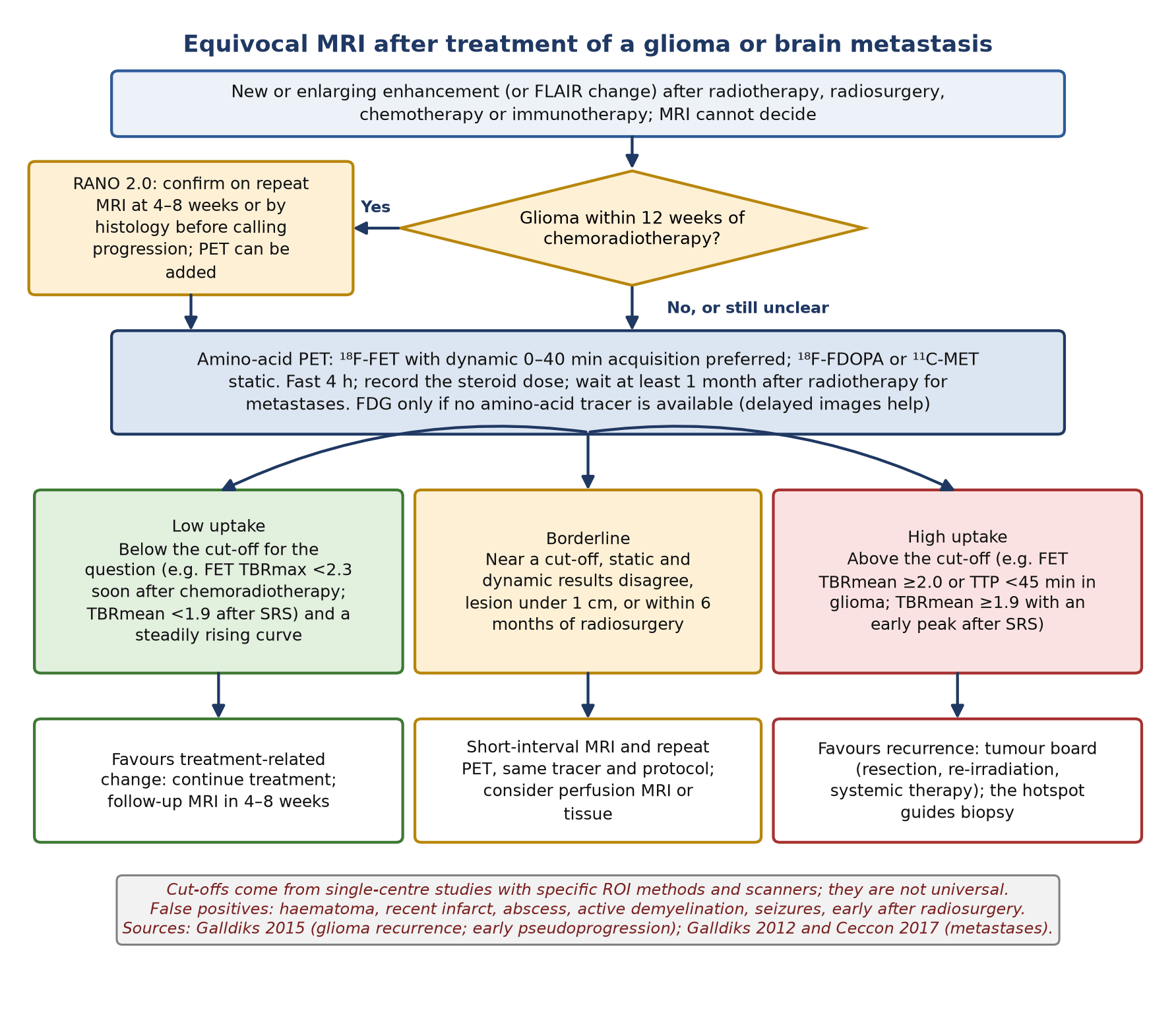
- Cut-offs belong to a question. FET TBRmax 2.5 separates tumour from non-tumour; TBRmean ≥2.0 or time to peak <45 min signals glioma recurrence; TBRmax 2.3 separates early pseudoprogression. Each came from one centre's method and none is universal.
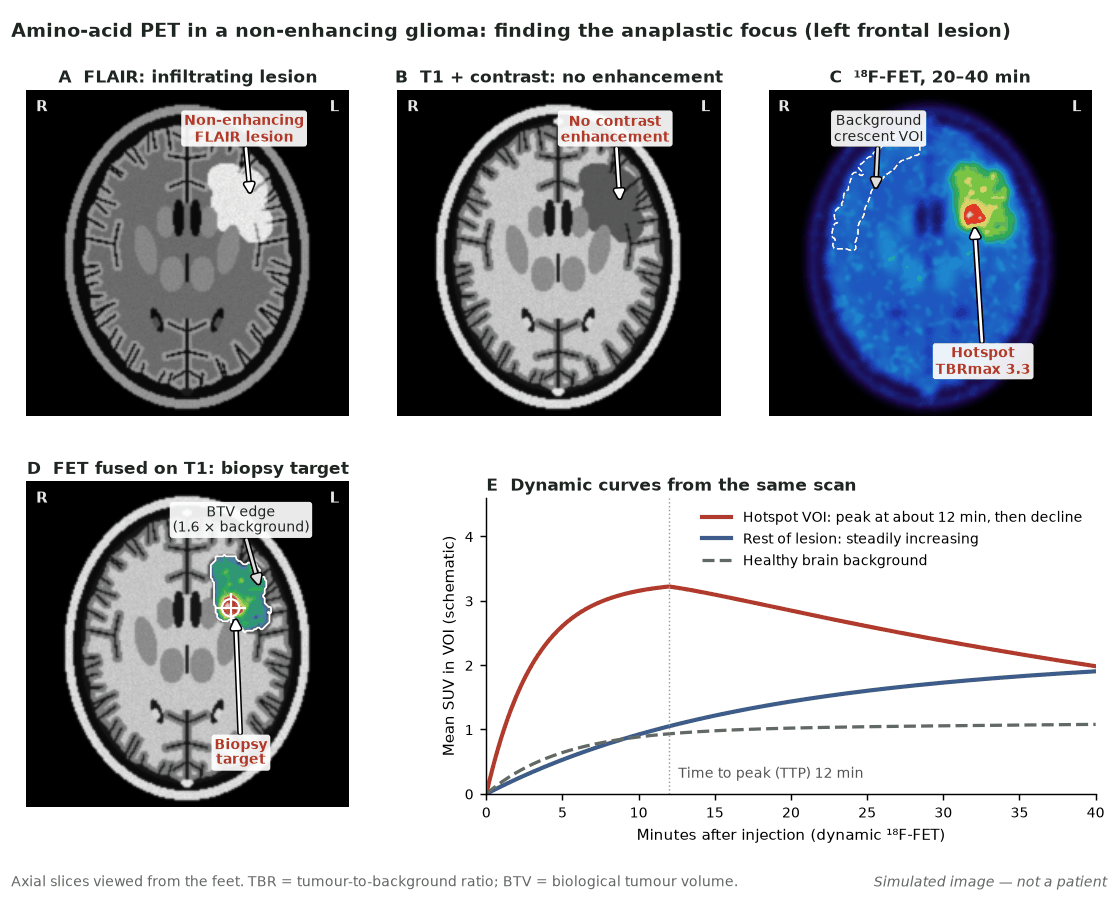
- Read the curve, not only the ratio. On dynamic FET, an early peak (before 20 min) with a plateau or fall favours high-grade tumour or recurrence; a curve that keeps rising favours lower-grade glioma or treatment-related change.
- A negative scan has limits. About one-third of WHO grade 2 gliomas take up little FET. A negative scan makes a grade 3–4 glioma, lymphoma or metastasis unlikely but does not exclude a low-grade glioma.
- PET RANO 1.0 defines response. PET-positive tissue is ≥1.6 × mean background; measurable disease exceeds 0.5 mL; progression is a rise of ≥30% in TBRmax, ≥10% in TBRmean or ≥40% in volume, or a new measurable lesion.
- Meningioma is an SSTR problem. ⁶⁸Ga-DOTATATE or ⁶⁸Ga-DOTATOC PET shows skull-base and bone extent that MRI misses; SUVmax 2.3 separated tumour from tumour-free tissue. PRRT remains investigational, with disease control in about two-thirds of treated patients.
1. The clinical problem
The neuro-oncology team asks five questions of imaging: is this a tumour, where is its most aggressive part, how far does it extend, is new enhancement after treatment tumour or treatment effect, and is treatment working? Magnetic resonance imaging (MRI) answers the anatomical part; positron emission tomography (PET) is added when the answer depends on biology.
Classification in force: WHO CNS5 (2021)
- Integrated diagnosis. The fifth edition of the World Health Organization (WHO) classification of central nervous system (CNS) tumours combines histology with molecular markers in a layered report, and grades tumours within each type using Arabic numerals.
- Adult-type diffuse gliomas. Three types remain: astrocytoma, IDH-mutant (CNS WHO grade 2, 3 or 4); oligodendroglioma, IDH-mutant and 1p/19q-codeleted (grade 2 or 3); and glioblastoma, IDH-wildtype (grade 4). IDH is isocitrate dehydrogenase.
- Molecular grading. Homozygous deletion of CDKN2A/B makes an IDH-mutant astrocytoma grade 4. An IDH-wildtype diffuse astrocytic glioma in an adult is a glioblastoma if it has TERT promoter mutation, EGFR amplification or combined gain of chromosome 7 and loss of chromosome 10, even without necrosis or microvascular proliferation.
- Why it matters for PET. Most cut-offs were derived in cohorts graded by the older histological system, and the joint EANM/EANO/RANO/SNMMI guideline notes that uptake data by molecular type are limited; apply grade-based thresholds to CNS5 diagnoses with care.
Response criteria in force: RANO 2.0 (2023)
- One set of criteria. Response Assessment in Neuro-Oncology (RANO) 2.0 replaces separate high-grade and low-grade criteria. For newly diagnosed glioma the post-radiotherapy MRI, not the post-operative MRI, is the baseline.
- Definitions. Partial response is a ≥50% decrease in the sum of products of perpendicular diameters (or ≥65% by volume) sustained for at least 4 weeks on stable or falling steroids. Progression is a ≥25% increase (or ≥40% by volume) from the smallest measurement.
- The 12-week rule. Pseudoprogression occurs in up to 30–40% of glioblastomas in the 12 weeks after chemoradiotherapy. In a clinically stable patient, progression in this window must be confirmed on repeat MRI (for example at 4 or 8 weeks) or by histology.
- Where PET stands. RANO 2.0 states that amino-acid PET may help separate progression from pseudoprogression but needs further validation before formal inclusion. The separate PET RANO 1.0 criteria (section 7) fill that gap for trials.
Why enhancement after treatment is non-specific
Gadolinium enhancement reflects vascular surface area and leakage across a disrupted blood–tumour barrier, not tumour cell number. Radiotherapy, surgery, inflammation and immunotherapy disrupt the barrier; corticosteroids and anti-angiogenic drugs such as bevacizumab restore it, so enhancement can fade while tumour grows (pseudoresponse). T2 and fluid-attenuated inversion recovery (FLAIR) change is no more specific: oedema, radiation injury, demyelination and ischaemia look the same. After stereotactic radiosurgery (SRS) for metastases, radiation injury and recurrence both appear as an enlarging ring-enhancing lesion.
- Before treatment: choosing the biopsy target in a non-enhancing glioma, and defining metabolically active tumour beyond the enhancing margin.
- After treatment: separating recurrence from pseudoprogression or radiation injury in glioma and in metastases after radiotherapy, and recognising pseudoresponse.
- Meningioma: defining skull-base and transosseous extent, distinguishing recurrence from scar, and selecting patients for peptide receptor radionuclide therapy (PRRT).
- Lymphoma: suggesting primary CNS lymphoma (PCNSL) rather than glioblastoma on ¹⁸F-FDG, ideally before steroids are started.
2. Tracers and why they work
¹⁸F-fluoroethyl-L-tyrosine (FET), ¹¹C-methyl-L-methionine (MET) and 3,4-dihydroxy-6-¹⁸F-fluoro-L-phenylalanine (FDOPA) are carried across the blood–brain barrier and into cells by system L amino-acid transport. Gliomas overexpress L-type amino-acid transporters, chiefly LAT1, so tumour-to-background contrast is high and uptake does not depend on barrier breakdown. This is why amino-acid PET shows the non-enhancing parts of a glioma and why it is less affected than MRI by treatment-induced leakage. ¹⁸F-fluorodeoxyglucose (FDG) reports glucose metabolism, which is intense in normal grey matter; this single fact explains most of its failures in the brain.
| Tracer | Mechanism | Physiological uptake | Practical points |
|---|---|---|---|
| ¹⁸F-FET | LAT1/LAT2 transport | Slight: vessels and venous sinuses, basal ganglia, cerebellum, skin, salivary glands; occasionally pineal, choroid plexus, clivus | Half-life 110 min allows central supply. The only tracer with established dynamic (kinetic) criteria. FDA-approved in 2026 for recurrence versus treatment-related change |
| ¹¹C-MET | System L transport | As for FET | 20-min half-life needs an on-site cyclotron. Kinetic analysis does not help grading |
| ¹⁸F-FDOPA | LAT1 transport, then decarboxylation | Moderate in striatum and pituitary; slight in cerebellum, skin, salivary glands; no vascular uptake | Striatal uptake can hide adjacent tumour; the guideline reference region is the contralateral striatum. No carbidopa needed |
| ¹⁸F-FDG | Glucose metabolism | High in cortex, deep grey nuclei and cerebellum; moderate in eye muscles | Poor contrast for glioma. Niche: PCNSL, high-grade tumours, and metastasis versus radiation injury with delayed images |
| ⁶⁸Ga-DOTATATE / ⁶⁸Ga-DOTATOC | Somatostatin receptor type 2 (SSTR2) binding | High in pituitary; slight in vessels, choroid plexus, salivary glands; almost none in brain | Tracer of choice for meningioma; ⁶⁴Cu-DOTATATE and ¹⁸F-SiTATE are alternatives |
Physiological uptake and practical points after the EANM/EANO/RANO/SNMMI glioma guideline (2019), the PET-RANO/EANO recommendations (2016) and the joint meningioma guideline (2024).
Avidity by tumour type
- High-grade glioma. More than 95% of WHO grade 3 and 4 gliomas show increased FET uptake.
- Low-grade glioma. About one-third of WHO grade 2 gliomas, and most dysembryoplastic neuroepithelial tumours, are FET-negative. Uptake overlaps widely between grades, and oligodendroglial tumours are often avid; about half of grade 2 oligodendroglial tumours show a decreasing curve, which mimics a high-grade pattern.
- Brain metastases. FET uptake in untreated metastases is highly variable, most of all in melanoma, and small lesions are underestimated because of limited resolution. PET adds little to MRI for detecting metastases; its value is after treatment.
- Meningioma. Meningiomas overexpress SSTR2, giving intense uptake on SSTR ligands against almost no brain background. A few meningiomas take up little tracer, and grade cannot be read from uptake intensity.
When FDG fails and what replaces it
In a meta-analysis of five histology-controlled studies (119 patients), FET detected brain tumours with a pooled sensitivity of 94% and specificity of 88%, against 38% and 86% for FDG; the authors concluded that FET should be preferred for a new isolated lesion. FDG fails because grey-matter uptake hides tumours in or near cortex, grade 2 gliomas are often hypometabolic, and inflammation (abscess, demyelination, fungal infection, neurosarcoidosis) is avid. It keeps three niches: PCNSL, which has a higher maximum standardised uptake value (SUVmax) than glioblastoma unless corticosteroids have reduced it; lymphoma versus opportunistic infection; and recurrence versus radiation injury when no amino-acid tracer is available, read against white matter (more sensitive) or grey matter (more specific), with delayed images at 3–5 h for metastases.
3. Indications by clinical scenario
The PET-RANO/EANO glioma recommendations (2016) included only Oxford level 1–3 studies but gave no grade per indication; the RANO/PET reports on metastases (2019) and meningioma (2017) did give levels.
| Scenario | Preferred tracer | What PET adds | Guideline position |
|---|---|---|---|
| New brain lesion: tumour or not? | FET or MET | Higher accuracy than MRI alone; a high TBR has a high positive predictive value | PET-RANO/EANO 2016: recommended when MRI is uncertain (histology-validated studies) |
| Grading and the anaplastic focus in a non-enhancing glioma | Dynamic FET | About 40% of MRI-'low-grade' non-enhancing gliomas contain an anaplastic focus; kinetic analysis detected grade 3–4 tumour with sensitivity and specificity of about 95% | PET-RANO/EANO 2016: dynamic FET improves grading; static values overlap |
| Biopsy targeting | FET, MET or FDOPA | The hotspot marks the most malignant part (Figure 1) | PET-RANO/EANO 2016: particularly helpful in non-enhancing gliomas |
| Extent for surgery and radiotherapy | FET or MET | Biological tumour volume extends 2–3.5 cm beyond enhancement in high-grade glioma | PET-RANO/EANO 2016: feasible, preliminary evidence. GLIAA randomised trial: no benefit for FET-guided re-irradiation; contrast MRI remains the standard for that target |
| Glioma: recurrence versus treatment-related change | FET (dynamic), FDOPA or MET | Higher accuracy than MRI; FET TBRmean ≥2.0 or TTP <45 min gave 93% accuracy | PET-RANO/EANO 2016 and EANM/EANO/RANO/SNMMI 2019: common indication. FDA-approved indication of ¹⁸F-FET (2026) |
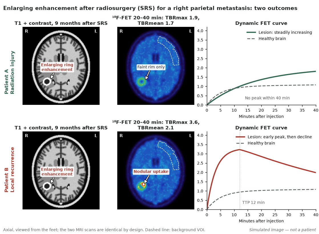
| Metastasis after radiotherapy or SRS: recurrence versus radiation injury | Amino-acid PET, dynamic FET best | High accuracy, highest with TBR plus curve shape | RANO/PET 2019: amino-acid PET useful (evidence level 2); FDG usable but variable (level 2); advanced MRI comparison limited (level 3). Scan at least 1 month after radiotherapy (2025 guideline) |
| Pseudoprogression after immunotherapy for metastases | FET | Pilot data only | RANO/PET 2019: evidence level 3 |
| Response assessment, including pseudoresponse | FET or FDOPA | Metabolic response precedes MRI change and predicts survival in some studies | PET RANO 1.0 (2024) for glioma trials; PET RANO BM 1.0 (2025) for metastasis trials; systemic therapy of metastases: level 3 |
| Meningioma: extent, recurrence versus scar, radiotherapy planning | ⁶⁸Ga-DOTATATE or DOTATOC | Skull-base and transosseous extent; detection better than contrast MRI | RANO/PET 2017: use when extent or recurrence is unclear (evidence class 3, recommendation level C); joint procedure guideline 2024 |
| PCNSL versus glioblastoma | FDG | Intense uptake favours lymphoma; steroids reduce it | PET-RANO/EANO 2016 and 2019 guideline: FDG role |
4. Evidence at a glance
| Study | Design and n | Key finding | What it changed |
|---|---|---|---|
| Rapp et al., J Nucl Med 2013 | Retrospective; 174 new lesions suggestive of glioma, histology in 168 | TBRmax 2.5 separated neoplastic from non-neoplastic lesions: sensitivity 57%, specificity 92%, PPV 98%, NPV 27%. TBRmax 2.5 separated high- from low-grade glioma: sensitivity 80%, specificity 65% | Defined FET cut-offs at diagnosis and showed that low uptake does not exclude tumour |
| Dunet et al., Neuro Oncol 2016 | Meta-analysis; 5 studies, 119 patients, histology | Brain tumour diagnosis: FET sensitivity 94%, specificity 88%; FDG 38% and 86%. Grading: similar for both | FET, not FDG, for a new isolated lesion |
| Galldiks et al., Neuro Oncol 2015 | Retrospective; 124 patients, 132 scans, suspected progression or recurrence; histology in 95% | TBRmean ≥2.0 or TTP <45 min: sensitivity 93%, specificity 100%, accuracy 93%, against 85% for MRI | Combined static and dynamic FET criteria for recurrence |
| Galldiks et al., Eur J Nucl Med Mol Imaging 2015 | 22 glioblastomas with new enhancement within 12 weeks of chemoradiotherapy | TBRmax 1.9 ± 0.4 in pseudoprogression versus 2.8 ± 0.5 in early progression; cut-off 2.3: sensitivity 100%, specificity 91%, accuracy 96% | FET for early pseudoprogression (small cohort) |
| Kebir et al., Clin Cancer Res 2016 | 26 glioblastomas with enlarging enhancement more than 3 months after chemoradiotherapy | Late pseudoprogression in 7. TBRmax cut-off 1.9: sensitivity 84%, specificity 86%; TTP 25 versus 40 min | Pseudoprogression is not confined to 12 weeks; FET helps later too |
| Yu et al., Front Oncol 2023 | Meta-analysis; 22 studies, 1514 patients | Glioma recurrence: FET sensitivity 84%, specificity 86%; FDOPA 95% and 90% (FDOPA data from small studies) | Both tracers accurate; head-to-head trials needed |
| Galldiks et al., J Nucl Med 2012 | 31 patients, 40 lesions after radiotherapy of brain metastases | TBRmean >1.9 plus curve pattern II or III: sensitivity 95%, specificity 91%, accuracy 93% | Dynamic FET for metastases after radiotherapy |
| Ceccon et al., Neuro Oncol 2017 | 62 patients, 76 lesions, mostly after SRS; histology in 34% | TBRmax 3.3 ± 1.0 in recurrence versus 2.2 ± 0.4 in radiation injury; TBR plus curve slope: accuracy 88% | Confirmed FET criteria for metastases |
| GLIAA / NOA-10 ARO2013-01 (Grosu et al., Lancet Oncol 2026) | Randomised; 200 patients with recurrent glioblastoma; re-irradiation 39 Gy in 13 fractions planned on FET PET or contrast MRI | Median progression-free survival 4.0 versus 4.9 months; adjusted hazard ratio 1.14 (95% CI 0.85–1.52) | FET-guided re-irradiation is not standard; contrast MRI remains the planning method |
| Rachinger et al., J Nucl Med 2015 | Prospective; 21 meningiomas, 115 neuronavigated samples | ⁶⁸Ga-DOTATATE SUVmax 2.3 separated tumour from tumour-free tissue; sensitivity 90% versus 79% for MRI | Histology-based threshold used in the 2024 guideline |
| Mirian et al., J Nucl Med 2021 | Individual-patient meta-analysis; 6 cohorts, 111 treatment-refractory meningiomas given PRRT | Disease control 63%; 6-month progression-free survival 94%, 48% and 0% for WHO grades 1, 2 and 3 | Best current estimate of PRRT benefit; no randomised data |
5. Patient preparation and acquisition
Only points that differ from routine FDG practice are listed (2019 glioma, 2025 metastasis and 2024 meningioma guidelines).
- Fasting. At least 4 h before amino-acid PET, because dietary amino acids compete for transport. The 2025 guideline advises postponing the scan if fasting was not respected, to keep serial studies comparable. For FDG it asks for a 6-h fast and postponement if glucose exceeds 8.9 mmol/L (160 mg/dL); the 2019 guideline asks for at least 4 h.
- FDOPA. Carbidopa premedication is not needed; most brain-tumour studies did not use it.
- Steroids. Record the dose. Corticosteroids may slightly lower amino-acid TBRs by raising uptake in healthy brain, and they reduce FDG uptake in lymphoma.
- MRI for fusion. Recent T1 with and without contrast and T2/FLAIR; check co-registration by displaying the scalp and nose.
- Positioning. Head holder, whole brain including the cerebellum in the field, patient watched throughout. A seizure during the uptake phase raises FET and FDG uptake in the affected cortex.
- Timing after treatment. Wait at least 1 month after radiotherapy or surgery for metastases. For a PET RANO baseline, scan within 14 days before treatment starts but as late as possible after surgery.
| Tracer | Adult activity | Acquisition | Quantification |
|---|---|---|---|
| ¹⁸F-FET | 185–200 MBq | Dynamic 0–40 or 0–50 min from injection: 12 × 5 s, 6 × 10 s, 6 × 30 s, 5 × 60 s, then 5-min frames; clinical read on the 20–40 min sum | TBRmax, TBRmean, biological tumour volume; time–activity curve |
| ¹¹C-MET | 370–555 MBq | 20-min static from 10 min | TBRmax, TBRmean |
| ¹⁸F-FDOPA | 185–200 MBq | 10–20 min static starting 10–30 min after injection | Tumour-to-striatum ratio (2019 guideline) or TBR (PET RANO 1.0) |
| ¹⁸F-FDG | 185–200 MBq | 10–20 min static at 45–60 min; optional delayed images at 3–5 h for metastases | Visual against white and grey matter |
| ⁶⁸Ga-DOTATATE | 150–200 MBq (DOTATOC 100–200 MBq) | 10–20 min static of the head at 60 min; whole body only if metastatic or spinal disease is suspected | SUVmax; ratio to superior sagittal sinus |
Activities and timing from the joint EANM/EANO/RANO/SNMMI procedure guidelines (glioma 2019, brain metastases 2025, meningioma 2024).
Reconstruction and the background region
- Reconstruction. Iterative reconstruction with all corrections, one reconstruction meeting EARL harmonisation standards, and optionally a high-resolution brain reconstruction (voxels under 3 mm, resolution under 6 mm). The 2019 glioma guideline advises against resolution modelling (point-spread-function) because of Gibbs artefacts; the 2024 meningioma guideline allows it with that caveat. Use the same scanner and protocol for serial scans, because TBR depends on resolution.
- Background. Draw a large crescent-shaped volume of interest (VOI) in healthy-appearing cortex, including grey and white matter, in the contralateral hemisphere; PET RANO 1.0 prefers the frontal lobe. Use its mean. TBRmax is the lesion SUVmax divided by the background mean; TBRmean is the mean of the tumour VOI divided by the background mean.
- Tumour VOI for curves. For the time–activity curve, use an isocontour around the tumour maximum giving 1–2 mL, or a 1.6-cm circle centred on the maximum, drawn on the 20–40 min (or 10–30 min) sum. Classify the curve as increasing, plateau or decreasing and note the time to peak (TTP).
6. Interpretation: quantification, variants and pitfalls
- Check motion and PET–MRI alignment before reading anything else.
- Look first: is uptake visibly above contralateral cortex, and where does it sit relative to enhancement and FLAIR change?
- Measure against the crescent background: TBRmax, TBRmean and, if needed, the biological tumour volume (voxels ≥1.6 × background).
- Read the dynamic curve (FET): pattern and TTP, with the healthy-brain curve plotted to exclude technical errors.
- Compare with the previous PET, done with the same tracer and protocol.
- Place the result against the clinical question and its own cut-off, the treatment timeline, steroids and MRI, then answer the question asked.
Cut-offs by clinical question
| Question | Tracer and parameter | Cut-off | Source |
|---|---|---|---|
| Neoplastic versus non-neoplastic | FET TBRmax / TBRmean | 2.5 / 1.9 | Rapp 2013 |
| Neoplastic versus non-neoplastic | MET TBRmax | 1.3–1.5 | 2019 guideline |
| Grade 2 versus grade 3–4 | FET TBRmean / TBRmax / TTP | 1.9–2.0 / 2.5–2.7 / <35 min | 2019 guideline |
| Tumour extent (biological volume) | FET / MET / FDOPA | 1.6 / 1.3 / 2.0 × background | 2019 guideline; PET RANO 1.0 uses 1.6 for all |
| Glioma recurrence | FET TBRmean (1.6-cm ROI) or TTP | ≥2.0 or <45 min | Galldiks 2015 (Neuro Oncol) |
| Glioma recurrence | MET TBRmax; FDOPA TSRmax / TSRmean | 1.6; 2.1 / 1.8 | 2019 guideline |
| Early pseudoprogression (≤12 weeks) | FET TBRmax | 2.3 | Galldiks 2015 (EJNMMI) |
| Late pseudoprogression (>12 weeks) | FET TBRmax / TBRmean | 1.9 / 1.9 | Kebir 2016; 2019 guideline |
| Malignant transformation of grade 2 glioma | FET TBRmax / TBRmean / TTP | Rise >33% / >13% / fall of 6 min | 2019 guideline |
| Metastasis after radiotherapy | FET TBRmean plus curve | ≥1.9 with TTP ≤20 min; or ≥1.95 with slope ≤0.37 SUV/h | Galldiks 2012; Ceccon 2017; 2025 guideline |
| Meningioma versus tumour-free tissue | ⁶⁸Ga-DOTATATE SUVmax | 2.3 | Rachinger 2015 |
Thresholds depend on the ROI method, scanner, reconstruction and clinical question. They were derived mostly in single centres on older scanners and are not universal; the metastasis guideline advises favouring specificity when choosing a cut-off. TSR, tumour-to-striatum ratio.
Time–activity curve patterns (dynamic FET)
- Pattern I: uptake increases steadily to the end of the acquisition. Seen in WHO grade 2 glioma and typical of treatment-related change (radiation injury, pseudoprogression); also seen in inflammatory lesions.
- Pattern II: early peak (TTP ≤20 min) followed by a plateau.
- Pattern III: early peak followed by a steady decline. Patterns II and III favour grade 3–4 glioma or recurrence.
- Change over time: a switch from an increasing curve to an early peak with decline in a grade 2 glioma indicates malignant transformation.
Normal variants
- FET and MET: slight uptake in venous sinuses and vessels, basal ganglia, cerebellum, skin and salivary glands; occasionally pineal body, choroid plexus and clivus marrow.
- FDOPA: moderate striatal and pituitary uptake. Tumour next to the basal ganglia is easily missed.
- SSTR ligands: high pituitary uptake, which varies with age and sex; slight uptake in vessels, choroid plexus and salivary glands.
Pitfalls and mimics
| Pitfall | Tracer | What happens | How to avoid it |
|---|---|---|---|
| Haematoma | FET; MET; FDG | Increased uptake for up to 14, 45 and 4 days after bleeding | Check MRI for blood products; wait or repeat |
| Recent infarct | FET; FDG | Increased uptake up to 7 and 14 days | Correlate with diffusion MRI and the clinical timeline |
| Abscess, inflammation, active demyelination | All | Moderate amino-acid uptake is possible (rare with FET); FDG uptake is variable and often high | Look at the pattern (rim), history and MRI; an increasing curve can indicate inflammation |
| Early after high-dose focused radiotherapy | FET | More PET-positive radiation-induced lesions in the first 6 months | Interpret cautiously; repeat PET |
| Vessels and venous sinuses | FET, MET | Blood-pool activity up to about 15 min looks like tumour; lesions near a sinus can show a falsely decreasing curve | Use late summed images; check curve against anatomy |
| Low reference uptake | Amino acids | Atrophy, infarct or reduced delivery in the background VOI inflates TBR | Keep the background away from damaged brain |
| Small lesions | All | Partial-volume loss under about 1 cm | Do not call a small lesion negative on TBR alone |
| FET-negative glioma | FET | About one-third of grade 2 gliomas show low uptake | A negative scan does not exclude a low-grade glioma |
| Oligodendroglial tumours | FET | A decreasing curve in about half of grade 2 tumours mimics high grade | Integrate with molecular diagnosis |
| Steroids | FDG; amino acids | Lower FDG uptake in lymphoma; slightly lower amino-acid TBR | Scan before steroids where possible; record the dose |
| Pituitary, inflammation, other tumours | SSTR | Physiological pituitary uptake; granulomatous disease and some metastases, gliomas or lymphomas take up tracer, usually less intensely | Read location and shape with MRI |
Occurrence and timings from the 2019 glioma guideline (Table 2), the 2025 metastasis guideline and the 2024 meningioma guideline.
How to word the conclusion
- Answer the question in the first sentence: for example, 'Findings favour treatment-related change rather than recurrent tumour.'
- Give the numbers and method (TBRmax, TBRmean, curve, background region) and the cut-off applied, with its source.
- State certainty ('favours', 'indeterminate: repeat in 4–8 weeks') and the limitation that matters: small lesion, recent bleed, steroids, recent radiosurgery, change of tracer or scanner.
7. Response assessment and follow-up
PET RANO 1.0 (Albert et al., 2024) is the first standard framework for amino-acid PET response in diffuse glioma. It was written for trials, rests largely on expert opinion, is intended first as a secondary or exploratory endpoint, and evaluates static PET alone, volumetrically, without MRI, steroid dose or clinical status.
| Term | PET RANO 1.0 definition |
|---|---|
| PET-positive disease | Voxels with SUV ≥1.6 × the mean of healthy background (crescent VOI, preferably frontal), for any amino-acid tracer; check the volume visually to exclude vessels, muscle, choroid plexus and (for FDOPA) striatum |
| Measurable disease | PET-positive volume >0.5 mL (equivalent to a diameter above 1 cm) |
| Non-measurable disease | Visible uptake below TBRmax 1.6, or a PET-positive volume under 0.5 mL |
| Target lesions | Highest uptake or largest volume; with several measurable lesions, 2–3 targets |
| Progressive disease (measurable baseline) | Increase of ≥30% in TBRmax, ≥10% in TBRmean or ≥40% in PET volume in any target lesion, or any new measurable lesion |
| Partial response | Decrease of ≥30% in TBRmax, ≥10% in TBRmean or ≥40% in volume, with no progression criterion met; a change from measurable to non-measurable disease also counts |
| Complete response | Disappearance of all PET-positive disease and no new lesion |
| Stable disease | None of the above |
| Non-measurable or PET-negative baseline | Progression only if a measurable lesion appears; stable disease is the best possible outcome |
Response is compared with baseline or with the nadir after a response.
- Timing. Baseline within 14 days before treatment; 4–6 weeks after surgery if no further treatment is planned. Follow-up every 2–3 months for grade 4 and every 3–6 months for grade 2–3 glioma, ideally with MRI. An optional scan 2–4 weeks after starting treatment assesses early metabolic response.
- Equivocal results. Repeat after one or two cycles with the same tracer; if progression is confirmed, backdate it to the first suspicious scan.
- Earlier response signals. The 2019 guideline lists a >20% fall in FET TBRmax 7–10 days into radiochemotherapy, and a >45% fall in biological tumour volume on bevacizumab, as markers of response.
- Brain metastases. PET RANO BM 1.0 (2025) uses the same progression thresholds (≥30% TBRmax, ≥10% TBRmean, ≥40% volume, or a new measurable lesion). In one study, a fall in FET TBR of at least 10% identified responders to immunotherapy or targeted therapy with 82% accuracy.
- Meningioma. No PET response framework exists; response is judged on MRI (RANO or modified Macdonald). After PRRT, the 2024 guideline asks for MRI and SSTR PET about 1 month after the second cycle and 2 months after the fourth.
8. Theranostics and emerging tracers
SSTR-targeted PRRT for meningioma
- Status. Investigational. It is not approved for meningioma by the FDA or the European Medicines Agency, and no randomised trial has reported. The 2024 joint guideline suggests considering it when surgery and radiotherapy are no longer options, after a multidisciplinary decision and preferably within a trial.
- Evidence. In an individual-patient meta-analysis of 111 treatment-refractory meningiomas, disease control was 63% and 6-month progression-free survival was 94%, 48% and 0% for WHO grades 1, 2 and 3. Higher total administered activity was associated with lower risk of progression.
- Regimen. Usually four cycles of 7.4 GBq ¹⁷⁷Lu-DOTATATE (or 3.7 GBq ⁹⁰Y-DOTATOC) 8 ± 2 weeks apart, with lysine–arginine infusion for renal protection. Stop long-acting somatostatin analogues 4–6 weeks before, and taper steroids where possible, because they may downregulate SSTR2.
- Trials. Randomised phase II trials of ¹⁷⁷Lu-DOTATATE against standard care are recruiting: EORTC (NCT06326190) and NRG/RTOG MOMENTUM-1 (NCT06955169). The SSTR antagonist ¹⁷⁷Lu-satoreotide is in phase I/II (NCT04997317).
| Agent | Use in brain tumours | Evidence level |
|---|---|---|
| ¹⁸F-FET | Glioma diagnosis, biopsy, extent, recurrence, response | FDA-approved for recurrence versus treatment-related change (September 2026, company announcement); guideline-endorsed |
| ¹⁸F-FDOPA | As for FET (static only) | Guideline-endorsed; European national authorisations include suspected recurrence or residual primary brain tumour; US label covers Parkinsonian syndromes only |
| ¹¹C-MET | As for FET (static only) | Guideline-endorsed; limited by on-site production |
| ⁶⁸Ga-DOTATATE, ⁶⁸Ga-DOTATOC, ⁶⁴Cu-DOTATATE, ¹⁸F-SiTATE | Meningioma extent, recurrence, PRRT selection | Guideline-endorsed (2024); in the USA ⁶⁸Ga- and ⁶⁴Cu-DOTATATE are the routinely available SSTR tracers |
| ¹⁸F-fluciclovine | Recurrence of glioma or metastasis | Investigational in the brain (repurposed); read on SUV rather than TBR |
| TSPO ligands | Glioblastoma imaging and prognosis | Investigational |
| PSMA ligands | Expressed in glioma neovasculature; imaging and possible therapy | Investigational |
| ¹⁸F-FLT, ¹¹C-choline, ¹⁸F-FMISO | Proliferation, membranes, hypoxia | Investigational; not ready for clinical use |
| ¹⁷⁷Lu-DOTATATE or ⁹⁰Y-DOTATOC PRRT | Refractory meningioma | Investigational; randomised trials recruiting |
TSPO, 18-kDa translocator protein; PSMA, prostate-specific membrane antigen; FLT, fluorothymidine; FMISO, fluoromisonidazole.
9. Structured report example
- Clinical question: IDH-wildtype glioblastoma, left temporal, resected and treated with chemoradiotherapy and temozolomide; radiotherapy finished 7 months ago. MRI shows new enhancement at the posterior cavity margin. Recurrence or treatment-related change? Dexamethasone 2 mg daily.
- Procedure: Fasted 5 h. 196 MBq ¹⁸F-FET intravenously; dynamic acquisition 0–40 min, low-dose CT for attenuation correction. Clinical read on 20–40 min sum, fused with the MRI of last week. No motion.
- Findings: Nodular uptake along the posterior cavity margin, corresponding to the new enhancement. Background: crescent VOI, right frontal. TBRmax 3.1, TBRmean 2.3 (1.6-cm ROI), PET-positive volume 4.2 mL. Time–activity curve: early peak at 15 min, then decline (pattern III). No other lesion. Physiological uptake in venous sinuses and basal ganglia.
- Comparison: No previous FET PET.
- Conclusion: Findings favour recurrent glioblastoma: TBRmean above 2.0 and TTP under 45 min, the criteria that gave 93% accuracy for recurrence (Galldiks et al., 2015). The low steroid dose is unlikely to affect interpretation. Suggest discussion at the neuro-oncology board; the hotspot is suitable for biopsy targeting if tissue is needed.
10. Take-home points
- Use amino-acid PET, not FDG, for glioma; FET has the widest evidence base and, from 2026, US approval for recurrence versus treatment-related change.
- Fast the patient for 4 h, record the steroid dose, acquire FET dynamically, and use a large contralateral crescent for background.
- Quote cut-offs for the question asked, with their source; TBRs change with scanner, reconstruction and ROI method.
- An early peak with washout favours tumour; a steadily rising curve favours treatment-related change or low-grade tissue.
- A negative FET scan makes high-grade glioma unlikely but does not exclude a grade 2 glioma.
- After SRS for metastases, combine TBRmean (about 1.9–1.95) with curve shape, and wait at least 1 month after treatment.
- PET RANO 1.0 defines response for trials: ≥1.6 × background, >0.5 mL measurable, and 30/10/40% changes in TBRmax, TBRmean and volume.
- In meningioma, read SSTR PET against the normal pituitary; PRRT is investigational and belongs in trials.
Test yourself
5 quick questions. Pick an answer to see the explanation.
References
- Louis DN, Perry A, Wesseling P, et al. The 2021 WHO Classification of Tumors of the Central Nervous System: a summary. Neuro Oncol. 2021;23(8):1231-51.
- Wen PY, van den Bent M, Youssef G, et al. RANO 2.0: update to the Response Assessment in Neuro-Oncology criteria for high- and low-grade gliomas in adults. J Clin Oncol. 2023;41(33):5187-99.
- Law I, Albert NL, Arbizu J, et al. Joint EANM/EANO/RANO practice guidelines/SNMMI procedure standards for imaging of gliomas using PET with radiolabelled amino acids and [18F]FDG: version 1.0. Eur J Nucl Med Mol Imaging. 2019;46(3):540-57.
- Albert NL, Weller M, Suchorska B, et al. Response Assessment in Neuro-Oncology working group and European Association for Neuro-Oncology recommendations for the clinical use of PET imaging in gliomas. Neuro Oncol. 2016;18(9):1199-208.
- Albert NL, Galldiks N, Ellingson BM, et al. PET-based response assessment criteria for diffuse gliomas (PET RANO 1.0): a report of the RANO group. Lancet Oncol. 2024;25(1):e29-41.
- Galldiks N, Langen KJ, Albert NL, et al. PET imaging in patients with brain metastasis: report of the RANO/PET group. Neuro Oncol. 2019;21(5):585-95.
- Verger A, Tolboom N, Cicone F, et al. Joint EANM/EANO/RANO/SNMMI practice guideline/procedure standard for PET imaging of brain metastases: version 1.0. Eur J Nucl Med Mol Imaging. 2025;52(5):1822-39.
- Albert NL, Galldiks N, Ellingson BM, et al. RANO criteria for response assessment of brain metastases based on amino acid PET imaging. Nat Med. 2025;31(5):1424-30.
- Galldiks N, Albert NL, Sommerauer M, et al. PET imaging in patients with meningioma: report of the RANO/PET Group. Neuro Oncol. 2017;19(12):1576-87.
- Albert NL, Preusser M, Traub-Weidinger T, et al. Joint EANM/EANO/RANO/SNMMI practice guideline/procedure standards for diagnostics and therapy (theranostics) of meningiomas using radiolabeled somatostatin receptor ligands: version 1.0. Eur J Nucl Med Mol Imaging. 2024;51(12):3662-79.
- Rapp M, Heinzel A, Galldiks N, et al. Diagnostic performance of 18F-FET PET in newly diagnosed cerebral lesions suggestive of glioma. J Nucl Med. 2013;54(2):229-35.
- Dunet V, Pomoni A, Hottinger A, Nicod-Lalonde M, Prior JO. Performance of 18F-FET versus 18F-FDG-PET for the diagnosis and grading of brain tumors: systematic review and meta-analysis. Neuro Oncol. 2016;18(3):426-34.
- Galldiks N, Stoffels G, Filss C, et al. The use of dynamic O-(2-18F-fluoroethyl)-L-tyrosine PET in the diagnosis of patients with progressive and recurrent glioma. Neuro Oncol. 2015;17(9):1293-300.
- Galldiks N, Dunkl V, Stoffels G, et al. Diagnosis of pseudoprogression in patients with glioblastoma using O-(2-[18F]fluoroethyl)-L-tyrosine PET. Eur J Nucl Med Mol Imaging. 2015;42(5):685-95.
- Kebir S, Fimmers R, Galldiks N, et al. Late pseudoprogression in glioblastoma: diagnostic value of dynamic O-(2-[18F]fluoroethyl)-L-tyrosine PET. Clin Cancer Res. 2016;22(9):2190-6.
- Yu P, Wang Y, Su F, Chen Y. Comparing [18F]FET PET and [18F]FDOPA PET for glioma recurrence diagnosis: a systematic review and meta-analysis. Front Oncol. 2023;13:1346951.
- Galldiks N, Stoffels G, Filss CP, et al. Role of O-(2-18F-fluoroethyl)-L-tyrosine PET for differentiation of local recurrent brain metastasis from radiation necrosis. J Nucl Med. 2012;53(9):1367-74.
- Ceccon G, Lohmann P, Stoffels G, et al. Dynamic O-(2-18F-fluoroethyl)-L-tyrosine positron emission tomography differentiates brain metastasis recurrence from radiation injury after radiotherapy. Neuro Oncol. 2017;19(2):281-8.
- Grosu AL, Weber WA, Graf E, et al. O-(2-[18F]fluoroethyl)-L-tyrosine-PET-guided versus contrast-enhanced T1-weighted MRI-guided re-irradiation in patients with recurrent glioblastoma (GLIAA/NOA-10 ARO2013-01): a multicentre, open-label, randomised trial. Lancet Oncol. 2026;27(2):169-80.
- Rachinger W, Stoecklein VM, Terpolilli NA, et al. Increased 68Ga-DOTATATE uptake in PET imaging discriminates meningioma and tumor-free tissue. J Nucl Med. 2015;56(3):347-53.
- Mirian C, Duun-Henriksen AK, Maier A, et al. Somatostatin receptor-targeted radiopeptide therapy in treatment-refractory meningioma: individual patient data meta-analysis. J Nucl Med. 2021;62(4):507-13.
- Pokharel B, Feng M, Yang Y, et al. Prostate-specific membrane antigen in malignant brain tumors: vascular heterogeneity, imaging promise, and therapeutic challenges. Front Oncol. 2026;16:1934007.
- Telix Pharmaceuticals. FDA approves Telix's brain cancer imaging drug Pixclara [press release]. 14 September 2026.
- Feinstein Institutes for Medical Research. Fluorodopa F 18 injection: prescribing information (NDA 200655). Silver Spring (MD): US Food and Drug Administration; 2019.
- Curium. IASOdopa (fluorodopa (18F)): therapeutic indications, product information. Accessed September 2026.