PET/CT in Oesophageal, Gastric and GI Stromal Tumours
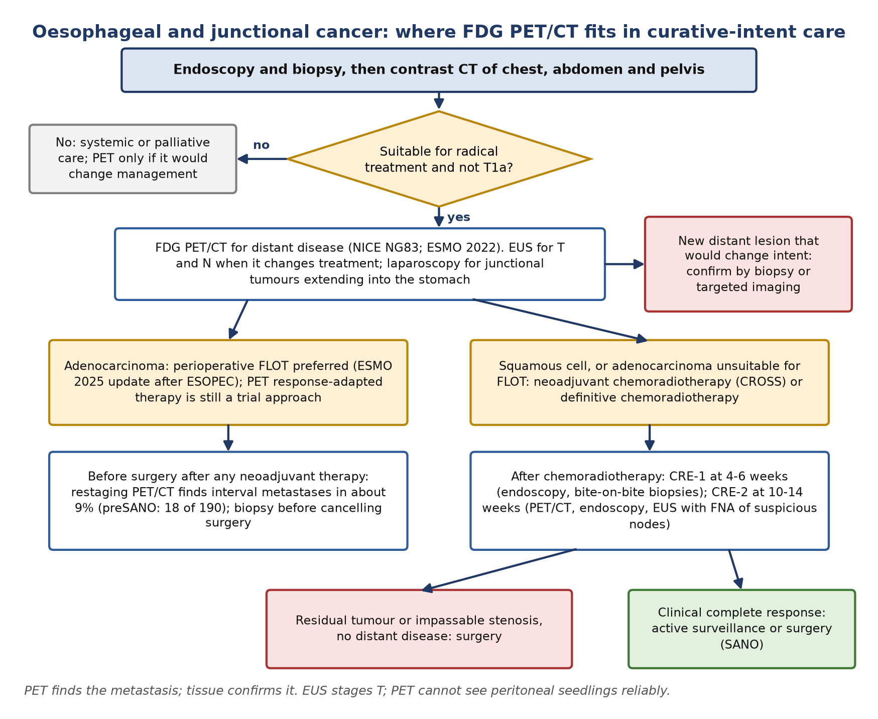
- PET/CT is the distant-disease test. NICE NG83 offers FDG PET/CT to everyone with an oesophageal or junctional cancer suitable for radical treatment, except T1a. In a meta-analysis its sensitivity for distant metastases was 71% against 52% for CT, with specificity 93%.
- It is not the T or N test. Endoscopic ultrasound (EUS) detects regional nodes more often (sensitivity 80% versus 57% for PET), while PET is more specific (85%). Coeliac nodes are regional (N) in oesophageal cancer, not M1.
- Gastric cancer is different. Diffuse, signet-ring and mucinous tumours are often poorly avid, and PET is blind to most peritoneal disease. In PLASTIC, PET found distant metastases in 3% of patients and staging laparoscopy found peritoneal or unresectable disease in 19%.
- Early metabolic response is real but not yet routine. A fall of 35% or more in tumour SUV after 14 days of chemotherapy predicted histological response in junctional adenocarcinoma (MUNICON). Switching chemotherapy in PET non-responders raised pathological complete response in CALGB 80803, but harmed patients with squamous cell carcinoma in the SCOPE2 substudy.
- Restaging before surgery pays. After neoadjuvant chemoradiotherapy, PET/CT found histologically proven interval metastases in 9% (preSANO). For organ preservation, PET/CT is combined with bite-on-bite biopsies and EUS; active surveillance was non-inferior to surgery at 2 years in SANO.
- GIST responds metabolically within days. Imatinib cut mean SUVmax from 14.2 to 5.5 after 1 week (ACRIN 6665). On CT, the Choi criteria (size down more than 10% or density down more than 15%) capture response that RECIST misses. Genotype first: PDGFRA D842V does not respond to imatinib.
- Know the mimics. Physiological gastric and junctional uptake, reflux or radiation oesophagitis, recent instrumentation, stents, anastomoses and radiation-induced liver injury in the caudate or left lobe after chemoradiotherapy.
1. The clinical problem
Oesophageal squamous cell carcinoma (SCC) and adenocarcinoma behave as two diseases that share an organ. SCC arises mainly in the upper and middle oesophagus and responds well to chemoradiotherapy; adenocarcinoma arises in the distal oesophagus and at the oesophagogastric junction (OGJ, also written GOJ), usually on a background of Barrett's oesophagus. Gastric adenocarcinoma is divided by the Lauren classification into intestinal and diffuse types, and the diffuse, poorly cohesive (signet-ring) tumours spread along the wall and across the peritoneum. Gastrointestinal stromal tumours (GISTs) are sarcomas driven, in most cases, by activating mutations in KIT or PDGFRA, and are treated with tyrosine kinase inhibitors such as imatinib.
At the tumour board the questions are practical. Is there distant disease that makes surgery or radical chemoradiotherapy futile? Which nodes are involved, and are they regional? Is the tumour responding to neoadjuvant treatment, and has anything new appeared before the operation? After chemoradiotherapy, is there any residual tumour, or can the patient avoid oesophagectomy? In GIST, is imatinib working, and how quickly can we know?
Staging system in force: AJCC/UICC eighth edition
The eighth edition of the American Joint Committee on Cancer (AJCC) and Union for International Cancer Control (UICC) classification, still in force for the oesophagus and stomach, introduced separate clinical (cTNM), pathological (pTNM) and post-neoadjuvant pathological (ypTNM) stage groups. The cTNM groups are what a PET/CT report feeds. They differ by histology, whereas the ypTNM groups are identical for SCC and adenocarcinoma.
| Element | Eighth-edition rule | What the PET/CT report must give |
|---|---|---|
| Regional nodes | Periesophageal nodes from the upper oesophageal sphincter to the coeliac artery; coeliac nodes are regional | Report a coeliac node as N disease, not M1 |
| N category | By number of involved regional nodes: N1 1–2, N2 3–6, N3 7 or more | Count the avid nodes and give their stations |
| cM1 | Any distant metastasis: cStage IVB for both histologies | Name each distant site and say whether tissue confirmation is needed |
| Adenocarcinoma cStage IVA | cT4b N0–1, or any cN2–N3 M0 | Three or more avid regional nodes move the patient into stage IVA |
| OGJ tumours | Epicentre no more than 2 cm into the gastric cardia: staged as oesophageal; more than 2 cm: staged as gastric | State the epicentre relative to the junction |
| Gastric cancer | Positive peritoneal cytology is M1 | PET cannot exclude it; laparoscopy with washings can |
Junctional adenocarcinomas are also described by the Siewert classification: type I has its centre 1–5 cm above the anatomical cardia (distal oesophageal adenocarcinoma), type II from 1 cm above to 2 cm below it (true cardia carcinoma), and type III from 2 to 5 cm below it (subcardial gastric carcinoma). The type guides the surgical approach and, for type III tumours, the need for staging laparoscopy. GISTs are not staged with TNM; the British Sarcoma Group (BSG) guideline recommends risk assessment by mitotic rate, tumour size, site and rupture instead.
Where nuclear medicine changes management
- Selection for radical treatment: detecting distant metastases that CT has missed spares patients futile oesophagectomy.
- Restaging after neoadjuvant therapy: finding interval metastases, and contributing to the clinical response evaluation that allows active surveillance.
- Radiotherapy planning: defining the length of the tumour and involved nodes.
- Response-adapted treatment: an early metabolic response scan in adenocarcinoma, currently a trial tool.
- GIST: early proof that imatinib is working, especially before surgery, and problem-solving when CT response is ambiguous.
2. Tracers and why they work
¹⁸F-fluorodeoxyglucose (FDG) is taken up by glucose transporters, phosphorylated by hexokinase and trapped. Most oesophageal SCCs and adenocarcinomas beyond the earliest stages are avid. Uptake reflects cell density and glycolysis, so tumours with abundant mucin, diffuse single-cell infiltration or small volume are harder to see, and inflammation in the oesophagus, junction and stomach produces the recurring false positives.
| Tumour | Usual FDG avidity | Practical point |
|---|---|---|
| Oesophageal SCC and adenocarcinoma (T2 and above) | High | PET stages distant disease; EUS stages T and N |
| Early (T1a) oesophageal cancer | Often low or not separable from normal wall | NICE NG83 excludes T1a from routine PET/CT |
| Gastric, intestinal (Lauren) type | Usually high | Detected in 83% of locally advanced tumours in one series |
| Gastric, diffuse, signet-ring or mucinous | Often low | Detected in only 41% of non-intestinal tumours; a negative scan does not exclude disease |
| GIST | Usually high before treatment | A small proportion have no FDG uptake: check baseline avidity before planning PET response assessment |
The Lauren effect is well documented. In 40 locally advanced gastric carcinomas, FDG PET detected 60% overall: 83% of intestinal-type tumours against 41% of non-intestinal tumours. Mean SUV was 6.7 against 4.8, and mucus-containing tumours averaged 3.9 against 7.2 for those without mucus. FDG uptake did not predict survival in that series. Newer scanners detect more primaries, but the direction of the difference holds, and the practical rule is unchanged: a faint or absent primary on PET does not reassure.
When FDG fails and what replaces it
- T stage: EUS, which images the wall layers; PET cannot measure depth of invasion.
- Regional nodes, especially those next to the primary: EUS with fine-needle aspiration (FNA); avid nodes blend into an avid primary.
- Peritoneum: staging laparoscopy with peritoneal washings; PET is insensitive to small peritoneal deposits.
- Locoregional residual disease after chemoradiotherapy: endoscopy with bite-on-bite biopsies and EUS with FNA of suspicious nodes.
- Fibroblast activation protein inhibitor (FAPI) PET: an investigational stromal tracer with promising results in the peritoneum (Section 8).
3. Indications by clinical scenario
| Scenario | Role of nuclear medicine | Guideline position |
|---|---|---|
| Oesophageal or OGJ cancer suitable for radical treatment | Whole-body FDG PET/CT after endoscopy and CT, for distant staging | NICE NG83 (2018): offer, except for T1a tumours. ESMO 2022: PET/CT in candidates for oesophagectomy, because unsuspected metastases avoid futile surgery |
| T and N staging | PET adds specificity; EUS is the T-staging test and the more sensitive nodal test | ESMO 2022 and NICE NG83: EUS when it will change management |
| Gastric cancer, potentially curable | PET/CT only if metastatic disease is suspected and the result will guide management | NICE NG83: consider only in that situation; offer staging laparoscopy to all with potentially curable gastric cancer. ESMO 2022: laparoscopy with peritoneal washings for stage IB–III disease considered resectable |
| Radiotherapy planning | PET/CT helps define tumour length and involved nodes | Systematic review: PET changes target volumes, but validation against pathology and outcome is limited |
| Early response during chemotherapy | Day-14 PET to identify metabolic non-responders | Research and trials; not a standard of care (MUNICON, CALGB 80803, SCOPE2) |
| After neoadjuvant therapy, before surgery | Restaging PET/CT for interval metastases | Supported by preSANO (9% interval metastases); tissue confirmation before cancelling surgery |
| After chemoradiotherapy, organ preservation | PET/CT as part of clinical response evaluation 10–14 weeks after treatment and during surveillance | SANO trial protocol. The ESMO 2025 interim update prefers perioperative FLOT for resectable adenocarcinoma, with chemoradiotherapy if FLOT is unsuitable, so this pathway now applies mainly to SCC |
| Suspected recurrence | Characterise a CT or clinical abnormality when salvage treatment is possible | No routine PET surveillance recommendation in the guidelines cited here |
| GIST, staging | Contrast CT is the investigation of choice; PET is sometimes useful | ESMO–EURACAN–GENTURIS 2022 and BSG: PET mainly when early detection of response is of special interest |
| GIST, neoadjuvant or advanced treatment | PET within days to weeks of starting a tyrosine kinase inhibitor | BSG: early response assessment is mandatory before surgery; PET shows response within a few weeks and helps when response is in doubt |
The two UK and European documents agree for oesophageal and junctional cancer and differ slightly in tone for gastric cancer. NICE restricts PET in gastric cancer to suspected metastatic disease; ESMO 2022 does not recommend it routinely and puts its weight on laparoscopy. Single-centre series argue for broader use: in a UK cohort of 105 patients who had PET/CT, 86% of gastric primaries were avid (93% of intestinal against 78% of non-intestinal), and PET/CT upstaged 19%, mostly by finding distant metastases. Local practice varies, and the report should say plainly what a negative scan can and cannot exclude.
4. Evidence at a glance
| Study | Design and size | Key finding | What it changed |
|---|---|---|---|
| van Vliet 2008 | Meta-analysis of EUS, CT and FDG PET in oesophageal cancer | Regional nodes: sensitivity EUS 80%, CT 50%, PET 57%; specificity 70%, 83%, 85%. Distant metastases: PET sensitivity 71% and specificity 93%, against 52% and 91% for CT | PET for distant disease, EUS for regional nodes |
| PLASTIC (Gertsen 2021) | Prospective multicentre cohort, 394 patients with locally advanced gastric cancer | Treatment intent changed in 16%. PET/CT found distant metastases in 12 (3%), sensitivity 33%, specificity 97%; laparoscopy found peritoneal or unresectable disease in 73 (19%). PET incidental findings in 22% | Laparoscopy, not routine PET, for gastric cancer |
| Stahl 2003 | 40 locally advanced gastric carcinomas | Detection 83% intestinal versus 41% non-intestinal; lower SUV in mucus-containing tumours | Explains false-negative gastric PET |
| Weber 2001 | 40 patients, junctional adenocarcinoma, PET at baseline and day 14 | A 35% fall in FDG uptake predicted clinical response with sensitivity 93% and specificity 95% | Defined the day-14, 35% threshold |
| MUNICON (Lordick 2007) | Phase II, 119 patients (110 evaluable), junctional adenocarcinoma | Non-responders went straight to surgery. Major histological remission 58% in responders, none in non-responders; median event-free survival 29.7 versus 14.1 months | Showed a PET-guided algorithm is feasible |
| MUNICON II (zum Büschenfelde 2011) | Phase II, 56 patients; non-responders given salvage chemoradiotherapy | Major histological remission 26% in non-responders, but R0 rate not improved (70% versus 82%) and 2-year survival 42% versus 71% | Poor prognosis of non-responders is partly biology |
| CALGB 80803 (Goodman 2021) | Randomised phase II, 241 patients, oesophageal and junctional adenocarcinoma | Non-responders (fall under 35% in SUVmax) who switched chemotherapy during chemoradiotherapy reached pCR in 18% and 20%. Responders to FOLFOX: pCR 40%, 5-year survival 53%. Median survival 48.8 months in responders versus 27.4 in non-responders | Supports PET-adapted chemotherapy in adenocarcinoma, pending phase III data |
| SCOPE2 PET substudy (Mukherjee 2023) | Randomised phase II, 103 patients, definitive chemoradiotherapy, mostly SCC | Closed for futility and possible harm. In SCC non-responders, switching to carboplatin and paclitaxel gave worse overall survival (20.4 versus 42.5 months) | Do not personalise chemotherapy by early PET in SCC |
| preSANO (Noordman 2018) | Prospective diagnostic cohort, 207 analysed after CROSS chemoradiotherapy | PET/CT missed 15% of substantial residual tumours; bite-on-bite biopsies with FNA missed 10%. PET/CT found proven interval metastases in 18 of 190 (9%) | Defined the response evaluation used in SANO |
| SANO (van der Wilk 2025) | Stepped-wedge cluster-randomised non-inferiority phase III, 309 patients with clinical complete response | 2-year overall survival 74% with active surveillance versus 71% with standard surgery | Active surveillance is an option after clinical complete response |
| ESOPEC (in ESMO 2025 update) | Phase III, 438 patients, resectable oesophageal adenocarcinoma | Perioperative FLOT versus CROSS: 3-year overall survival 57.4% versus 50.7% (HR 0.70) | FLOT preferred for adenocarcinoma |
| Choi 2007 | 40 patients with metastatic GIST, CT and PET at 2 months | Size fall over 10% or density fall over 15% identified PET responders with sensitivity 97% and specificity 100% (RECIST 52%) | Choi criteria for GIST on CT |
| ACRIN 6665 (Van den Abbeele 2012) | Phase II, neoadjuvant imatinib, PET at baseline, 1–7 days and 4 or 8 weeks | Mean SUVmax 14.2 at baseline, 5.5 after 1 week, 3.0 before surgery; metabolic response in 36 of 44 at week 1 against RECIST partial response in 2 of 39 | PET response precedes size change by weeks |
5. Patient preparation and acquisition
Standard oncological FDG preparation applies: fasting, glucose check, a quiet uptake period of about 60 minutes, and the same scanner, protocol and reconstruction whenever scans will be compared quantitatively. Points specific to these tumours:
- Order of tests: for oesophageal and junctional cancer, PET/CT follows endoscopy and contrast CT and is reserved for patients still suitable for radical treatment (NICE NG83). Record the date of any endoscopy, EUS, biopsy, dilatation or stent insertion, because instrumentation adds inflammatory uptake.
- Coverage: skull base to mid-thigh, which includes the neck; for cervical and upper-thoracic SCC make sure the whole neck is in the field of view.
- Early response studies: the trial scans were at baseline and 14 days after starting chemotherapy (Weber, MUNICON, CALGB 80803), with the percentage change in SUV as the measure. Use the same scanner and uptake time, or the percentage change is meaningless.
- After chemoradiotherapy: in the SANO protocol the first response evaluation (endoscopy with bite-on-bite biopsies) is 4–6 weeks after treatment and the second, including PET/CT, at 10–14 weeks. Earlier scans are dominated by radiation oesophagitis.
- Radiotherapy planning: a planning scan in the treatment position lets PET and planning CT register without deformation; say so in the request.
- GIST: the baseline scan must precede the first imatinib dose, and a baseline without uptake makes PET useless for response. A response scan can be informative within the first week (ACRIN 6665) and within a few weeks in practice (BSG). Record the mutation if known.
6. Interpretation
Oesophagus and junction
Describe the primary by level, length of the avid segment and relation to the junction, and give SUVmax as a baseline for any later response assessment. Do not assign a T category from PET. For nodes, count the avid regional nodes and name their stations, because the N category rests on the number (N1 1–2, N2 3–6, N3 7 or more). Periesophageal nodes beside the primary often merge with it on PET, one reason nodal sensitivity is low; say when a node cannot be separated. Remember that coeliac nodes are regional. Distant disease is usually in the liver, lungs, bone, adrenals or non-regional nodes; a single lesion that would remove curative options needs tissue or targeted imaging before treatment is changed.
Stomach
The normal stomach takes up FDG to a variable and sometimes marked degree, so a primary is called when uptake is focal, clearly above adjacent gastric wall and matched by wall thickening on CT. A poorly avid primary predicts poor sensitivity for its nodes and metastases. State explicitly that PET does not exclude peritoneal disease: in PLASTIC, PET/CT had a sensitivity of 33% for distant metastases, and laparoscopy found peritoneal or locally unresectable disease in almost one patient in five.
GIST
Most untreated GISTs are avid, often heterogeneous with a photopenic necrotic or cystic centre. Report SUVmax of the primary and representative metastases at baseline. On treatment, compare the same lesions on the same scanner. Because response on CT is recognised by falling density as well as size, read the PET together with the contrast CT, and look for the progression pattern of a new enhancing, avid nodule inside a previously responding mass.
Criteria in use
| Setting | Criterion | Exact definition |
|---|---|---|
| Early response, oesophagogastric adenocarcinoma | Metabolic response (MUNICON, CALGB 80803) | Fall in tumour SUV of 35% or more between baseline and day 14 (CALGB 80803 used SUVmax after induction chemotherapy); less than 35% is non-response |
| After chemoradiotherapy | Clinical complete response (SANO) | No residual tumour on endoscopy with bite-on-bite biopsies, EUS, and PET/CT at the two response evaluations; an impassable stenosis is not a complete response |
| GIST on CT | Choi partial response | Decrease in size of more than 10% or decrease in density (Hounsfield units) of more than 15% |
| GIST on CT | Progression patterns (BSG) | Increasing density or enhancement within a previously responding low-density lesion ('nodule within the mass'); apparent new liver lesions may be responding lesions that have become hypodense |
Pitfalls and mimics
| Finding | Why it misleads | How to avoid the error |
|---|---|---|
| Physiological gastric and junctional uptake | Normal mucosa, a contracted fundus or a hiatus hernia can look like a tumour | Require focal uptake with CT wall thickening; compare with endoscopy |
| Reflux oesophagitis and Barrett's segment | Linear or junctional uptake without a mass | Look for diffuse, low-grade, linear uptake; the endoscopy report decides |
| Radiation oesophagitis | Diffuse uptake along the treated segment for weeks after chemoradiotherapy | Time the response scan at 10–14 weeks; residual tumour is proved by biopsy and EUS, not PET alone (preSANO) |
| Radiation-induced liver injury | Focal uptake in the caudate or left lobe after chemoradiotherapy, mimicking an interval metastasis | Match the lesion to the high-dose region of the radiotherapy plan; 3% in one series and about 8% in the literature; confirm before cancelling surgery |
| Stents, dilatation, recent biopsy, anastomosis | Inflammatory uptake at the site | Record dates of instrumentation and surgery; interpret focal nodular uptake with CT |
| Brown fat | Symmetrical supraclavicular, paravertebral and mediastinal uptake that can hide or mimic nodes | Fat density on CT; warm the patient and repeat if it obscures a key station |
| Diffuse, signet-ring or mucinous gastric cancer | Primary and metastases poorly avid | Report as non-diagnostic, not negative; rely on laparoscopy and CT |
| Peritoneal disease | Small, flat deposits below PET resolution | Staging laparoscopy with washings |
| GIST without FDG uptake | No baseline signal to follow | Use CT (Choi) or MRI for response |
Wording the conclusion
- Give the clinical TNM elements PET can support and say what it cannot, for example: Avid distal oesophageal primary; one avid coeliac node (regional, cN1 by count); two liver lesions consistent with metastases (cM1, cStage IVB). T category requires EUS.
- Name the lesion that would change intent and how to confirm it.
- For gastric cancer, state the avidity of the primary and that peritoneal disease is not excluded.
- For response scans, give the percentage change in SUV, the measure used (SUVmax or SUVpeak) and whether acquisition matched the baseline.
7. Response assessment and follow-up
Early metabolic response in adenocarcinoma
The Munich group established the principle. In 40 junctional adenocarcinomas, FDG uptake fell by a mean of 54% at day 14 in tumours that went on to respond and by 15% in those that did not; a 35% threshold predicted clinical response with sensitivity 93% and specificity 95%. MUNICON then used the threshold to act: 54 of 110 evaluable patients (49%) were metabolic responders and completed 12 weeks of chemotherapy before surgery, while non-responders stopped after 2 weeks and went straight to surgery. Major histological remission (less than 10% residual tumour) occurred in 58% of responders and in no non-responder, and median event-free survival was 29.7 against 14.1 months. MUNICON II gave non-responders salvage chemoradiotherapy: histological response improved (26%), but the R0 rate and survival did not, suggesting that metabolic non-response marks aggressive biology as well as drug resistance.
CALGB 80803 tested switching rather than stopping. After induction with FOLFOX (oxaliplatin, leucovorin and fluorouracil) or carboplatin and paclitaxel, non-responders (SUVmax fall under 35%) crossed to the other regimen during chemoradiotherapy. Their pathological complete response (pCR) rates were 18% and 20%, higher than historically expected for non-responders; responders to FOLFOX who continued it reached 40% pCR and a 5-year survival of 53%. Median survival was 48.8 months for responders and 27.4 months for non-responders.
The picture is different in SCC. In the SCOPE2 PET substudy of definitive chemoradiotherapy, 61% of patients were non-responders at day 14 of cisplatin and capecitabine. Switching SCC non-responders to carboplatin and paclitaxel gave worse treatment-failure-free survival and worse overall survival (20.4 against 42.5 months), and early metabolic response was not prognostic. The substudy was closed for futility and possible harm. The lesson for the report: give the percentage change, but do not imply that it should change treatment outside a protocol.
Restaging and organ preservation after chemoradiotherapy
After the CROSS regimen (carboplatin, paclitaxel and 41.4 Gy), roughly half of SCCs and a quarter of adenocarcinomas have no residual tumour in the primary. preSANO asked how to find the rest. Endoscopy with regular biopsies and FNA missed 31% of tumours with more than 10% residual carcinoma; bite-on-bite biopsies with FNA missed 10%; EUS measurement of maximum tumour thickness missed 28%; PET/CT missed 15%. PET/CT's distinctive contribution was distant: it found histologically proven interval metastases in 18 of 190 patients (9%), 17 of them with adenocarcinoma.
SANO then compared active surveillance with standard oesophagectomy in 309 patients with a clinical complete response, defined by biopsies, EUS and PET/CT. Two-year overall survival was 74% with surveillance and 71% with surgery, within the non-inferiority margin. During surveillance, response evaluations are repeated every 3 months in the first year, every 4 months in the second, every 6 months in the third and yearly in the fourth and fifth; PET/CT was built into the sixth and ninth evaluations (about 16 and 30 months). The nuclear medicine role in this pathway is specific: detect distant disease and flag locoregional regrowth, while tissue decides locoregional status.
GIST
Imatinib switches off glycolysis in sensitive GIST within days. In ACRIN 6665, 36 of 44 patients had a complete or partial metabolic response at week 1, and 23 of 27 with KIT exon 11 mutations had responded metabolically by then; by contrast RECIST showed a partial response in only 2 of 39. On CT, a falling density often precedes shrinkage, and lesions may even grow transiently. The Choi criteria were built against PET: in 40 patients a size fall over 10% or a density fall over 15% at 2 months identified PET responders with sensitivity 97% and specificity 100%. BSG adds that absence of progression at 6 months is equivalent to response, and that the 'nodule within the mass' is the typical pattern of secondary progression.
Genotype shapes the question. Mutational analysis is mandatory before neoadjuvant imatinib: PDGFRA exon 18 D842V GISTs are insensitive to imatinib (avapritinib is the most active drug), KIT exon 9 tumours need 800 mg daily, and NF1-related and many SDH-deficient 'wild-type' GISTs respond poorly. BSG notes that in the absence of molecular analysis, FDG PET/CT can show response very rapidly, within a few weeks, so that surgery is not delayed in non-responding disease.
8. Theranostics and emerging tracers
| Tracer or approach | Target | Evidence level (2026) | What it may add |
|---|---|---|---|
| ¹⁸F-FDG | Glucose metabolism | Guideline-endorsed for oesophageal and junctional staging; selective in gastric cancer and GIST | Distant staging, restaging, early response |
| ⁶⁸Ga-FAPI PET | Fibroblast activation protein on cancer-associated fibroblasts | Investigational | In a systematic review of 10 studies of pancreatic, gastric and biliary cancer, FAPI outperformed FDG for primary, nodes, metastases and peritoneal carcinomatosis in every study, with higher SUVmax and tumour-to-background ratios |
| Response-adapted treatment by FDG PET | Early change in glycolysis | Phase II evidence only | Positive in adenocarcinoma (CALGB 80803); harmful in SCC during definitive chemoradiotherapy (SCOPE2) |
| Radioligand therapy | None established | None of the guidelines cited here includes a radioligand therapy for these tumours | FAP-directed therapy remains a research question |
FAPI is attractive precisely where FDG is weakest: diffuse and signet-ring gastric cancer and small peritoneal deposits, because its signal comes from the tumour stroma rather than tumour glycolysis. The studies so far are small, mostly single-centre and at high pre-test probability, and fibrosis and inflammation also take up FAPI. It should be described to referrers as a research test until prospective data show that it changes outcome.
9. Structured report example
- Indication: 69-year-old; biopsy-proven adenocarcinoma of the distal oesophagus, Siewert type I; CT shows no metastases. Being considered for perioperative chemotherapy and oesophagectomy.
- Technique: ¹⁸F-FDG 280 MBq, uptake 61 min, glucose 6.1 mmol/L; skull base to mid-thigh; low-dose CT. Endoscopy with biopsy 9 days earlier; no stent.
- Primary: avid distal oesophageal tumour over 5 cm, SUVmax 12.4, ending at the junction; no gastric extension beyond the cardia.
- Nodes: one avid coeliac axis node, 11 mm short axis, SUVmax 7.1 (regional). No other avid regional or non-regional nodes.
- Distant: focal lesion in liver segment VII, 16 mm, SUVmax 8.9, above background liver (2.4); corresponding hypodensity on the diagnostic CT. No lung, bone or adrenal lesion.
- Conclusion: Avid distal oesophageal adenocarcinoma with a regional coeliac nodal metastasis (at least cN1) and a solitary liver lesion suspicious for metastasis (cM1, cStage IVB if confirmed). T category requires EUS.
- Recommendation: Tissue confirmation of the liver lesion (or liver MRI) before curative intent is abandoned; discuss at the upper gastrointestinal multidisciplinary meeting. Baseline SUVmax recorded for any response assessment on the same scanner.
10. Take-home points
- FDG PET/CT belongs in the staging of every oesophageal or junctional cancer being considered for radical treatment (except T1a): its job is distant disease.
- EUS stages T and is more sensitive for regional nodes; coeliac nodes are regional, and the N category depends on the number of involved nodes.
- In gastric cancer, use PET selectively. Diffuse, signet-ring and mucinous tumours are often poorly avid, and staging laparoscopy, not PET, finds peritoneal disease.
- A 35% fall in SUV at day 14 predicts response in adenocarcinoma, and switching chemotherapy helped non-responders in CALGB 80803; the same strategy harmed SCC patients in SCOPE2. Response-adapted treatment is still a trial tool.
- Restage with PET/CT before surgery: about 1 in 11 patients has interval metastases after chemoradiotherapy. Confirm before cancelling surgery, remembering radiation-induced liver injury.
- After chemoradiotherapy, clinical complete response needs biopsies, EUS and PET/CT together; active surveillance was non-inferior at 2 years in SANO.
- In GIST, check the genotype first, record baseline avidity, and expect metabolic response within days; on CT use Choi, not RECIST alone.
Test yourself
5 quick questions. Pick an answer to see the explanation.
References
- Rice TW, Ishwaran H, Ferguson MK, Blackstone EH, Goldstraw P. Cancer of the esophagus and esophagogastric junction: an eighth edition staging primer. J Thorac Oncol. 2017;12(1):36-42.
- Siewert JR, Stein HJ. Classification of adenocarcinoma of the oesophagogastric junction. Br J Surg. 1998;85(11):1457-9.
- Obermannová R, Alsina M, Cervantes A, et al. Oesophageal cancer: ESMO Clinical Practice Guideline for diagnosis, treatment and follow-up. Ann Oncol. 2022;33(10):992-1004.
- Obermannová RL, Leong T, Smyth EC, et al. ESMO Clinical Practice Guideline interim update on the treatment of locally advanced oesophageal and oesophagogastric junction adenocarcinoma and metastatic squamous-cell carcinoma. ESMO Open. 2025;10(2):104134.
- Lordick F, Carneiro F, Cascinu S, et al. Gastric cancer: ESMO Clinical Practice Guideline for diagnosis, treatment and follow-up. Ann Oncol. 2022;33(10):1005-20.
- National Institute for Health and Care Excellence. Oesophago-gastric cancer: assessment and management in adults. NICE guideline NG83. London: NICE; 2018.
- Casali PG, Blay JY, Abecassis N, et al. Gastrointestinal stromal tumours: ESMO–EURACAN–GENTURIS Clinical Practice Guidelines for diagnosis, treatment and follow-up. Ann Oncol. 2022;33(1):20-33.
- Judson I, Jones RL, Wong NACS, et al. Gastrointestinal stromal tumour (GIST): British Sarcoma Group clinical practice guidelines. Br J Cancer. 2025;132(1):1-10.
- van Vliet EP, Heijenbrok-Kal MH, Hunink MG, Kuipers EJ, Siersema PD. Staging investigations for oesophageal cancer: a meta-analysis. Br J Cancer. 2008;98(3):547-57.
- Gertsen EC, Brenkman HJF, van Hillegersberg R, et al. 18F-fludeoxyglucose-positron emission tomography/computed tomography and laparoscopy for staging of locally advanced gastric cancer: a multicenter prospective Dutch cohort study (PLASTIC). JAMA Surg. 2021;156(12):e215340.
- Stahl A, Ott K, Weber WA, et al. FDG PET imaging of locally advanced gastric carcinomas: correlation with endoscopic and histopathological findings. Eur J Nucl Med Mol Imaging. 2003;30(2):288-95.
- Bosch KD, Chicklore S, Cook GJ, et al. Staging FDG PET-CT changes management in patients with gastric adenocarcinoma who are eligible for radical treatment. Eur J Nucl Med Mol Imaging. 2020;47(4):759-67.
- Weber WA, Ott K, Becker K, et al. Prediction of response to preoperative chemotherapy in adenocarcinomas of the esophagogastric junction by metabolic imaging. J Clin Oncol. 2001;19(12):3058-65.
- Lordick F, Ott K, Krause BJ, et al. PET to assess early metabolic response and to guide treatment of adenocarcinoma of the oesophagogastric junction: the MUNICON phase II trial. Lancet Oncol. 2007;8(9):797-805.
- zum Büschenfelde CM, Herrmann K, Schuster T, et al. 18F-FDG PET-guided salvage neoadjuvant radiochemotherapy of adenocarcinoma of the esophagogastric junction: the MUNICON II trial. J Nucl Med. 2011;52(8):1189-96.
- Goodman KA, Ou FS, Hall NC, et al. Randomized phase II study of PET response-adapted combined modality therapy for esophageal cancer: mature results of the CALGB 80803 (Alliance) trial. J Clin Oncol. 2021;39(25):2803-15.
- Mukherjee S, Hurt CN, Adams R, et al. Efficacy of early PET-CT directed switch to carboplatin and paclitaxel based definitive chemoradiotherapy in patients with oesophageal cancer who have a poor early response to induction cisplatin and capecitabine in the UK: a multi-centre randomised controlled phase II trial. EClinicalMedicine. 2023;61:102059.
- Noordman BJ, Spaander MCW, Valkema R, et al. Detection of residual disease after neoadjuvant chemoradiotherapy for oesophageal cancer (preSANO): a prospective multicentre, diagnostic cohort study. Lancet Oncol. 2018;19(7):965-74.
- Eyck BM, van der Wilk BJ, Noordman BJ, et al. Updated protocol of the SANO trial: a stepped-wedge cluster randomised trial comparing surgery with active surveillance after neoadjuvant chemoradiotherapy for oesophageal cancer. Trials. 2021;22(1):345.
- van der Wilk BJ, Eyck BM, Wijnhoven BPL, et al. Neoadjuvant chemoradiotherapy followed by active surveillance versus standard surgery for oesophageal cancer (SANO trial): a multicentre, stepped-wedge, cluster-randomised, non-inferiority, phase 3 trial. Lancet Oncol. 2025;26(4):425-36.
- Choi H, Charnsangavej C, Faria SC, et al. Correlation of computed tomography and positron emission tomography in patients with metastatic gastrointestinal stromal tumor treated at a single institution with imatinib mesylate: proposal of new computed tomography response criteria. J Clin Oncol. 2007;25(13):1753-9.
- Van den Abbeele AD, Gatsonis C, de Vries DJ, et al. ACRIN 6665/RTOG 0132 phase II trial of neoadjuvant imatinib mesylate for operable malignant gastrointestinal stromal tumor: monitoring with 18F-FDG PET and correlation with genotype and GLUT4 expression. J Nucl Med. 2012;53(4):567-74.
- Voncken FEM, Aleman BMP, van Dieren JM, et al. Radiation-induced liver injury mimicking liver metastases on FDG-PET-CT after chemoradiotherapy for esophageal cancer: a retrospective study and literature review. Strahlenther Onkol. 2018;194(2):156-63.
- Muijs CT, Beukema JC, Pruim J, et al. A systematic review on the role of FDG-PET/CT in tumour delineation and radiotherapy planning in patients with esophageal cancer. Radiother Oncol. 2010;97(2):165-71.
- Veldhuijzen van Zanten SEM, Pieterman KJ, Wijnhoven BPL, et al. FAPI PET versus FDG PET, CT or MRI for staging pancreatic-, gastric- and cholangiocarcinoma: systematic review and head-to-head comparisons of diagnostic performances. Diagnostics (Basel). 2022;12(8):1958.