PET/CT in Head and Neck Cancer
- Staging language. AJCC/UICC 8th edition stages HPV-associated (p16-positive) oropharyngeal cancer separately, adds depth of invasion to oral cavity T and extranodal extension to N for non-viral cancers. Nasopharynx moved to version 9 in January 2025, where the number of distant lesions (up to 3, or more than 3) splits M1.
- Where FDG changes management. Distant metastases and second primaries (pooled sensitivity 89%, specificity 95%), the unknown primary before panendoscopy, and the response scan after chemoradiotherapy.
- The clinically N0 neck. FDG misses about half of occult nodal disease (pooled sensitivity 50%). A negative PET does not replace sentinel node biopsy or elective neck dissection.
- PET-NECK. In 564 patients with N2–N3 disease, PET/CT at 12 weeks with neck dissection only for incomplete or equivocal response gave survival no worse than planned dissection (2-year overall survival 84.9% vs 81.5%) with 54 instead of 221 operations.
- Timing and predictive values. Scan at 12 weeks or later. Negative predictive value is about 95%; positive predictive value only about 50–60%, so a positive primary site needs biopsy.
- Score it. Hopkins criteria (internal jugular vein and liver as references; scores 1–3 negative, 4–5 positive) or ACR NI-RADS (categories 1–4, each with a management step). Pick one and use it every time.
- Sentinel node in early oral cancer. ⁹⁹ᵐTc-nanocolloid lymphoscintigraphy with SPECT/CT: in SENT the sentinel node was found in 99.5% (sensitivity 86%, negative predictive value 95%); two randomised trials showed outcomes equivalent or non-inferior to elective neck dissection with less morbidity.
- Emerging. FAPI PET for the FDG-negative unknown primary and FMISO hypoxia PET for dose painting remain investigational.
1. The clinical problem: staging and where PET changes management
Head and neck squamous cell carcinoma (HNSCC) arises in the oral cavity, oropharynx, larynx and hypopharynx; nasopharyngeal carcinoma (NPC) is a distinct, often Epstein–Barr virus (EBV)-related disease with its own staging chapter. Most patients present with locoregionally advanced disease. The team needs four answers from imaging: the local extent of the primary (endoscopy, CT and MRI do most of this), the nodal status by level, distant metastases and synchronous second primaries, and later whether treatment has worked. ¹⁸F-fluorodeoxyglucose (FDG) PET/CT contributes most to the last three.
Staging system in force
Head and neck sites other than the nasopharynx are staged with the American Joint Committee on Cancer (AJCC) and Union for International Cancer Control (UICC) 8th edition unless local policy has adopted a newer version. The changes that matter when you report PET/CT are summarised below.
| Change (AJCC/UICC 8th edition unless stated) | What it means for the PET/CT reader |
|---|---|
| HPV-associated (p16-positive) oropharyngeal cancer has its own chapter. Clinical N1: one or more ipsilateral nodes, none larger than 6 cm; N2: contralateral or bilateral nodes, none larger than 6 cm; N3: any node larger than 6 cm | Report laterality and the largest node size; node number no longer changes clinical N. Stage I is T0–2 N0–1, stage II T0–2 N2 or T3 N0–2, stage III T4 or N3, stage IV M1 |
| Extranodal extension (ENE) enters N for non-viral cancers (oral cavity, larynx, hypopharynx, p16-negative oropharynx, unknown primary). Any clinical ENE is cN3b; pathological ENE raises pN by one category | Clinical ENE needs unambiguous clinical signs (skin invasion, muscle infiltration or dense tethering, cranial nerve, brachial plexus, sympathetic or phrenic nerve dysfunction) supported by imaging. Imaging alone is not enough, and doubt means ENE-negative |
| Oral cavity T combines size with depth of invasion (DOI); for example a tumour of 2 cm or less with DOI over 5 mm is T2, and a tumour over 4 cm with DOI over 10 mm is T4a | DOI is measured clinically and on MRI or ultrasound, not on PET. DOI also informs the sentinel node decision |
| Unknown primary: T0 is used only when the node is p16-positive (staged as oropharynx) or EBV-positive (staged as nasopharynx); other cases use the cervical nodes and unknown primary chapter | The pathology result, not a guess about the likely primary, decides the chapter |
| Nasopharynx, version 9 (from January 2025): advanced radiological ENE (into adjacent muscle, skin or neurovascular bundles) becomes N3; non-metastatic disease is regrouped into stages I–III; M1a is 3 or fewer metastatic lesions and M1b more than 3 | Count and list distant lesions: the count now separates stage IVA from IVB |
Where FDG PET/CT changes management
- Distant metastases and second primaries: a meta-analysis of 12 studies gave pooled sensitivity 88.8% and specificity 95.1%, with similar performance in NPC and in other sites.
- Unknown primary: FDG PET/CT is done before panendoscopy and tonsillectomy so that biopsies can be directed (see the Nucpaedia article on cancer of unknown primary for the whole pathway).
- Nodal staging: sensitivity 79% and specificity 86% overall, but only 50% sensitivity in the clinically N0 neck.
- Response after chemoradiotherapy: the largest effect on practice: a negative 12-week scan replaces planned neck dissection.
- Recurrence: restaging before salvage surgery or re-irradiation, where finding distant disease changes the intent of treatment.
2. Tracers and why they work
- ¹⁸F-FDG: squamous carcinoma has high glucose uptake and phosphorylation, so primaries and nodes are usually intensely avid. In a cohort of 562 patients, median baseline SUVmax was 11.0 in the primary and 8.1 in nodes.
- Why FDG fails: micrometastases and small nodes are below resolution (hence the 50% sensitivity in the N0 neck); necrotic or cystic nodes may show little uptake; tumour next to intensely avid tissue (Waldeyer ring, brain, active muscle) can be hidden; dental metal degrades the CT and attenuation correction; and inflammation, reactive nodes and treatment effects are also avid.
- HPV-positive disease: after chemoradiotherapy FDG is slightly less accurate in HPV-positive tumours (nodal sensitivity 75% vs 89% and specificity 87% vs 95% in a meta-analysis of 20 studies).
- What replaces FDG: MRI for local extent, perineural and skull-base spread and the nasopharyngeal primary; sentinel node biopsy for the clinically N0 neck in early oral cancer; endoscopy and biopsy for a mucosal abnormality.
- ⁹⁹ᵐTc-nanocolloid: a radiocolloid injected around the tumour, carried in lymphatics and retained in the first-echelon (sentinel) node. It is the most used sentinel node tracer in Europe.
- ⁹⁹ᵐTc-tilmanocept: a small receptor-targeted agent that binds the mannose receptor (CD206) on nodal macrophages and dendritic cells. It is approved in the USA for guiding sentinel node biopsy in clinically node-negative oral cavity SCC, and the EANM guideline notes its potential for levels I–II in floor-of-mouth tumours. A hybrid indocyanine green–⁹⁹ᵐTc-nanocolloid tracer adds intraoperative fluorescence.
3. Indications by clinical scenario
| Scenario | Role of nuclear medicine | Guideline position (strength where given) |
|---|---|---|
| Neck node SCC, primary not found | FDG PET/CT after clinical examination and contrast CT, before examination under anaesthesia, directed biopsies and tonsillectomy | ASCO 2020: PET/CT is the next step if the primary is not evident on examination and contrast CT (strong). NICE NG36: consider PET/CT as the first investigation to find the primary |
| Staging of locoregionally advanced HNSCC | Whole-body FDG PET/CT for distant metastases and second primaries | NICE: offer PET/CT for N3 disease and for T4 hypopharynx or nasopharynx; chest CT for others who need systemic staging (all except T1N0 and T2N0). EHNS–ESMO–ESTRO 2020: chest imaging with CT and/or FDG-PET strongly recommended [IV, A] |
| Nasopharyngeal carcinoma | FDG PET/CT for nodal and distant staging; MRI for the primary | ESMO–EURACAN 2021: FDG-PET is the best method for distant metastases and is recommended at least in locally advanced disease [III, B] |
| Clinically N0 neck | FDG PET/CT can upstage but cannot clear the neck | EANM 2019: FDG PET/CT does not replace sentinel node biopsy in oral cancer |
| Early oral cancer, cT1–T2 N0 | Lymphoscintigraphy with SPECT/CT for sentinel node biopsy | NICE: offer sentinel node biopsy instead of elective neck dissection unless the neck must be opened anyway (for example free-flap reconstruction). EHNS–ESMO–ESTRO: a valid option when DOI is under 10 mm |
| Radiotherapy planning | FDG PET/CT in the treatment position to help define gross tumour and nodal volumes | Widely used but not mandated by these guidelines; supported by imaging–pathology comparison, not by outcome trials |
| Response after chemoradiotherapy, node-positive | FDG PET/CT at 12 weeks or later to decide on neck dissection | EHNS–ESMO–ESTRO: PET/CT 3 months after chemoradiotherapy [I, A]; no neck dissection if PET is negative and nodes are normal-sized at 12 weeks [I, A]. ASCO 2019: scan at 12 weeks or later (strong, high-quality evidence). NICE (2018 update): scan 3–6 months after treatment |
| Surveillance and suspected recurrence | FDG PET/CT for symptoms, equivocal findings or restaging before salvage | EHNS–ESMO–ESTRO: imaging for symptoms or abnormal examination [III, A]. ACR NI-RADS provides a surveillance reporting template |
Guidelines differ on the response-scan window: PET-NECK and ASCO use 12 weeks or later, EHNS–ESMO–ESTRO says 3 months, NICE allows 3–6 months, and the NI-RADS white paper places the first post-treatment PET/CT and contrast CT at 8–12 weeks. In practice, aim for 12 weeks and do not scan earlier to answer the neck-dissection question.
Radiotherapy planning deserves an honest note. In a study validated against laryngectomy specimens, FDG gross tumour volumes were smaller than CT or MRI volumes and closest to the specimen, but no modality showed superficial mucosal extension. FDG therefore refines, and does not replace, clinical and endoscopic delineation.
4. Evidence at a glance
| Study | Design and size | Key finding | What it changed |
|---|---|---|---|
| PET-NECK (Mehanna 2016) | Randomised non-inferiority trial, 564 patients, N2–N3 (84% oropharynx, 75% p16-positive), 37 UK centres | PET/CT 12 weeks after chemoradiotherapy with dissection only for incomplete or equivocal response: 2-year overall survival 84.9% vs 81.5% (HR 0.92, 95% CI 0.65–1.32); 54 vs 221 neck dissections; saving £1,492 per patient | Ended routine planned neck dissection |
| Gupta 2011 | Meta-analysis, 51 studies, 2,335 patients | Post-treatment FDG: primary site NPV 95.1%, PPV 58.6%; neck NPV 94.5%, PPV 52.1%; better accuracy at 12 weeks or later | Set the 12-week rule and the reliance on NPV |
| Helsen 2018 | Meta-analysis, 20 studies, 1,293 patients, nodal response within 6 months | Sensitivity 85%, specificity 93%; lower in HPV-positive tumours (75% vs 89%; 87% vs 95%) | Caution with residual nodes in HPV-positive disease |
| Hopkins criteria (Marcus 2014) | Retrospective, 214 patients, scans 5–24 weeks after treatment, 3 readers | Sensitivity 68.1%, specificity 92.2%, PPV 71.1%, NPV 91.1%; overall kappa 0.69–0.79; score predicted survival | First validated qualitative 5-point scale |
| Zhong 2020 | Retrospective, 562 patients, four criteria compared | Hopkins, NI-RADS, Porceddu and Deauville performed alike (AUC 0.75–0.76); indeterminate primary-site calls: NI-RADS 91, Porceddu 25, Deauville 20, Hopkins 13 | Any validated scale works; use one consistently |
| Kyzas 2008 | Meta-analysis, 32 studies, 1,236 patients | Nodal sensitivity 79%, specificity 86%; cN0 neck sensitivity 50%, specificity 87% | PET cannot clear the N0 neck |
| ACRIN 6685 (Lowe 2019) | Prospective multicentre, 287 enrolled; 270 N0 neck sides with dissection | Visual NPV for the clinically N0 side 0.868; PET/CT changed the surgical plan in 22% | PET/CT informs, but does not replace, neck surgery |
| Xu 2011 | Meta-analysis, 12 studies | Distant metastases and second primaries: sensitivity 88.8%, specificity 95.1% | Basis for whole-body PET in advanced disease |
| Rusthoven 2004 | Review, 16 studies, 302 patients with a cervical unknown primary | PET found 24.5% of primaries missed by conventional work-up; sensitivity 88.3%, specificity 74.9%; 39.3% false-positive rate in the tonsils; sensitivity 81.5% in the tongue base | PET before panendoscopy; tonsillar uptake needs histology |
| SENT (Schilling 2015) | Prospective multicentre observational, 415 patients, T1–T2 N0 oral SCC | Sentinel node found in 99.5%; 23% positive; sensitivity 86%, NPV 95%; disease-specific survival 94% at 3 years | Validated SLNB as a staging operation |
| Garrel 2020 (France) | Randomised equivalence trial, 307 patients (279 analysed), T1–T2 N0 oral and oropharyngeal | 2-year neck recurrence-free survival 90.7% after SLNB vs 89.6% after neck dissection; less morbidity for 6 months | SLNB as a standard of care |
| Hasegawa 2021 (Japan) | Randomised non-inferiority trial, 271 patients, T1–T2 N0 oral SCC | 3-year overall survival 87.9% with SLNB-guided dissection vs 86.6% with elective dissection; better neck function | Confirmed SLNB in a second population |
A US trial, NRG-HN006 (NCT04333537), is randomising early oral cavity cancer to SLNB or elective neck dissection (planned 686 patients) and was still recruiting in 2026.
5. Patient preparation and acquisition
FDG PET/CT: what differs in the head and neck
- Standard preparation: fasting, glucose check and 60-minute uptake as for any oncological FDG study.
- Quiet uptake: no talking or chewing during uptake, because speech and chewing raise laryngeal, tongue and masticatory muscle uptake; published response-assessment series use a silence protocol. Keep the patient warm to limit brown fat.
- Dedicated neck acquisition: many centres add a high-resolution head and neck acquisition with contrast-enhanced CT to the whole-body study; fine anatomy is what separates a node from a vessel or muscle.
- Radiotherapy planning: scan in the treatment position with the thermoplastic mask, so that PET can be registered to the planning CT.
- Response scan: book for 12 weeks after the last fraction and ask for the clinical and endoscopic findings, so that ulcers and recent biopsies are known.
Sentinel node lymphoscintigraphy in early oral cancer (EANM 2019, SNMMI-endorsed)
- Who: biopsy-proven early (T1–T2) oral SCC with a neck that is N0 on palpation and imaging (ultrasound, CT or MRI).
- Activity: ⁹⁹ᵐTc-nanocolloid 40–50 MBq for a same-day protocol or 70–120 MBq for a 2-day protocol, in a total volume of 0.4–0.5 mL (0.1–0.2 mL per syringe).
- Injection: two to four submucosal injections around the tumour at 3, 6, 9 and 12 o'clock (four is optimal), after local anaesthetic spray or gel or a lingual nerve block. The patient rinses the mouth without swallowing.
- Imaging: dynamic imaging, early and delayed static images and SPECT/CT. The report states lymphatic ducts, order of appearance, nodal level and intensity, and relates each node to the jugular vein and the digastric and omohyoid muscles. Nodes behind the jugular vein are hard to reach through a small incision, so say so.
- Calling a sentinel node: a node with its own afferent lymphatic, or a single node in a basin, is a definite sentinel node; a node between the injection and the first node, or with rising uptake on delayed images, is highly probable; a later second-echelon node is less probable. A contralateral node on SPECT/CT, even if faint, should be treated as a highly probable sentinel node.
- Pitfalls: in floor-of-mouth tumours level I nodes lie close to the injection and can be hidden by shine-through; previous neck surgery or radiotherapy distorts drainage; widespread field change makes the injection margin uncertain; staged biopsy after resection of the primary is unreliable. If no node migrates and SPECT/CT is negative, consider reinjection.
- Dose: low: a worst-case estimate of about 0.04 mSv effective dose after 20 MBq, plus the CT of SPECT/CT (about 2.4 mSv reported for one low-dose bed position).
6. Interpretation: criteria, variants and pitfalls
Staging read
- Confirm the primary and its extent, but remember that no modality shows superficial mucosal spread well; the endoscopist sees it.
- Report nodes by level: level I submental and submandibular; level II upper jugular (skull base to the lower border of the hyoid); level III mid-jugular (to the cricoid); level IV lower jugular; level V posterior triangle; level VI central compartment; level VII upper mediastinum. Name retropharyngeal nodes separately.
- Report the side and largest size of involved nodes (both drive N in p16-positive disease) and any imaging features suggesting ENE, while leaving the clinical ENE call to the team.
- Search lungs, bone and liver for metastases and the lung and oesophagus for second primaries; give each distant lesion a location, as the count matters for NPC version 9.
Response criteria in use
| Hopkins score | Definition (reference: internal jugular vein, IJV, and liver) | Meaning |
|---|---|---|
| 1 | Focal uptake less than IJV | Complete metabolic response (negative) |
| 2 | Focal uptake greater than IJV but less than liver | Likely complete metabolic response (negative) |
| 3 | Diffuse uptake greater than IJV or liver | Likely post-radiation inflammation (negative) |
| 4 | Focal uptake greater than liver | Likely residual tumour (positive) |
| 5 | Focal and intense uptake greater than liver | Residual tumour (positive) |
Score the primary site, right neck and left neck separately; the overall score is the highest of the three. A new lesion not present at baseline is progressive disease (Marcus et al., 2014).
| NI-RADS category | PET/CT features | Management |
|---|---|---|
| 0 Incomplete | Prior imaging unavailable but will be obtained | Obtain prior imaging |
| 1 No evidence of recurrence | Expected post-treatment change; no abnormal FDG uptake, or diffuse curvilinear mucosal uptake (radiation mucositis) | Routine surveillance |
| 2a Low suspicion, superficial (primary) | Focal non-mass-like mucosal enhancement with mild focal mucosal uptake | Direct visual inspection |
| 2b Low suspicion, deep (primary) | Ill-defined deep soft tissue with mild enhancement and mild uptake | PET or short-interval (3-month) follow-up |
| 2 Low suspicion (neck) | Enlarging node without specific abnormal features, or a node with mild to moderate uptake | PET or short-interval follow-up |
| 3 High suspicion | New or enlarging discrete mass, or enlarging node with new necrosis or gross ENE, with intense focal uptake | Biopsy |
| 4 Known recurrence | Pathologically proven or definite radiological or clinical progression | Treat |
ACR NI-RADS categories for the primary site and neck, applied to contrast CT or PET/CT (Aiken et al., 2018).
- Deauville-style scales: some centres transfer the lymphoma 5-point scale, using mediastinal blood pool and liver as references (scores 1–2 effectively a complete response). The Porceddu criteria are a simpler 3-point call (negative, equivocal, positive) based on uptake relative to adjacent normal tissue and/or liver.
- Head to head: in 562 patients all four systems predicted outcome similarly; NI-RADS labelled the most primary-site cases indeterminate and Hopkins the fewest, while Porceddu and Deauville offered the best balance of few indeterminate calls and high NPV. Positive predictive value for indeterminate uptake was poor with every scale (36% at the primary, 48% in nodes).
- Why NPV matters more than PPV: with an NPV around 95% a negative scan safely avoids surgery; with a PPV around 50–60% a positive primary site needs examination under anaesthesia and biopsy before salvage surgery. In the neck, a positive node usually goes to dissection, which is both diagnostic and therapeutic.
Normal variants, pitfalls and mimics
| Finding | Why it happens | How to tell |
|---|---|---|
| Muscle uptake (tongue, masticators, larynx, sternocleidomastoid, scalenes) | Talking, chewing, swallowing or tension during uptake | Symmetric and linear along a muscle on CT; prevent with a quiet uptake |
| Brown fat | Cold or anxious patient | Symmetric uptake in cervical and supraclavicular fat density on CT |
| Unilateral vocal cord uptake | Recurrent laryngeal nerve palsy: the normal cord works harder and is hot, the paralysed cord is cold | Check cord position and a dilated ventricle or pyriform sinus on CT, and the history (thyroid, neck or chest surgery, tumour along the nerve) |
| Asymmetric Waldeyer ring (palatine or lingual tonsil, adenoid) | Normal lymphoid tissue varies; tonsillitis | Low specificity in the tonsils (39% false positives in the unknown-primary setting); correlate with endoscopy, CT and, when needed, tonsillectomy |
| Dental and periodontal inflammation; metal artefact | Infection; streak artefact and overcorrection from dental hardware | Gum-line location; review non-attenuation-corrected images; metal can also hide a lesion |
| Post-radiotherapy mucositis and post-surgical change | Inflammation after treatment or biopsy | Diffuse, curvilinear and mucosal (Hopkins 3, NI-RADS 1); avoid scanning before 12 weeks |
| Reactive nodes | Infection, recent biopsy | Preserved fatty hilum, symmetric; use ultrasound or fine-needle aspiration if it changes treatment |
| Focal thyroid uptake | Benign nodule or incidental thyroid cancer | Needs ultrasound; do not assume it is nodal disease |
| False negatives | Micrometastases, necrotic or cystic nodes, lesions next to hot structures, low-avidity tumours | Read the CT: node size, shape, necrosis and loss of hilum |
Wording the conclusion
- Give the location by subsite and nodal level, the comparison with baseline, the score and the system used (for example Hopkins overall 5, left level IIa).
- Attach the action the score implies (no further action, short-interval imaging, direct inspection, biopsy or neck dissection) and avoid unqualified phrases such as 'cannot be excluded'.
- Separate the primary site from the neck, as the next step differs.
7. Response assessment and follow-up
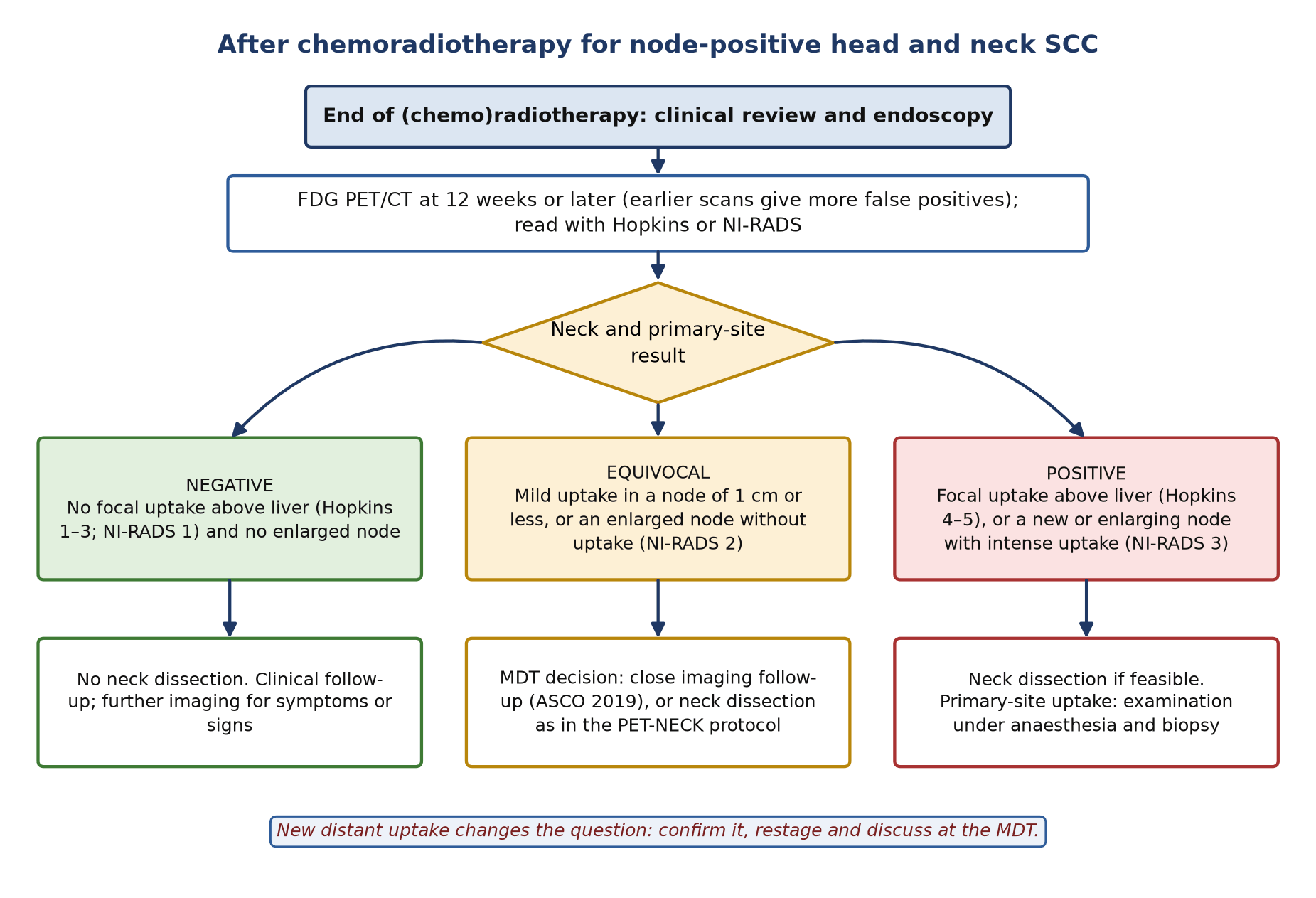
- Timing: 12 weeks or later. Earlier scans are less accurate because radiation inflammation is still active (Gupta meta-regression); the PET-NECK and ASCO pathways both use 12 weeks.
- Negative scan: no focal uptake above liver and no enlarged node: no neck dissection (ASCO 2019 recommendation 6.1a, strong; EHNS–ESMO–ESTRO [I, A]).
- Intense nodal uptake: neck dissection if feasible (ASCO 6.1a).
- Equivocal: ASCO 2019 (recommendation 6.2, moderate) allows close observation with serial cross-sectional imaging or PET/CT for mild uptake in a node of 1 cm or less, or a persistently enlarged node of 1 cm or more without uptake. PET-NECK treated exactly these findings as equivocal and dissected them. This is a real difference between a trial protocol and a guideline: document which your MDT follows. A repeat PET/CT 4–6 weeks later is another published option.
- Primary site uptake: focal residual uptake at the primary calls for examination under anaesthesia and biopsy, because PPV is low (58.6% in the meta-analysis).
- HPV-positive disease: FDG accuracy is lower, but PET-NECK found no difference in its survival result by p16 status.
- After a negative response scan: EHNS–ESMO–ESTRO advise clinical review with flexible endoscopy every 2–3 months for 2 years and every 6 months to year 5 [III, A], with imaging for symptoms or examination findings. NI-RADS offers a structured template and category-linked actions for any surveillance imaging.
- Suspected recurrence: restage with whole-body FDG PET/CT before salvage; distant disease changes the intent of treatment.
8. Theranostics and emerging tracers
There is no approved radioligand therapy for HNSCC. The PET tracers below are research tools; none is recommended in a guideline for routine care.
| Tracer | Target and idea | Best evidence | Status in 2026 |
|---|---|---|---|
| ⁶⁸Ga-FAPI | Fibroblast activation protein on cancer-associated fibroblasts; low salivary and mucosal background | Prospective single-centre comparison in 91 patients with an unknown primary: FAPI found 46 primaries vs 17 with FDG (sensitivity 51% vs 25%, PPV 98% vs 43%) and changed treatment in 24% | Investigational; no multicentre validation |
| ¹⁸F-FMISO | Hypoxia, a cause of radioresistance; a target for dose painting | Randomised phase II (53 patients, closed early): non-hypoxic tumours 100% 5-year local control vs 74% if hypoxic (p = 0.039); escalation to 77 Gy on the hypoxic volume gave 25% higher local control (16/19 vs 10/17), not significant (p = 0.150) | Trial use only; prognostic value is clearer than therapeutic benefit |
| ⁹⁹ᵐTc-tilmanocept and hybrid ICG–⁹⁹ᵐTc-nanocolloid | Receptor-targeted or fluorescent sentinel node mapping | Technical advantages noted in the EANM guideline, especially for floor-of-mouth tumours | Tilmanocept approved in the USA; hybrid tracer in use at some centres |
9. Structured report example
- Indication: p16-positive left tonsillar SCC, cT2 N1 (AJCC 8th), chemoradiotherapy completed 12 weeks ago. Question: residual disease; is neck dissection needed?
- Technique: ¹⁸F-FDG with 60-minute quiet uptake; skull vertex to mid-thigh with a dedicated head and neck acquisition and contrast-enhanced CT. Compared with the baseline PET/CT.
- Primary site: diffuse, symmetric curvilinear uptake in the oropharyngeal mucosa above blood pool, no focal uptake at the left tonsil (Hopkins 3; NI-RADS 1).
- Right neck: no abnormal uptake, no enlarged node (Hopkins 1).
- Left neck: the level IIa node has decreased from 32 mm to 14 mm and shows no uptake above blood pool (Hopkins 1). The level III node is no longer seen.
- Elsewhere: no FDG-avid distant disease or second primary.
- Conclusion: complete metabolic response at the primary site and neck; Hopkins overall score 3 (negative). The persistently enlarged but non-avid left level IIa node is suitable for close imaging follow-up rather than dissection (ASCO 2019, recommendation 6.2); for MDT decision.
10. Take-home points
- Stage with AJCC/UICC 8th edition (nasopharynx: version 9 from 2025); report laterality and size for p16-positive nodes and count distant lesions in NPC.
- Use whole-body FDG PET/CT for distant disease and second primaries in advanced disease, and before panendoscopy for the unknown primary.
- A negative PET does not clear the clinically N0 neck; early oral cancer needs sentinel node biopsy or elective dissection.
- Scan 12 weeks or later after chemoradiotherapy; a negative scan avoids neck dissection, a positive primary site needs biopsy.
- Score every response scan with one system (Hopkins or NI-RADS) and state the action it implies.
- Know the pitfalls: muscle, brown fat, vocal cord palsy, Waldeyer ring asymmetry, dental change, mucositis and metal.
- Sentinel node lymphoscintigraphy needs peritumoral injections (four is optimal), dynamic and static imaging and SPECT/CT, and a report that gives levels and anatomy.
Test yourself
5 quick questions. Pick an answer to see the explanation.
References
- Lydiatt WM, Patel SG, O'Sullivan B, et al. Head and neck cancers—major changes in the American Joint Committee on Cancer eighth edition cancer staging manual. CA Cancer J Clin. 2017;67(2):122-37.
- Pan JJ, Mai HQ, Ng WT, et al. Ninth version of the AJCC and UICC nasopharyngeal cancer TNM staging classification. JAMA Oncol. 2024;10(12):1627-35.
- Machiels JP, René Leemans C, Golusinski W, et al. Squamous cell carcinoma of the oral cavity, larynx, oropharynx and hypopharynx: EHNS-ESMO-ESTRO Clinical Practice Guidelines for diagnosis, treatment and follow-up. Ann Oncol. 2020;31(11):1462-75.
- Bossi P, Chan AT, Licitra L, et al. Nasopharyngeal carcinoma: ESMO-EURACAN Clinical Practice Guidelines for diagnosis, treatment and follow-up. Ann Oncol. 2021;32(4):452-65.
- National Institute for Health and Care Excellence. Cancer of the upper aerodigestive tract: assessment and management in people aged 16 and over. NICE guideline NG36. London: NICE; 2016 (updated 2018).
- Koyfman SA, Ismaila N, Crook D, et al. Management of the neck in squamous cell carcinoma of the oral cavity and oropharynx: ASCO clinical practice guideline. J Clin Oncol. 2019;37(20):1753-74.
- Maghami E, Ismaila N, Alvarez A, et al. Diagnosis and management of squamous cell carcinoma of unknown primary in the head and neck: ASCO guideline. J Clin Oncol. 2020;38(22):2570-96.
- Mehanna H, Wong WL, McConkey CC, et al. PET-CT surveillance versus neck dissection in advanced head and neck cancer. N Engl J Med. 2016;374(15):1444-54.
- Gupta T, Master Z, Kannan S, et al. Diagnostic performance of post-treatment FDG PET or FDG PET/CT imaging in head and neck cancer: a systematic review and meta-analysis. Eur J Nucl Med Mol Imaging. 2011;38(11):2083-95.
- Helsen N, Van den Wyngaert T, Carp L, Stroobants S. FDG-PET/CT for treatment response assessment in head and neck squamous cell carcinoma: a systematic review and meta-analysis of diagnostic performance. Eur J Nucl Med Mol Imaging. 2018;45(6):1063-71.
- Marcus C, Ciarallo A, Tahari AK, et al. Head and neck PET/CT: therapy response interpretation criteria (Hopkins criteria)—interreader reliability, accuracy, and survival outcomes. J Nucl Med. 2014;55(9):1411-6.
- Aiken AH, Rath TJ, Anzai Y, et al. ACR Neck Imaging Reporting and Data Systems (NI-RADS): a white paper of the ACR NI-RADS Committee. J Am Coll Radiol. 2018;15(8):1097-108.
- Zhong J, Sundersingh M, Dyker K, et al. Post-treatment FDG PET-CT in head and neck carcinoma: comparative analysis of 4 qualitative interpretative criteria in a large patient cohort. Sci Rep. 2020;10(1):4086.
- Kyzas PA, Evangelou E, Denaxa-Kyza D, Ioannidis JP. 18F-fluorodeoxyglucose positron emission tomography to evaluate cervical node metastases in patients with head and neck squamous cell carcinoma: a meta-analysis. J Natl Cancer Inst. 2008;100(10):712-20.
- Lowe VJ, Duan F, Subramaniam RM, et al. Multicenter trial of [18F]fluorodeoxyglucose positron emission tomography/computed tomography staging of head and neck cancer and negative predictive value and surgical impact in the N0 neck: results from ACRIN 6685. J Clin Oncol. 2019;37(20):1704-12.
- Xu GZ, Guan DJ, He ZY. 18FDG-PET/CT for detecting distant metastases and second primary cancers in patients with head and neck cancer. A meta-analysis. Oral Oncol. 2011;47(7):560-5.
- Rusthoven KE, Koshy M, Paulino AC. The role of fluorodeoxyglucose positron emission tomography in cervical lymph node metastases from an unknown primary tumor. Cancer. 2004;101(11):2641-9.
- Giammarile F, Schilling C, Gnanasegaran G, et al. The EANM practical guidelines for sentinel lymph node localisation in oral cavity squamous cell carcinoma. Eur J Nucl Med Mol Imaging. 2019;46(3):623-37.
- Schilling C, Stoeckli SJ, Haerle SK, et al. Sentinel European Node Trial (SENT): 3-year results of sentinel node biopsy in oral cancer. Eur J Cancer. 2015;51(18):2777-84.
- Garrel R, Poissonnet G, Moyà Plana A, et al. Equivalence randomized trial to compare treatment on the basis of sentinel node biopsy versus neck node dissection in operable T1-T2N0 oral and oropharyngeal cancer. J Clin Oncol. 2020;38(34):4010-8.
- Hasegawa Y, Tsukahara K, Yoshimoto S, et al. Neck dissections based on sentinel lymph node navigation versus elective neck dissections in early oral cancers: a randomized, multicenter, and noninferiority trial. J Clin Oncol. 2021;39(18):2025-36.
- Daisne JF, Duprez T, Weynand B, et al. Tumor volume in pharyngolaryngeal squamous cell carcinoma: comparison at CT, MR imaging, and FDG PET and validation with surgical specimen. Radiology. 2004;233(1):93-100.
- Purohit BS, Ailianou A, Dulguerov N, et al. FDG-PET/CT pitfalls in oncological head and neck imaging. Insights Imaging. 2014;5(5):585-602.
- Gu B, Yang Z, Du X, et al. Imaging of tumor stroma using 68Ga-FAPI PET/CT to improve diagnostic accuracy of primary tumors in head and neck cancer of unknown primary: a comparative imaging trial. J Nucl Med. 2024;65(3):365-71.
- Welz S, Paulsen F, Pfannenberg C, et al. Dose escalation to hypoxic subvolumes in head and neck cancer: a randomized phase II study using dynamic [18F]FMISO PET/CT. Radiother Oncol. 2022;171:30-6.