PET/CT in Testicular Germ-Cell Tumours
- One validated indication. ¹⁸F-fluorodeoxyglucose positron emission tomography/computed tomography (FDG PET/CT) is guideline-endorsed for a residual mass larger than 3 cm after chemotherapy for pure seminoma, and for little else.
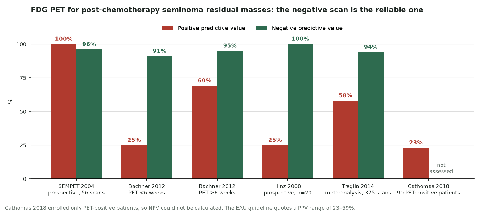
- Timing matters. Scan at least 6 weeks (ESMO–EURACAN), 8 weeks (ESMO consensus) or 2 months (EAU) after the last cycle; in one validation series the positive predictive value (PPV) rose from 25% before 6 weeks to 69% after.
- Trust the negative scan. The negative predictive value (NPV) is above 90%: a PET-negative residual seminoma mass needs follow-up only.
- Do not treat a positive scan on its own. In the largest registry of PET-positive residual masses, PPV was only 23%; repeat PET or obtain histology before radiotherapy, surgery or salvage chemotherapy.
- No PET for non-seminoma residual masses. Teratoma takes up little or no FDG, so a negative scan does not exclude it; residual masses over 1 cm are resected.
- No PET for primary staging. Contrast-enhanced CT of the chest, abdomen and pelvis stages the disease (EAU, strong); in high-risk stage I non-seminoma a third of PET-negative men on surveillance relapsed within about a year.
- Know the landing zones. Right testicular tumours drain first to interaortocaval and paracaval nodes, left ones to left para-aortic nodes just below the renal vein.
- Relapse with rising markers and negative CT is the one other setting where the ESMO consensus says FDG PET/CT may be useful (level IV, grade B).
1. The clinical problem
Testicular germ-cell tumours (TGCT) mainly affect young men and are among the most curable solid cancers. Cisplatin-based chemotherapy cures most men even with metastases, so the aim of imaging is as much to avoid overtreatment as to find disease. The oncologist needs imaging for three decisions: the extent of disease at diagnosis, whether a mass left after chemotherapy still contains tumour, and where the disease is when markers rise during follow-up.
Seminoma and non-seminoma
Pure seminoma and non-seminomatous germ-cell tumours (NSGCT: embryonal carcinoma, yolk sac tumour, choriocarcinoma, teratoma, and mixed tumours) behave differently, and this is the single most important fact for the PET reader. Seminoma is a sheet of uniform, glycolytic cells and is intensely FDG-avid; its post-chemotherapy residual masses are usually fibrosis. NSGCT residual masses often contain teratoma, which is FDG-poor, grows slowly, can transform, and must be removed. Stage I disease at diagnosis is found in 75–80% of men with seminoma and 55–64% with non-seminoma (EAU).
Serum tumour markers
- Alpha-fetoprotein (AFP): produced by yolk sac elements; a clearly raised AFP in a man with 'seminoma' implies a non-seminomatous component and changes management. Half-life 5–7 days.
- Human chorionic gonadotrophin (hCG): from syncytiotrophoblast; raised in up to 30% of pure seminomas and in most choriocarcinomas. Half-life 1–3 days.
- Lactate dehydrogenase (LDH): a marker of tumour burden rather than histology.
- Timing: markers are measured before and after orchidectomy and followed to normalisation; a plateau or rise after orchidectomy means metastatic disease (stage IS) even when CT is normal. Teratoma produces none of these markers.
Staging systems in force
The EAU guideline recommends the Union for International Cancer Control (UICC) TNM classification, 8th edition (2016, with the 2024 errata). It is unusual in adding a serum marker (S) category to T, N and M. Nodal categories are by size: N1 up to 2 cm, N2 more than 2 cm to 5 cm, N3 more than 5 cm. M1a is non-regional nodes or lung, M1b any other distant site. The American Joint Committee on Cancer (AJCC) 8th edition differs in details (for example, it splits pT1 seminoma at 3 cm), so ESMO–EURACAN asks that the system used is always stated.
For metastatic disease, treatment intensity is set by the International Germ Cell Cancer Collaborative Group (IGCCCG) classification. The 1997 groups were revalidated in 2021 on about 12,000 men treated between 1990 and 2013; the groups did not change, but survival did, and new adverse factors were identified.
| IGCCCG group | Non-seminoma (all criteria for good; any for intermediate or poor) | Seminoma | 5-year PFS / OS (2021 update) |
|---|---|---|---|
| Good | Testis or retroperitoneal primary, no non-pulmonary visceral metastases (NPVM), AFP <1,000 ng/ml, hCG <5,000 IU/l, LDH <1.5 × upper limit of normal (ULN) | Any primary site, no NPVM, normal AFP, any hCG, any LDH | NSGCT 90% / 96%; seminoma 89% / 95% |
| Intermediate | AFP 1,000–10,000 ng/ml, or hCG 5,000–50,000 IU/l, or LDH 1.5–10 × ULN | NPVM present (liver, bone, brain), normal AFP | NSGCT 78% / 89%; seminoma 79% / 88% |
| Poor | Mediastinal primary, or NPVM, or AFP >10,000 ng/ml, or hCG >50,000 IU/l, or LDH >10 × ULN | No poor-prognosis seminoma | NSGCT 54% / 67% |
PFS, progression-free survival; OS, overall survival. 2021 refinements: in NSGCT, LDH above 2.5 × ULN, older age and lung metastases are additional adverse factors (online calculator); in good-prognosis seminoma, LDH above 2.5 × ULN lowered 3-year PFS from 92% to 80%.
Lymphatic drainage: where to look first
The testis develops near the kidney and its lymphatics follow the gonadal vessels back to the renal hilum. In a surgical mapping series of 214 men with stage II non-seminoma, solitary deposits from right-sided tumours lay most often in the interaortocaval zone, then in the paracaval and precaval zones; deposits from left-sided tumours lay predominantly in the upper para-aortic zone, below the left renal vein. Hilar and suprahilar deposits were infrequent, and even multiple metastases spared the external iliac regions. The same template logic guides surgery: left-sided residual masses need para-aortic resection, right-sided ones paracaval and interaortocaval resection (EAU). CT staging rests on this map, and so does PET reading: a 9 mm node in the right landing zone matters more than a 12 mm node elsewhere.
Where nuclear medicine changes management
- Post-chemotherapy seminoma mass >3 cm: a negative FDG PET/CT allows surveillance instead of surgery or radiotherapy.
- Marker-positive relapse with negative CT: PET may find the site of disease (ESMO consensus, weak evidence).
- Everything else (primary staging, stage I, non-seminoma residual masses, routine surveillance) is covered by CT or MRI, markers and surgery, and guidelines advise against routine PET.
2. Tracers and why they work
Only ¹⁸F-FDG is in clinical use. Glucose-transporter expression and glycolysis are high in seminoma and in embryonal carcinoma and choriocarcinoma, so these tumours are usually avid. The problem is not detecting active tumour but separating it from what else lies in a residual mass.
| Tissue | Typical FDG uptake | Consequence |
|---|---|---|
| Seminoma | High | Viable residual seminoma is usually visible; a negative scan is reliable if timed correctly |
| Embryonal carcinoma, choriocarcinoma | Usually high | Detectable, but these men are followed mainly by markers |
| Yolk sac tumour | Variable | AFP is the better marker |
| Teratoma (post-pubertal type) | Low or absent | False-negative PET; resection is required |
| Necrosis and fibrosis | Absent | True negative |
| Post-chemotherapy inflammation, granulomatous reaction | Mild to intense | False-positive PET, especially before 6–8 weeks |
When FDG fails and what replaces it
- Teratoma: no tracer reliably shows it; the answer is resection of every residual non-seminoma mass above 1 cm.
- Sub-centimetre nodes in stage I: below the resolution needed; surveillance with CT or MRI and markers is the standard.
- Early after chemotherapy: inflammation and desmoplastic reaction; wait, or repeat later.
- Occult marker-producing disease: markers are more sensitive than any scan; the microRNA-371a-3p (M371) test had 90.1% sensitivity and 94.0% specificity for the primary diagnosis in a prospective study of 616 patients, but, like AFP and hCG, is not expressed by teratoma and is not yet standard.
3. Indications by clinical scenario
| Scenario | Role of FDG PET/CT | Guideline position (strength where given) |
|---|---|---|
| Primary staging, any histology | Not indicated | EAU: do not use PET-CT or bone scan for staging (strong). ESMO–EURACAN: no evidence for routine use [III, D]. ESMO consensus: not routinely recommended (level I, grade B) |
| Stage I (normal CT, normalised markers) | Not indicated | As above; FDG PET cannot see micrometastases in sub-centimetre nodes (ESMO consensus) |
| Equivocal node on staging CT, markers negative | Not recommended | ESMO consensus: repeat contrast CT after 6–8 weeks (V, B), not PET (V, C) |
| Negative CT, raised markers at staging | Not useful | ESMO consensus recommendation 4.7 (V, C) |
| Radiotherapy planning (stage IIA/B seminoma) | No defined role | Not addressed by EAU or ESMO; target volumes are defined on CT |
| Interim response during chemotherapy | Investigational | EAU: de-escalation guided by interim PET not recommended for routine use |
| Seminoma residual mass >3 cm after chemotherapy | Indicated | ESMO–EURACAN: recommended ≥6 weeks after chemotherapy [III, B]. ESMO consensus: may be helpful ≥8 weeks after (III, B). EAU: should be considered, not before 2 months; NPV >90%, PPV 23–69% (summary of evidence level 2b) |
| Seminoma residual mass ≤3 cm | Not needed | Follow-up with imaging and markers (EAU, ESMO). PET sensitivity for lesions <3 cm was 47% in a meta-analysis |
| Non-seminoma residual mass | Not indicated | EAU: restaging with FDG-PET not indicated; resect residual masses >1 cm (strong). ESMO consensus: PET cannot separate teratoma from necrosis |
| Marker-positive relapse, negative CT | May help | ESMO consensus 4.12: repeat FDG PET/CT may be useful (IV, B) |
| Routine surveillance | Not recommended | EAU: FDG-PET-CT not recommended during surveillance; MRI of the abdomen may replace CT in experienced centres |
- Timing: ESMO–EURACAN 2022 says at least 6 weeks after chemotherapy; the ESMO consensus conference and EAU say at least 8 weeks or 2 months. Scanning at 8 weeks satisfies all three.
- The positive scan: EAU advises a repeat PET 6 weeks later if the mass is not growing; ESMO–EURACAN recommends biopsy before radiotherapy or resection, or further follow-up with biopsy if the lesion stays positive or grows. The ESMO consensus advises serial PET until resolution or progression, with resection as an alternative for a persistent nodular mass.
- Raised markers and negative CT: the ESMO consensus says PET is not useful for staging at diagnosis in this situation (4.7) but may be useful at relapse (4.12). The EAU makes no recommendation for relapse.
4. Evidence at a glance
| Study | Design and n | Key finding | What it changed |
|---|---|---|---|
| SEMPET (De Santis 2004) | Prospective multicentre, 51 men with seminoma residual masses, 56 scans | PET: specificity 100%, sensitivity 80%, PPV 100%, NPV 96%; CT size cut-off (>3 cm): 74%, 70%, 37%, 92%. All 19 masses >3 cm correctly classified | Made PET the standard test for seminoma residual masses |
| Hinz 2008 | Prospective, 20 men, all with histology | No false negatives (NPV 100%), but 9 false positives: PPV 25% | First warning that a positive scan is unreliable |
| Bachner 2012 | Retrospective validation, 127 eligible scans | Before 6 weeks: sensitivity 50%, specificity 77%, PPV 25%, NPV 91%. After 6 weeks: 82%, 90%, 69%, 95%; accuracy 73% to 88% (P=0.032) | Established the 6-week minimum interval |
| Treglia 2014 | Meta-analysis, 9 studies, 375 scans | Pooled sensitivity 78%, specificity 86%, PPV 58%, NPV 94%. Sensitivity 89% for lesions >3 cm versus 47% for <3 cm | Supports restricting PET to masses >3 cm |
| Cathomas 2018 (Global Germ Cell Cancer Group registry) | Retrospective, 90 men with PET-positive residual masses (median 4.9 cm) | Median time to PET 6.9 weeks. Of 26 resected masses, 81% necrosis, 19% seminoma; no biopsy showed seminoma. PPV 23% | Positive PET alone should not trigger treatment |
| Oechsle 2008 | Prospective, 121 men with NSGCT before residual-mass surgery, all with histology | PET accuracy 56%, no better than CT (55%) or markers (56%); sensitivity 70%, specificity 48% | PET abandoned for non-seminoma residual masses |
| de Wit 2008 | Prospective, 72 men with clinical stage I/II NSGCT (target 169) | Nodal staging: PET sensitivity 66%, specificity 98%, NPV 78% (target 90% not reached); CT 41% and 95% | No routine PET staging |
| MRC TE22 (Huddart 2007) | Prospective, 111 high-risk stage I NSGCT scanned | 88 (79%) PET-negative; 33 of 87 on surveillance relapsed, 1-year relapse-free rate 63%; trial stopped early | PET cannot select stage I patients for surveillance |
| GETUG SEMITEP (Loriot 2022) | Phase 2, 102 men with good-prognosis seminoma | 68.4% PET-negative after 2 cycles of etoposide–cisplatin (EP) received 1 cycle of carboplatin; 3-year PFS 90.8% versus 90.0% with EP ×4 | Promising de-escalation; not yet routine (EAU) |
5. Patient preparation and acquisition
Standard oncological FDG practice applies (EANM procedure guideline version 2.0): fasting for at least 4 hours, blood glucose checked, imaging at about 60 minutes, and the same scanner and reconstruction for serial studies. What is specific to germ-cell tumours is mostly about timing and history.
- Timing after chemotherapy: book the residual-mass scan at least 8 weeks after the last cycle when possible. Ask for the date of day 1 and day 21 of the last cycle, not the date of the last clinic visit.
- Growth factors: granulocyte colony-stimulating factor (G-CSF) causes diffuse marrow and splenic uptake; in one study marrow uptake stayed above baseline for up to 4 weeks after the last dose. Record the date.
- Brown fat: these are young, often slim men; keep the waiting room warm to reduce supraclavicular and paravertebral brown-fat uptake, which lies in fat on CT.
- Field of view: skull base to mid-thigh is standard; include the inguinal regions and scrotum, because the remaining testis and the orchidectomy bed can be relevant.
- CT component: a diagnostic contrast-enhanced CT in the same session saves a separate study and helps measure the mass; oral contrast is optional.
- Request information: histology (pure seminoma or not), IGCCCG group, chemotherapy regimen and dates, current AFP, hCG and LDH, previous surgery (orchidectomy side, retroperitoneal lymph node dissection) and the size of the residual mass on the latest CT.
6. Interpretation
Criteria in use
There is no validated standardised uptake value (SUV) threshold for residual seminoma. The studies in the Treglia meta-analysis all read scans visually, three with added SUV measurements. No guideline defines a positive scan. Readers compare the mass with the surrounding background and the blood pool: focal uptake clearly above them suggests viable tumour, and uptake no higher than blood pool is read as negative, but neither reference level has been validated as a cut-off. Faint uptake at the rim of a mass, rather than a focal nodule, is a common pattern of post-treatment inflammation. Record SUVmax so that a repeat scan can be compared, but base the conclusion on the pattern.
Pitfalls and mimics
| Pitfall | What it looks like | How to handle it |
|---|---|---|
| Early scan after chemotherapy | Uptake in the residual mass from inflammation and desmoplastic reaction | Check the interval; if under 6–8 weeks, say so and suggest repeating |
| Granulomatous or sarcoid-like reaction | Symmetric mediastinal and hilar nodes, sometimes within the mass | Symmetry and distribution; histology if it would change treatment |
| Teratoma (non-seminoma or mixed tumour) | Growing or stable mass with little or no uptake | Never call a non-seminoma mass benign on PET; resection decides |
| Growing teratoma syndrome | Masses enlarge during or after chemotherapy while markers normalise; low uptake | Recognise the pattern; early surgery (EAU) |
| Small residual (<1–3 cm) | No uptake because of partial-volume effect | PET sensitivity is low below 3 cm; follow with CT or MRI |
| Brown fat | Symmetric supraclavicular, axillary and paravertebral uptake in fat | Fat density on CT; warming before injection |
| Bleomycin lung toxicity | Bilateral, often basal and subpleural, parenchymal uptake with ground-glass or reticular change | Alert the clinical team; not metastases |
| G-CSF effect | Diffuse marrow and splenic uptake | Diffuse rather than focal; check the date of the last dose |
| Thymic rebound after chemotherapy | Bilobed anterior mediastinal soft tissue with mild uptake in a young man | Shape and location; follow-up |
| Remaining testis and ureter | Normal testicular uptake; ureteric urine activity crossing a retroperitoneal mass | Correlate with CT; late or prone images if needed |
Wording the conclusion
- State the histology, the size of the mass and the interval since chemotherapy in the first line of the conclusion.
- Negative: 'No FDG uptake above blood pool in the 4.6 cm residual mass, 9 weeks after chemotherapy: no evidence of viable seminoma.'
- Positive: 'Focal uptake (SUVmax 7.8) in the lateral part of the mass. Viable seminoma is possible, but false positives are common after chemotherapy; suggest repeat FDG PET/CT in about 6 weeks or biopsy before any treatment.'
- Non-seminoma: if asked to report PET, state that a negative scan does not exclude teratoma or viable tumour.
7. Response assessment and follow-up
After chemotherapy for seminoma
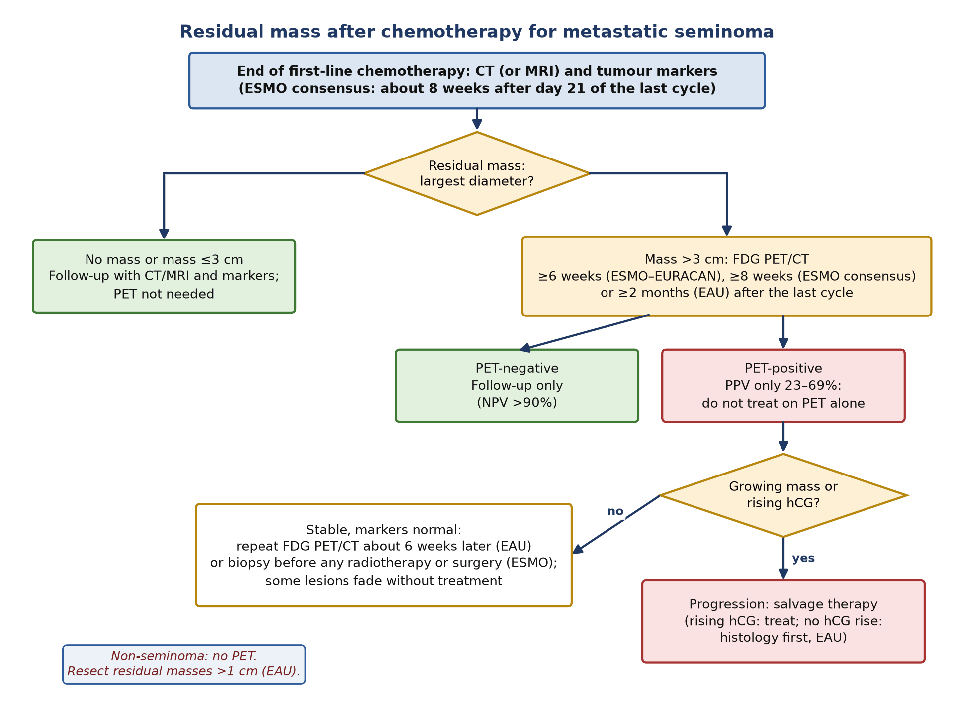
Restaging with CT or MRI and markers comes first. The ESMO consensus sets this at about 8 weeks after day 21 of the last cycle. A complete response, or a residual mass of 3 cm or less, needs only follow-up. A mass larger than 3 cm goes to FDG PET/CT at the right interval. Negative scans with a stable or shrinking mass are followed routinely. Positive scans need caution: in the Cathomas registry most PET-positive masses were necrosis and relapse occurred in 17% overall, almost all successfully salvaged. A growing mass that enhances on CT or is FDG-avid is progression and needs salvage therapy (EAU); rising hCG alone justifies salvage chemotherapy, whereas progression without an hCG rise should be confirmed histologically first. Surgery for seminoma residual masses is difficult because of dense fibrosis and belongs in referral centres.
Interim PET during chemotherapy
Two phase 2 studies have used PET to shorten treatment in good-prognosis seminoma. In GETUG SEMITEP, men with a negative scan after 2 cycles of EP received 1 cycle of carboplatin instead of 2 more cycles of EP, with similar 3-year PFS and less neuropathy and ototoxicity. In a trial of carboplatin AUC 10, a Deauville score of 3 or less after 1 cycle allowed 44% of 48 men to stop after 3 cycles. There are no agreed interpretation criteria for interim PET in germ-cell tumours, and the EAU does not recommend PET-guided de-escalation outside trials.
After chemotherapy for non-seminoma
Restaging is by CT and markers 3–4 weeks after the start of the last cycle (EAU; 4–6 weeks in the ESMO consensus). Any residual retroperitoneal mass above 1 cm with normal or normalising markers is resected, whatever its FDG uptake, because after first-line BEP (bleomycin, etoposide, cisplatin) fewer than one in ten contain viable cancer, about a third to a half contain teratoma and about 40% contain necrosis or fibrosis only.
Follow-up and relapse
- Surveillance: CT or, in experienced centres, MRI of the abdomen with markers; the EAU does not recommend FDG PET/CT for routine surveillance, to limit radiation in young men.
- Rising markers with negative CT: first exclude false-positive marker rises and repeat the test; if the rise is confirmed, FDG PET/CT may locate disease (ESMO consensus 4.12, IV, B). A negative PET does not end the search: repeat imaging as markers rise further.
- Late relapse: recurrence more than 2 years after complete response occurs in 2–3% and often contains yolk sac tumour or teratoma (ESMO–EURACAN), both of which may be FDG-poor; surgery is the mainstay.
- Real-world data: in a 20-year single-centre series of 249 men with seminoma, PET done for clinical suspicion of relapse after treatment of advanced disease had PPV 100% and NPV 94%; retrospective and not guideline-endorsed.
8. Theranostics and emerging tracers
| Agent or approach | Setting | Evidence level (2026) |
|---|---|---|
| ¹⁸F-FDG, residual seminoma mass >3 cm | Post-chemotherapy | Guideline-endorsed (ESMO–EURACAN III, B; EAU; ESMO consensus) |
| ¹⁸F-FDG, interim response in seminoma | De-escalation of chemotherapy | Phase 2 trials; not recommended routinely |
| ¹⁸F-FDG, staging before primary retroperitoneal surgery | Clinical stage IIA/B seminoma | Research: in 26 men in the SEMS trial, sensitivity 95% and specificity 71% against surgical pathology |
| 3′-deoxy-3′-¹⁸F-fluorothymidine (FLT) | Proliferation; residual masses | Investigational: in 11 men with metastatic GCT it did not improve on FDG, and both teratomas were missed by both tracers |
| ⁶⁸Ga-labelled fibroblast activation protein inhibitor (FAPI) | Mediastinal seminoma | Case reports only |
| Radioligand therapy | Any | None approved or in routine trials |
The next change in this field is more likely to come from blood than from tracers. The M371 microRNA test detects viable seminoma and non-teratomatous NSGCT and may one day help decide what to do with a residual mass, but it misses teratoma and awaits standardisation (EAU).
9. Structured report example
- Clinical question: Pure seminoma of the left testis, stage IIC, IGCCCG good prognosis, 3 cycles of BEP completed 9 weeks ago (day 21). Markers normal. 4.6 × 3.8 cm left para-aortic residual mass on CT. Viable tumour?
- Technique: ¹⁸F-FDG 250 MBq after a 6-hour fast (glucose 5.1 mmol/l); imaging at 62 minutes, skull base to mid-thigh, with contrast-enhanced CT. No G-CSF in the last 6 weeks.
- Findings: The left para-aortic mass lies below the left renal vein, lateral and anterior to the aorta, and measures 4.6 × 3.8 cm (5.9 × 4.9 cm before chemotherapy). Its uptake does not exceed aortic blood pool (SUVmax 1.9; blood pool 1.8); no focal nodule. No other abnormal uptake. Symmetric uptake in the right testis, bone marrow and bowel is physiological. Lungs clear.
- Conclusion: FDG-negative residual mass, 9 weeks after chemotherapy: no evidence of viable seminoma. A negative scan at this interval has a high negative predictive value.
- Recommendation: Follow-up with CT or MRI and markers according to the local schedule; no further imaging with PET needed unless the mass grows or markers rise.
10. Take-home points
- Ask first: pure seminoma or not? PET helps with seminoma residual masses; it misleads with non-seminoma because teratoma is FDG-poor.
- Scan residual seminoma masses larger than 3 cm, and scan them late: at least 6 weeks, ideally 8 weeks, after the last cycle.
- A negative scan is reliable (NPV above 90%); a positive scan is not (PPV as low as 23%). Repeat it or get histology before treatment.
- Do not use PET for primary staging, stage I disease or routine surveillance; CT or MRI and markers do this job.
- Read nodes against the landing zones: interaortocaval for right-sided tumours, left para-aortic for left-sided tumours.
- Rising markers with negative CT is the one relapse scenario where PET may help.
- Always state the histology, the interval since chemotherapy and whether G-CSF was given, because each changes the meaning of uptake.
Test yourself
5 quick questions. Pick an answer to see the explanation.
References
- Nicol D, Berney DM, Boormans JL, et al. EAU Guidelines on Testicular Cancer. Arnhem: EAU Guidelines Office; 2025 (limited update; section 6.3.2 unchanged in the 2026 online edition).
- Patrikidou A, Cazzaniga W, Berney D, et al. European Association of Urology guidelines on testicular cancer: 2023 update. Eur Urol. 2023;84(3):289-301.
- Oldenburg J, Berney DM, Bokemeyer C, et al. Testicular seminoma and non-seminoma: ESMO-EURACAN Clinical Practice Guideline for diagnosis, treatment and follow-up. Ann Oncol. 2022;33(4):362-75.
- Honecker F, Aparicio J, Berney D, et al. ESMO Consensus Conference on testicular germ cell cancer: diagnosis, treatment and follow-up. Ann Oncol. 2018;29(8):1658-86.
- International Germ Cell Cancer Collaborative Group. International Germ Cell Consensus Classification: a prognostic factor-based staging system for metastatic germ cell cancers. J Clin Oncol. 1997;15(2):594-603.
- Gillessen S, Sauvé N, Collette L, et al. Predicting outcomes in men with metastatic nonseminomatous germ cell tumors (NSGCT): results from the IGCCCG Update Consortium. J Clin Oncol. 2021;39(14):1563-74.
- Beyer J, Collette L, Sauvé N, et al. Survival and new prognosticators in metastatic seminoma: results from the IGCCCG-Update Consortium. J Clin Oncol. 2021;39(14):1553-62.
- Weissbach L, Boedefeld EA. Localization of solitary and multiple metastases in stage II nonseminomatous testis tumor as basis for a modified staging lymph node dissection in stage I. J Urol. 1987;138(1):77-82.
- De Santis M, Becherer A, Bokemeyer C, et al. 2-18fluoro-deoxy-D-glucose positron emission tomography is a reliable predictor for viable tumor in postchemotherapy seminoma: an update of the prospective multicentric SEMPET trial. J Clin Oncol. 2004;22(6):1034-9.
- Hinz S, Schrader M, Kempkensteffen C, et al. The role of positron emission tomography in the evaluation of residual masses after chemotherapy for advanced stage seminoma. J Urol. 2008;179(3):936-40.
- Bachner M, Loriot Y, Gross-Goupil M, et al. 2-18fluoro-deoxy-D-glucose positron emission tomography (FDG-PET) for postchemotherapy seminoma residual lesions: a retrospective validation of the SEMPET trial. Ann Oncol. 2012;23(1):59-64.
- Treglia G, Sadeghi R, Annunziata S, et al. Diagnostic performance of fluorine-18-fluorodeoxyglucose positron emission tomography in the postchemotherapy management of patients with seminoma: systematic review and meta-analysis. Biomed Res Int. 2014;2014:852681.
- Cathomas R, Klingbiel D, Bernard B, et al. Questioning the value of fluorodeoxyglucose positron emission tomography for residual lesions after chemotherapy for metastatic seminoma: results of an international Global Germ Cell Cancer Group registry. J Clin Oncol. 2018;36(34):3381-7.
- Oechsle K, Hartmann M, Brenner W, et al. [18F]Fluorodeoxyglucose positron emission tomography in nonseminomatous germ cell tumors after chemotherapy: the German multicenter positron emission tomography study group. J Clin Oncol. 2008;26(36):5930-5.
- de Wit M, Brenner W, Hartmann M, et al. [18F]-FDG-PET in clinical stage I/II non-seminomatous germ cell tumours: results of the German multicentre trial. Ann Oncol. 2008;19(9):1619-23.
- Huddart RA, O'Doherty MJ, Padhani A, et al. 18fluorodeoxyglucose positron emission tomography in the prediction of relapse in patients with high-risk, clinical stage I nonseminomatous germ cell tumors: preliminary report of MRC Trial TE22 - the NCRI Testis Tumour Clinical Study Group. J Clin Oncol. 2007;25(21):3090-5.
- Loriot Y, Texier M, Culine S, et al. The GETUG SEMITEP trial: de-escalating chemotherapy in good-prognosis seminoma based on fluorodeoxyglucose positron emission tomography/computed tomography. Eur Urol. 2022;82(2):172-9.
- Shamash J, Syed R, Sarker SJ, et al. A phase II study of carboplatin AUC-10 guided by positron emission tomography-defined metabolic response in metastatic seminoma. Eur J Cancer. 2019;115:128-35.
- Hu B, Alsyouf M, Farkouh A, et al. Accuracy of 18F-fluorodeoxyglucose positron emission tomography in early metastatic testicular seminoma: analysis from the SEMS trial. J Urol. 2025;214(3):272-9.
- Conduit C, Koh TT, Hofman MS, et al. Two decades of FDG-PET/CT in seminoma: exploring its role in diagnosis, surveillance and follow-up. Cancer Imaging. 2022;22(1):58.
- Dieckmann KP, Radtke A, Geczi L, et al. Serum levels of microRNA-371a-3p (M371 test) as a new biomarker of testicular germ cell tumors: results of a prospective multicentric study. J Clin Oncol. 2019;37(16):1412-23.
- Pfannenberg C, Aschoff P, Dittmann H, et al. PET/CT with 18F-FLT: does it improve the therapeutic management of metastatic germ cell tumors? J Nucl Med. 2010;51(6):845-53.
- Boellaard R, Delgado-Bolton R, Oyen WJ, et al. FDG PET/CT: EANM procedure guidelines for tumour imaging: version 2.0. Eur J Nucl Med Mol Imaging. 2015;42(2):328-54.
- Sugawara Y, Fisher SJ, Zasadny KR, et al. Preclinical and clinical studies of bone marrow uptake of fluorine-1-fluorodeoxyglucose with or without granulocyte colony-stimulating factor during chemotherapy. J Clin Oncol. 1998;16(1):173-80.
- Cohade C, Osman M, Pannu HK, Wahl RL. Uptake in supraclavicular area fat ("USA-Fat"): description on 18F-FDG PET/CT. J Nucl Med. 2003;44(2):170-6.