Nuclear Medicine in Gynaecological Cancers
- Cervix: PET is the staging test for locally advanced disease. The 2023 European guideline (ESGO/ESTRO/ESP) recommends ¹⁸F-fluorodeoxyglucose (FDG) positron emission tomography with computed tomography (PET/CT), or chest and abdominal CT if PET is unavailable, for T1b3 and higher tumours, for any tumour with suspicious nodes, and before curative chemoradiotherapy [III, B].
- Nodes change the stage and the radiotherapy field. In the 2018 staging of the International Federation of Gynaecology and Obstetrics (FIGO), pelvic nodes make a cervical cancer stage IIIC1 and para-aortic nodes IIIC2, with 'r' when imaging assigns the stage and 'p' when pathology does. Para-aortic disease means extended-field treatment.
- A negative PET is not N0. Pooled sensitivity for para-aortic nodes is about 71% (specificity 97%), and 12% of women with a negative para-aortic PET/CT had para-aortic metastases at laparoscopic dissection.
- The 3-month scan after chemoradiotherapy predicts outcome. Three-year progression-free survival was 78%, 33% and 0% after complete metabolic response, partial response and progression.
- Early cervical, endometrial and vulvar cancer are staged by the sentinel node. Radiocolloid is mandatory in vulvar cancer; in the uterus indocyanine green is preferred and radiocolloid with single photon emission CT (SPECT/CT) is the partner or alternative.
- Know the menstrual cycle. Endometrial uptake peaks at menstruation and ovulation, and ovarian uptake is often functional mid-cycle. Focal ovarian or endometrial uptake after the menopause is abnormal until proven otherwise.
- Ovary: CT first, PET for the rising CA-125 (cancer antigen 125). FDG PET/CT has the highest pooled sensitivity for recurrence (91%) but under-reads small peritoneal implants. FAPI and oestrogen-receptor PET remain investigational.
1. The clinical problem
Gynaecological cancers ask nuclear medicine four different questions. In cervical and vulvar squamous cell carcinoma, spread is lymphatic and orderly, so the job is to find nodes: their presence decides between surgery and chemoradiotherapy, and their level decides the radiotherapy field. In endometrial cancer, most women are cured by surgery, staging is surgical and molecular, and imaging matters mainly for high-risk disease and recurrence. In ovarian cancer, spread is peritoneal, the questions are resectability and the site of relapse, and CT carries most of the load.
| Cancer | Staging system in force | How imaging enters the stage | Where nuclear medicine changes management |
|---|---|---|---|
| Cervix | FIGO 2018. ESGO/ESTRO/ESP 2023 asks for TNM (tumour, node, metastasis) staging with the FIGO stage also documented [IV, A] | Imaging and pathology may both assign stage. Pelvic nodes = IIIC1, para-aortic nodes = IIIC2; 'r' (imaging) or 'p' (pathology) records the method | PET/CT for locally advanced disease and before chemoradiotherapy; sentinel lymph node (SLN) biopsy in early disease; response at 3 months; recurrence |
| Endometrium | FIGO 2023, with a molecular annotation 'm' when the class is known (two molecularly defined stages: IAm POLEmut, POLE-mutated, and IICm p53abn, p53-abnormal) | Surgical and pathological; imaging is used to look for extrauterine disease | SLN biopsy for all presumed uterus-confined disease [II, A]; PET/CT for high-risk histology, suspected advanced disease and recurrence |
| Ovary, tube, peritoneum | FIGO 2014 | Surgical and pathological; CT maps disease and resectability | PET/CT for the rising CA-125 with negative conventional imaging and for suspected extra-abdominal disease |
| Vulva | FIGO 2021 and TNM 8th edition. ESGO 2023 advises TNM because the two are poorly aligned [V, B] | Groin status is pathological (SLN or lymphadenectomy) | Radiocolloid SLN mapping for small unifocal tumours; CT or FDG PET/CT for systemic staging of larger tumours |
ESGO (European Society of Gynaecological Oncology) guidelines, written with ESTRO (European Society for Radiotherapy and Oncology) and ESP (European Society of Pathology), grade each recommendation by level of evidence (I, randomised trials, to V, expert opinion) and strength (A, strongly recommended; B, generally recommended; C, optional; D, generally not recommended; E, never recommended). Those gradings are quoted in square brackets throughout.
- Para-aortic or distant disease in cervical cancer turns pelvic chemoradiotherapy into extended-field treatment, or curative intent into systemic treatment.
- A sentinel node instead of a lymphadenectomy cuts morbidity: after vulvar SLN biopsy alone, leg lymphoedema fell to 1.9% from 25.2% after lymphadenectomy.
- A metabolic response scan selects women with persistent disease after chemoradiotherapy for salvage surgery.
- A whole-body restaging scan at recurrence separates oligometastatic disease, which can be treated radically, from disseminated disease, which cannot.
2. Tracers and why they work
¹⁸F-fluorodeoxyglucose (FDG) enters cells through glucose transporters and is trapped after phosphorylation by hexokinase. Squamous cell carcinomas of the cervix and vulva are usually intensely avid, as are high-grade endometrial carcinomas and high-grade serous ovarian carcinoma. Low-grade and mucinous tumours are less predictable, so a faint primary tumour is not reassuring and a negative scan in these subtypes means less.
FDG positron emission tomography (PET) fails for two physical reasons. The first is size: in uterine cancers, node-based sensitivity was 100% for metastatic nodes of 10 mm or more, 67% for nodes of 5 to 9 mm and 13% for nodes of 4 mm or less. Micrometastases (up to 2 mm) and isolated tumour cells are beyond any PET scanner, which is why early cervical, endometrial and vulvar cancer are staged surgically with the sentinel node. The second is geometry: thin, flat peritoneal implants next to physiological bowel activity are easily missed, and in a meta-analysis of ovarian cancer FDG detected peritoneal metastases with a pooled sensitivity of only 70%.
⁹⁹ᵐTc-nanocolloid is injected into tissue, taken up by lymphatic capillaries and retained by macrophages in the first draining node or nodes. It gives a preoperative map (planar images and single photon emission computed tomography with CT, SPECT/CT) and lets the surgeon find the node with a gamma probe. Indocyanine green (ICG) is a fluorescent dye seen with a near-infrared camera during surgery; it is not a radiotracer and gives no preoperative map. A hybrid ICG-⁹⁹ᵐTc-nanocolloid tracer combines both signals.
| Tracer | Target | Established use in gynaecological cancer | Status (September 2026) |
|---|---|---|---|
| ¹⁸F-FDG | Glucose metabolism | Staging locally advanced cervical cancer, radiotherapy planning, response, recurrence in all four cancers | Guideline-endorsed |
| ⁹⁹ᵐTc-nanocolloid | Lymphatic drainage | SLN mapping in vulvar, cervical and endometrial cancer | Guideline-endorsed (mandatory in vulvar cancer) |
| ⁶⁸Ga-FAPI (fibroblast activation protein inhibitor) | Cancer-associated fibroblasts in tumour stroma | Peritoneal and nodal staging in ovarian and cervical cancer | Investigational |
| ¹⁸F-FES (fluoroestradiol) | Oestrogen receptor | Receptor status in low-grade serous ovarian and endometrial cancer | Investigational in gynaecology; approved only for breast cancer |
3. Indications by clinical scenario
3.1 Cervical cancer
Pelvic magnetic resonance imaging (MRI) is mandatory for local extent [II, A]; FDG PET/CT answers the nodal and distant question. In early disease managed by surgery, surgical and pathological staging of the pelvic nodes is the standard [III, A] and PET adds little, because the nodes that matter are microscopic. In locally advanced cervical cancer (LACC: T1b3 and higher, except T2a1) the risk of disease outside the pelvic radiotherapy field is real, and PET/CT is recommended.
| Scenario | Role of nuclear medicine | ESGO/ESTRO/ESP 2023 recommendation |
|---|---|---|
| Early stage (T1a1 with lymphovascular space invasion to T2a1), nodes not suspicious | SLN mapping before pelvic lymphadenectomy; radiocolloid with blue dye if ICG is not used | SLN biopsy should be performed; ICG preferred [III, A]; blue dye plus radiocolloid an alternative [IV, B] |
| Locally advanced, or any stage with suspicious nodes on imaging | FDG PET/CT for nodal and distant disease | PET/CT, or chest and abdominal CT if PET is unavailable [III, B]; PET/CT before curative chemoradiotherapy [III, B] |
| Para-aortic nodes negative on imaging | Recognise PET's false-negative rate | Para-aortic lymph node dissection, at least up to the inferior mesenteric artery, may be considered for staging [IV, C] |
| Equivocal extrauterine finding | Direct the biopsy to the most accessible avid lesion | Biopsy to avoid inappropriate treatment [IV, B] |
| Radiotherapy planning | PET/CT in the treatment position for contouring the primary and involved nodes | 3D imaging, preferably both MRI and PET/CT, in the treatment position, and a boost to pathological nodes of 60 Gy EQD2 (equivalent dose in 2 Gy fractions) [III, B]; elective para-aortic irradiation for any node at common iliac level or above, and considered for more than two level 1 nodes [IV, B] |
| Response after chemoradiotherapy | PET/CT (or MRI with or without CT) at 3 months or later | Not earlier than 3 months after treatment [IV, B]; repeat after a further 2 to 3 months if response is uncertain [IV, B] |
| Symptomatic suspected recurrence | PET/CT for systemic assessment and to select women for exenteration or radical treatment of oligometastatic disease | MRI or ultrasound for the pelvis, CT or PET/CT for systemic assessment [IV, A]; histological confirmation if feasible [IV, B] |
| Pregnancy | Avoid PET | PET/CT should be avoided in pregnancy; whole-body diffusion-weighted MRI instead [IV, B] |
How good is PET for the para-aortic nodes? A meta-analysis of 14 studies found a pooled sensitivity of 71% and specificity of 97%. Specificity is the dependable number: an avid para-aortic node on PET is usually real. Sensitivity is the weak point. In a French prospective series of 237 women with LACC and a negative para-aortic PET/CT, laparoscopic para-aortic lymphadenectomy found nodal metastases in 29 (12%). Three-year event-free survival was 74% with no para-aortic disease, 69% when the metastasis measured 5 mm or less (treated with extended fields), and 17% when it was larger than 5 mm.
Surgical staging versus imaging. The randomised Uterus-11 trial (255 women, FIGO 2009 stage IIB to IVA) compared laparoscopic staging with clinical and CT or MRI staging before chemoradiotherapy. Surgery upstaged 33% but did not improve disease-free survival overall (p=0.084); it did so in stage IIB (hazard ratio 0.51) and improved cancer-specific survival in a post-hoc analysis (hazard ratio 0.61). PET/CT was not mandated in either arm. This is why ESGO states only that para-aortic dissection 'may be considered' [IV, C]: the extra 12% found by the surgeon after a negative PET must be weighed against a surgical procedure that has not been shown to improve survival overall.
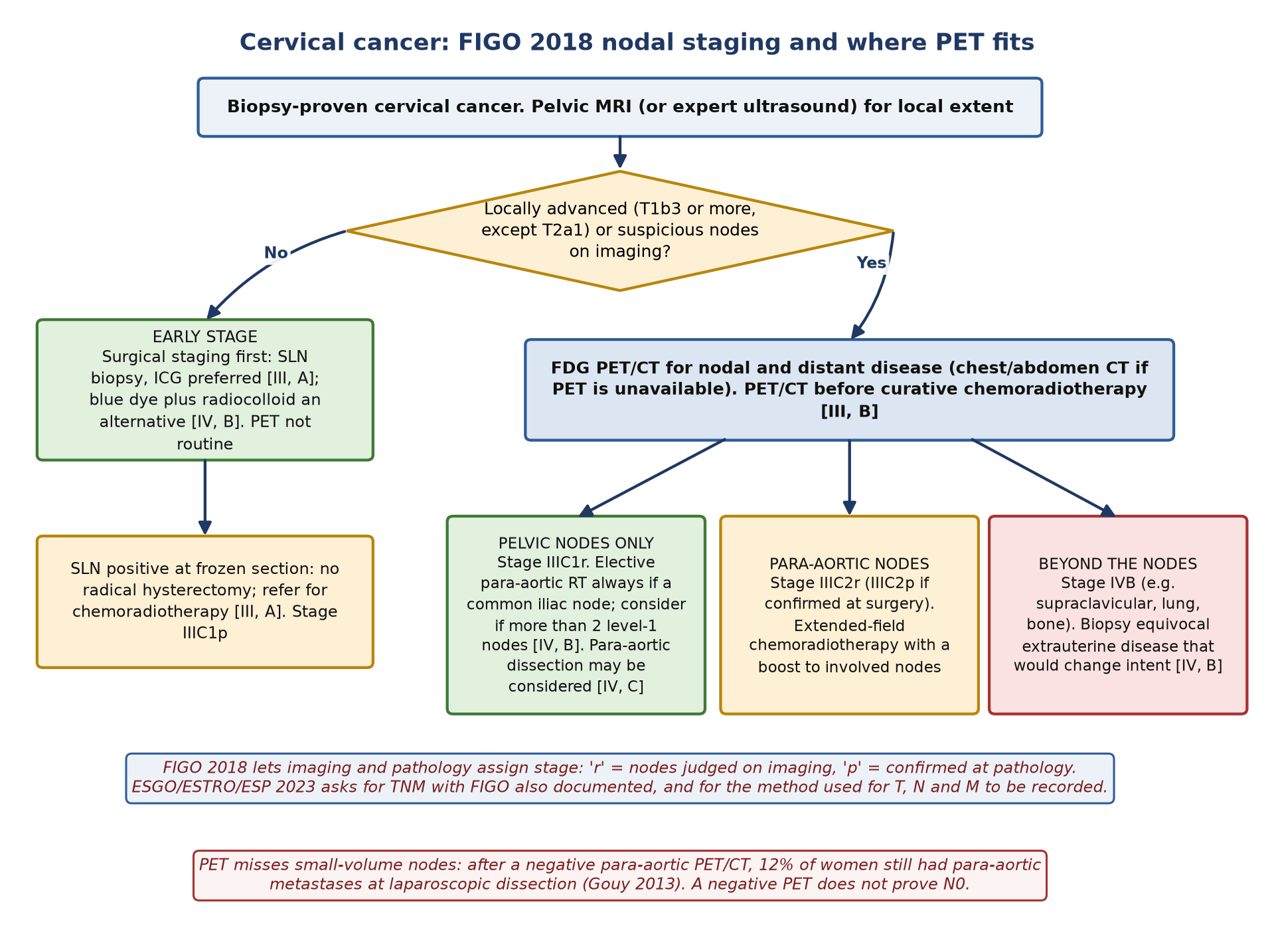
Sentinel nodes in early disease. In SENTICOL (139 women, stage IA1 with lymphovascular invasion to IB1), combined radiocolloid and blue dye found at least one SLN in 97.8%, with sensitivity 92.0% and negative predictive value 98.2%; there were no false negatives when SLNs were found on both sides. SENTIX then tested SLN biopsy without pelvic lymphadenectomy (FIGO 2018 IA1 with lymphovascular invasion to IB2): of 594 women in the intention-to-treat cohort, 82 (12%) had SLN metastases, 43.9% of them found only by ultrastaging, and the 2-year recurrence rate was 6.1%, non-inferior to the 7% reference.
3.2 Endometrial cancer
The 2025 ESGO/ESTRO/ESP update puts the sentinel node at the centre of staging and molecular classification (POLE-mutated, mismatch-repair deficient, no specific molecular profile or p53-abnormal) at the centre of risk assignment; molecular classification should be done for all endometrial carcinomas [IV, A]. Its printed recommendations contain no PET statement for primary staging. In practice FDG PET/CT is used selectively: high-grade or serous histology, carcinosarcoma, clinically advanced disease, and suspected recurrence.
| Scenario | Role of nuclear medicine | Guideline position or best evidence |
|---|---|---|
| Presumed uterus-confined disease | SLN mapping; radiocolloid with SPECT/CT where ICG is unavailable or as a partner | SLN biopsy for all presumed uterus-confined disease [II, A]; ICG by cervical injection preferred; if a side fails to map, side-specific lymphadenectomy in high-intermediate and high risk [II, A]; ultrastaging of every SLN [II, A] (ESGO 2025) |
| High-risk histology or suspected extrauterine disease | FDG PET/CT for nodal and distant disease | Meta-analysis: nodal sensitivity 72%, specificity 94%. No specific ESGO 2025 recommendation |
| Suspected recurrence | FDG PET/CT to localise disease and define oligometastatic status | Meta-analysis: sensitivity 95%, specificity 91%. ESGO 2025 defines oligometastatic disease as one to five metastases in up to three regions |
The evidence for SLN mapping came from two prospective studies. In SENTI-ENDO (125 eligible women, FIGO I to II) cervical injection of ⁹⁹ᵐTc and patent blue detected an SLN in 111; per hemipelvis the negative predictive value was 100%, but per patient sensitivity was 84% and all three false negatives were in type 2 (high-grade) cancers. Ultrastaging found 9 of the 19 node-positive women (47%). FIRES (385 women, cervical ICG, robotic surgery) mapped at least one SLN in 86%, with sensitivity 97.2% and negative predictive value 99.6%. SPECT/CT earns its place because uterine drainage is deep: it localises nodes near the injection site and in the para-aortic region that planar imaging misses.
3.3 Ovarian cancer
Ovarian cancer is staged at surgery. The ESGO-ESMO-ESP (European Society for Medical Oncology) consensus lists contrast-enhanced CT, MRI and PET/CT, with a structured report, as options for the initial evaluation of advanced disease [III, A]; in practice contrast CT is the workhorse and PET/CT is a problem-solving test, for example for suspected disease above the diaphragm. FDG's weakness is the peritoneum: small, flat implants on bowel serosa and mesentery sit next to physiological bowel uptake, and surgical exploration remains the reference standard for peritoneal spread.
The clearest indication is the rising CA-125 with negative or equivocal CT. In a meta-analysis of 34 studies of recurrent ovarian cancer, PET/CT had the highest pooled sensitivity (91%) and CA-125 the highest specificity (93%); the authors singled out women with a rising CA-125 and negative CT or MRI as those most likely to benefit. Before secondary cytoreduction, PET/CT can help to exclude disease beyond the abdomen.
3.4 Vulvar cancer
| Scenario | Role of nuclear medicine | ESGO 2023 recommendation |
|---|---|---|
| Unifocal tumour under 4 cm, deeper than T1a, no suspicious groin nodes | Groin ultrasound first [III, B], then SLN mapping with ⁹⁹ᵐTc-nanocolloid and lymphoscintigraphy | SLN procedure recommended [II, B]; radioactive tracer mandatory [II, A]; combine with blue dye or ICG [II, B]; preoperative lymphoscintigraphy advised [III, C] |
| Midline tumour | Show drainage to both groins | Bilateral SLN detection is mandatory; if only one side maps, contralateral lymphadenectomy [I, A] |
| SLN not found | Report method failure clearly | Inguinofemoral lymphadenectomy [I, A] |
| Larger tumours (beyond pT1a and not SLN candidates) | Systemic staging | CT of chest, abdomen and pelvis or FDG PET/CT [III, B] |
| Before primary chemoradiotherapy | Tumour and nodal extent | MRI and/or FDG PET/CT [IV, A] |
| Suspected recurrence | Systemic restaging | Ultrasound, MRI and/or CT (or FDG PET) of thorax, abdomen and pelvis; biopsy nodal or distant sites if feasible [V, B] |
GROINSS-V I (403 women, 623 groins) established safety: among 259 women with a unifocal tumour and a negative SLN, the groin recurrence rate was 2.3% and 3-year survival 97%, with far less morbidity than lymphadenectomy. GROINSS-V II then treated a positive SLN with inguinofemoral radiotherapy instead of lymphadenectomy. For micrometastases (2 mm or less) the 2-year isolated groin recurrence rate was 1.6%; for macrometastases it was 22% after radiotherapy against 6.9% after lymphadenectomy. The nuclear medicine report therefore feeds a pathology report whose metastasis size decides the next operation.
4. Evidence at a glance
| Study | Design and n | Key finding | What it changed |
|---|---|---|---|
| Grigsby 2004 | Retrospective, 152 women, PET before and after chemoradiotherapy | 5-year cause-specific survival 80% with no abnormal post-treatment uptake, 32% with persistent uptake, 0% with new sites | Established the post-treatment PET as a prognostic test |
| Schwarz 2007 (JAMA) | Prospective, 92 women, PET 2 to 4 months after chemoradiotherapy | 3-year PFS 78% (complete), 33% (partial), 0% (progression); hazard ratio 32.6 for progression, stronger than pretreatment nodal status | Validated the 3-month scan |
| Gouy 2013 | Prospective, 237 women with LACC and negative para-aortic PET/CT | 12% had para-aortic metastases at laparoscopy; 3-year EFS 17% if the metastasis was over 5 mm | Defined PET's false-negative rate; supports optional surgical staging |
| Uterus-11 (Marnitz 2020) | Randomised, 255 women, FIGO IIB to IVA | Surgical staging upstaged 33%; no overall DFS difference (p=0.084); benefit in IIB (HR 0.51) | Surgical para-aortic staging stays optional [IV, C] |
| Yu 2019 | Meta-analysis, 14 studies | Para-aortic PET/CT sensitivity 71%, specificity 97% | Quantified PET's para-aortic performance |
| SENTICOL (Lécuru 2011) | Prospective, 139 women, early cervical cancer | Detection 97.8%, sensitivity 92.0%; no false negatives with bilateral SLNs | Bilateral mapping became the quality standard |
| SENTIX (Cibula 2025) | Prospective single-arm, 594 in intention-to-treat | SLN biopsy without lymphadenectomy: 2-year recurrence 6.1%, non-inferior to 7% | Supports SLN-only staging in early cervical cancer |
| SENTI-ENDO (Ballester 2011) | Prospective, 125 women, FIGO I to II endometrial | Per-hemipelvis NPV 100%; per-patient sensitivity 84%; ultrastaging found 47% of node-positive cases | Established cervical dual-tracer injection |
| FIRES (Rossi 2017) | Prospective, 385 women, ICG mapping | Sensitivity 97.2%, NPV 99.6% | SLN replaced lymphadenectomy in endometrial staging |
| Bollineni 2016 | Meta-analysis, 21 studies (endometrial) | Nodes: sensitivity 72%, specificity 94%. Recurrence: 95% and 91% | PET is good for recurrence, limited for nodes |
| Gu 2009 | Meta-analysis, 34 studies (recurrent ovarian) | PET/CT pooled sensitivity 91%; CA-125 specificity 93% | PET/CT for the rising CA-125 with negative CT |
| GROINSS-V I (Van der Zee 2008) | Observational, 403 women | Groin recurrence 2.3% after negative SLN; lymphoedema 1.9% vs 25.2% | SLN biopsy became standard in small vulvar cancers |
| GROINSS-V II (Oonk 2021) | Single-arm phase II, 1535 registered, 322 SLN positive | Radiotherapy for micrometastases: 1.6% groin recurrence; for macrometastases 22% vs 6.9% with lymphadenectomy | Radiotherapy replaces lymphadenectomy only for micrometastases |
| Florit 2025 | Meta-analysis, FAPI vs FDG, gynaecological cancers | Ovarian peritoneal metastases: sensitivity 97% (FAPI) vs 70% (FDG) | Signals a role for FAPI; not yet practice |
PFS, progression-free survival; EFS, event-free survival; DFS, disease-free survival; HR, hazard ratio; NPV, negative predictive value.
5. Patient preparation and acquisition
Standard FDG preparation applies. What differs in gynaecological practice is the menstrual cycle, the bladder, radiotherapy positioning and the sentinel node protocols.
- Record the menstrual history. Note the first day of the last period, cycle regularity, contraception and hormone therapy on the request. When the timing is elective, a healthy-volunteer study suggested scanning within a week before or a few days after menstrual flow to avoid physiological endometrial and ovarian uptake.
- Exclude pregnancy. ESGO advises that PET/CT be avoided in pregnancy [IV, B]; whole-body diffusion-weighted MRI can stage without ionising radiation.
- Manage urine. Ask the patient to void immediately before the pelvic bed positions. Urine in the bladder, a ureter or a vaginal fornix can mimic or hide disease; late pelvic images after further voiding help when a focus is equivocal.
- Radiotherapy planning scans. Acquire in the treatment position (flat couch, immobilisation) so the images can be registered for contouring, as ESGO recommends for 3D planning.
Sentinel node lymphoscintigraphy
| Cervix and endometrium | Vulva | |
|---|---|---|
| Tracer | ⁹⁹ᵐTc-nanocolloid (ICG added in the theatre, or a hybrid tracer) | ⁹⁹ᵐTc-nanocolloid (mandatory), with blue dye or ICG |
| Injection | Four quadrants of the cervix, peri-orificial (around the scar after conisation); about 110 MBq in 2 mL is the commonest dose | Three or four intradermal injections around the tumour or excision scar, after topical lidocaine |
| Timing | The day before or the day of surgery | The day before or the day of surgery |
| Imaging | Anterior and lateral planar images (3 to 5 min) at about 30 min and 60 to 120 min; SPECT/CT after the delayed images | Dynamic images from injection, static images at 15 min and 2 h, then SPECT/CT |
| Why SPECT/CT | Drainage is deep; nodes near the injection site (parametrial) and para-aortic nodes are missed on planar images | Refines groin localisation and exposes contamination or lymphatic-vessel activity |
In a meta-analysis of eight studies in cervical cancer, SPECT/CT detected SLNs in 98.6% against 85.3% with planar imaging. Mark the skin over each node, report each node's basin (external iliac, obturator, common iliac, para-aortic, inguinal) and number, and state plainly when a side has not mapped: in endometrial cancer that side then needs lymphadenectomy if the risk is high-intermediate or high, and in a midline vulvar cancer the unmapped groin needs lymphadenectomy.
6. Interpretation
What to read and how to report it
- Primary tumour. Size, extension to the uterine body, vagina, parametria, bladder or rectum is MRI's job; note it on PET only when it is obvious, and record the maximum standardised uptake value (SUVmax) as a baseline.
- Nodes, by station. Report each avid node by station (external iliac, internal iliac, obturator, common iliac, para-aortic below or above the inferior mesenteric artery and the renal vein, inguinal, supraclavicular), with short-axis size. A node counts when its uptake is clearly above background and it corresponds to a node on CT; there is no validated SUV threshold, and a normal-sized avid node is still abnormal.
- Assign the stage. State the FIGO 2018 stage that the scan supports (for example IIIC2r), give the TNM categories alongside it as ESGO asks, and say which findings, if confirmed, would change the stage.
- Distant disease. Look at supraclavicular nodes, lung, liver, bone and peritoneum. Solitary unexpected findings that would change intent need a biopsy.
Post-treatment categories
The response studies in cervical cancer used simple visual categories rather than numeric thresholds: complete metabolic response (no abnormal FDG uptake), partial metabolic response (persistent abnormal uptake at treated sites, reduced from baseline) and progressive disease (new sites of abnormal uptake). Grigsby separated persistent uptake inside the irradiated region from new uptake outside it, and the distinction mattered: no woman with new sites was alive at 5 years.
Normal variants, pitfalls and mimics
| Finding | Why it happens | How to tell |
|---|---|---|
| Endometrial uptake in a premenopausal woman | Cyclical: mean SUV 5.0 during menstruation and 3.7 at ovulation, against 2.6 in the proliferative and 2.5 in the secretory phase | Thin, central, linear uptake in a normal uterus that fits the cycle day. After the menopause the mean is 1.7, and focal endometrial uptake needs investigation |
| Ovarian uptake in a premenopausal woman | Follicle or corpus luteum: in 26 of 32 women scanned from the late follicular to the early luteal phase (SUV 3.9) | Unilateral, rim-like or round, at a thin-walled cyst, mid-cycle. Ultrasound or a repeat scan in another phase settles it |
| Ovarian uptake after the menopause | No physiological ovarian uptake was seen in 55 postmenopausal volunteers | Treat as abnormal. An ovarian SUV above 7.9 separated malignant from benign uptake with sensitivity 57% and specificity 95% |
| Endometrium above a cervical tumour | Retained secretions or reactive change: adjacent endometrial SUV about 4.8 without invasion | Do not call corpus invasion on PET; MRI decides |
| Bowel and sigmoid colon | Physiological uptake, often focal | Follow the loop on the CT; peristalsis changes it on late images |
| Ureter, bladder, vaginal urine | Excreted FDG | Trace the ureter on the CT; void and re-image the pelvis |
| After surgery | Inflammation, granulation tissue, lymphocele wall | Timing, CT morphology (lymphoceles are fluid and cold centrally) |
| After radiotherapy | Cervicitis, proctitis, cystitis; sacral insufficiency fractures | Diffuse or linear pattern; fractures follow the sacral ala on CT. Wait at least 3 months |
| Leiomyoma (fibroid) | Variable uptake, occasionally high | Well-defined myometrial mass on CT or MRI; stable over time |
| Intrauterine device | Device in the cavity; endometrial uptake around it has other causes too | Identify it on CT and do not let it explain away focal uptake without correlation |
- Lead with the stage-defining finding: 'FDG-avid left para-aortic nodes below the renal vein; imaging stage FIGO IIIC2r.'
- Say what the scan cannot exclude: 'No avid para-aortic nodes. PET does not exclude small-volume para-aortic metastases.'
- Name physiology as physiology and give the reason: 'Right ovarian uptake at a thin-walled cyst on cycle day 14, in keeping with a corpus luteum.'
- Recommend a biopsy only when a result would change treatment, and name the target.
7. Response assessment and follow-up
- Cervix after chemoradiotherapy. Use the same imaging method as at baseline [V, B], no earlier than 3 months after treatment [IV, B]. If complete remission is uncertain at 3 months, repeat after a further 2 to 3 months, with biopsy if indicated [IV, B]. Women with persistent tumour 3 to 6 months after treatment and no regional or distant disease should be referred to a specialist centre to consider salvage surgery [IV, B].
- Why 3 months. Earlier scans are confounded by radiation inflammation. In the prospective JAMA study, PET at 2 to 4 months (mean 3) showed complete response in 70%, partial response in 16% and progression in 13%; partial response carried a hazard ratio for recurrence of 6.3 and progression 32.6.
- Cervix, routine follow-up. Imaging and blood tests are driven by risk of recurrence, symptoms or findings [V, A]; routine surveillance PET is not recommended.
- Vulva after chemoradiotherapy. Assess response at 12 weeks, clinically, with imaging and with biopsy if residual tumour is suspected [III, B].
- Endometrium. No routine surveillance imaging; PET/CT when recurrence is suspected, particularly to decide between local treatment of oligometastatic disease and systemic therapy.
- Ovary. Surveillance is clinical, with CA-125 and CT; PET/CT is for the rising marker with unhelpful conventional imaging and for planning secondary surgery.
8. Theranostics and emerging tracers
No radioligand therapy is approved for a gynaecological cancer. The emerging agents below are diagnostic, and their evidence is early.
| Agent or technique | What the evidence shows | Evidence level |
|---|---|---|
| ⁶⁸Ga-FAPI PET | Meta-analysis of head-to-head studies: in ovarian cancer, per-lesion peritoneal sensitivity 97% against 70% for FDG, nodal sensitivity 97% against 88% (specificity 83% against 41%); better nodal detection in cervical cancer. Mostly small single-centre studies | Investigational |
| ¹⁸F-FES PET | Approved by the US Food and Drug Administration in May 2020 only for oestrogen receptor-positive recurrent or metastatic breast cancer. In 15 women with ovarian cancer, SUVmax above 1.8 identified oestrogen receptor-alpha-positive lesions with sensitivity 79% and specificity 100%. The rationale is selection for endocrine therapy in low-grade serous ovarian and oestrogen receptor-positive endometrial cancer | Investigational in gynaecology |
| HER2 imaging | HER2-directed drugs are used in some serous endometrial and other gynaecological cancers, but HER2 PET has no validated role in selecting patients | Investigational |
| PET/MRI of the pelvis | Early data suggest FDG PET/MRI matches MRI and outperforms PET/CT for local staging of cervical cancer, with nodal staging comparable to PET/CT; costlier and slower, and not yet tested in large prospective studies | Small single-centre studies; no guideline recommends routine use |
| Hybrid ICG-⁹⁹ᵐTc-nanocolloid | One tracer gives the preoperative map, the probe signal and fluorescence in theatre | Clinical use in some centres; limited comparative data |
9. Structured report example
- Clinical question: Squamous cell carcinoma of the cervix, 5 cm on MRI with left parametrial invasion. Nodal and distant staging before chemoradiotherapy.
- Technique: Blood glucose 5.4 mmol/L. 250 MBq ¹⁸F-FDG; imaging from skull base to mid-thigh at 60 min in the radiotherapy position. Voided before the pelvic bed. Cycle day not applicable (postmenopausal).
- Primary: Intensely avid cervical mass (SUVmax 18) extending into the left parametrium, as on MRI.
- Nodes: Avid left external iliac (short axis 14 mm) and left common iliac (9 mm) nodes. Avid 8 mm left para-aortic node at L3, below the inferior mesenteric artery, separate from the ureter. No inguinal or supraclavicular disease.
- Distant: No avid lung, liver, bone or peritoneal disease.
- Physiological: Renal and ureteric excretion; bowel uptake without CT correlate.
- Conclusion: Imaging stage FIGO IIIC2r, with TNM categories recorded: pelvic and para-aortic nodal disease. The para-aortic node supports extended-field chemoradiotherapy with a nodal boost. No distant metastases.
10. Take-home points
- In cervical cancer, FDG PET/CT is recommended for locally advanced disease, suspicious nodes and before curative chemoradiotherapy; it is not needed for surgically staged early disease.
- FIGO 2018 lets imaging assign nodal stage: pelvic nodes IIIC1r, para-aortic nodes IIIC2r. Report nodes by station, because the level sets the radiotherapy field.
- An avid para-aortic node is usually real (specificity 97%); a negative para-aortic PET still misses about one woman in eight.
- Scan the response no earlier than 3 months after chemoradiotherapy; new sites of uptake carry the worst prognosis.
- Sentinel node mapping stages early cervical, endometrial and vulvar cancer. Radiocolloid is mandatory in vulvar cancer, and SPECT/CT maps deep pelvic and para-aortic drainage.
- Time elective scans in premenopausal women away from menstruation and ovulation; after the menopause, focal ovarian or endometrial uptake is abnormal.
- In ovarian cancer, PET/CT is the test for a rising CA-125 with negative CT; small peritoneal implants remain its blind spot. FAPI and FES are research tools for now.
Test yourself
5 quick questions. Pick an answer to see the explanation.
References
- Bhatla N, Berek JS, Cuello Fredes M, et al. Revised FIGO staging for carcinoma of the cervix uteri. Int J Gynaecol Obstet. 2019;145(1):129-35.
- Cibula D, Raspollini MR, Planchamp F, et al. ESGO/ESTRO/ESP guidelines for the management of patients with cervical cancer - update 2023. Int J Gynecol Cancer. 2023;33(5):649-66.
- Fischerova D, Frühauf F, Burgetova A, et al. The role of imaging in cervical cancer staging: ESGO/ESTRO/ESP guidelines (update 2023). Cancers (Basel). 2024;16(4):775.
- Yu W, Kou C, Bai W, et al. The diagnostic performance of PET/CT scans for the detection of para-aortic metastatic lymph nodes in patients with cervical cancer: a meta-analysis. PLoS One. 2019;14(7):e0220080.
- Gouy S, Morice P, Narducci F, et al. Prospective multicenter study evaluating the survival of patients with locally advanced cervical cancer undergoing laparoscopic para-aortic lymphadenectomy before chemoradiotherapy in the era of positron emission tomography imaging. J Clin Oncol. 2013;31(24):3026-33.
- Marnitz S, Tsunoda AT, Martus P, et al. Surgical versus clinical staging prior to primary chemoradiation in patients with cervical cancer FIGO stages IIB-IVA: oncologic results of a prospective randomized international multicenter (Uterus-11) intergroup study. Int J Gynecol Cancer. 2020;30(12):1855-61.
- Grigsby PW, Siegel BA, Dehdashti F, et al. Posttherapy [18F] fluorodeoxyglucose positron emission tomography in carcinoma of the cervix: response and outcome. J Clin Oncol. 2004;22(11):2167-71.
- Schwarz JK, Siegel BA, Dehdashti F, Grigsby PW. Association of posttherapy positron emission tomography with tumor response and survival in cervical carcinoma. JAMA. 2007;298(19):2289-95.
- Lécuru F, Mathevet P, Querleu D, et al. Bilateral negative sentinel nodes accurately predict absence of lymph node metastasis in early cervical cancer: results of the SENTICOL study. J Clin Oncol. 2011;29(13):1686-91.
- Cibula D, Marnitz S, Jarkovský J, et al. Sentinel lymph node biopsy without systematic pelvic lymphadenectomy in females with early-stage cervical cancer: final outcome of the SENTIX prospective, single-arm, noninferiority, international trial. Nat Cancer. 2025;6(9):1585-94.
- Concin N, Matias-Guiu X, Cibula D, et al. ESGO-ESTRO-ESP guidelines for the management of patients with endometrial carcinoma: update 2025. Lancet Oncol. 2025;26(8):e423-35.
- Rossi EC, Kowalski LD, Scalici J, et al. A comparison of sentinel lymph node biopsy to lymphadenectomy for endometrial cancer staging (FIRES trial): a multicentre, prospective, cohort study. Lancet Oncol. 2017;18(3):384-92.
- Ballester M, Dubernard G, Lécuru F, et al. Detection rate and diagnostic accuracy of sentinel-node biopsy in early stage endometrial cancer: a prospective multicentre study (SENTI-ENDO). Lancet Oncol. 2011;12(5):469-76.
- Bollineni VR, Ytre-Hauge S, Bollineni-Balabay O, et al. High diagnostic value of 18F-FDG PET/CT in endometrial cancer: systematic review and meta-analysis of the literature. J Nucl Med. 2016;57(6):879-85.
- Prat J; FIGO Committee on Gynecologic Oncology. Staging classification for cancer of the ovary, fallopian tube, and peritoneum. Int J Gynaecol Obstet. 2014;124(1):1-5.
- Ledermann JA, Matias-Guiu X, Amant F, et al. ESGO-ESMO-ESP consensus conference recommendations on ovarian cancer: pathology and molecular biology and early, advanced and recurrent disease. Ann Oncol. 2024;35(3):248-66.
- Gu P, Pan LL, Wu SQ, et al. CA 125, PET alone, PET-CT, CT and MRI in diagnosing recurrent ovarian carcinoma: a systematic review and meta-analysis. Eur J Radiol. 2009;71(1):164-74.
- Florit A, de Koster EJ, Sassano S, et al. Head-to-head comparison of fibroblast activation protein inhibitors (FAPI) radiopharmaceuticals and [18F]FDG in gynaecological malignancies: systematic literature review and meta-analysis. Eur J Nucl Med Mol Imaging. 2025;52(11):3975-89.
- Oonk MHM, Planchamp F, Baldwin P, et al. European Society of Gynaecological Oncology guidelines for the management of patients with vulvar cancer - update 2023. Int J Gynecol Cancer. 2023;33(7):1023-43.
- Van der Zee AG, Oonk MH, De Hullu JA, et al. Sentinel node dissection is safe in the treatment of early-stage vulvar cancer. J Clin Oncol. 2008;26(6):884-9.
- Oonk MHM, Slomovitz B, Baldwin PJW, et al. Radiotherapy versus inguinofemoral lymphadenectomy as treatment for vulvar cancer patients with micrometastases in the sentinel node: results of GROINSS-V II. J Clin Oncol. 2021;39(32):3623-32.
- Collarino A, Vidal-Sicart S, Perotti G, Valdés Olmos RA. The sentinel node approach in gynaecological malignancies. Clin Transl Imaging. 2016;4(5):411-20.
- Lerman H, Metser U, Grisaru D, et al. Normal and abnormal 18F-FDG endometrial and ovarian uptake in pre- and postmenopausal patients: assessment by PET/CT. J Nucl Med. 2004;45(2):266-71.
- Nishizawa S, Inubushi M, Okada H. Physiological 18F-FDG uptake in the ovaries and uterus of healthy female volunteers. Eur J Nucl Med Mol Imaging. 2005;32(5):549-56.
- van Kruchten M, de Vries EF, Arts HJ, et al. Assessment of estrogen receptor expression in epithelial ovarian cancer patients using 16α-18F-fluoro-17β-estradiol PET/CT. J Nucl Med. 2015;56(1):50-5.
- Postcoital bleeding in a 44-year-old · Beginner