PET/CT in Bone and Soft-Tissue Sarcoma
- FDG uptake follows grade, not the label 'sarcoma'. In a meta-analysis of 441 soft-tissue lesions every intermediate- or high-grade sarcoma was FDG-positive, but only about three-quarters of low-grade sarcomas were, and 39% of benign lesions were too.
- Use PET to choose where to biopsy. In a heterogeneous mass (a dedifferentiated liposarcoma, a chondrosarcoma, a plexiform neurofibroma) the hottest solid part is the most likely to hold the highest grade.
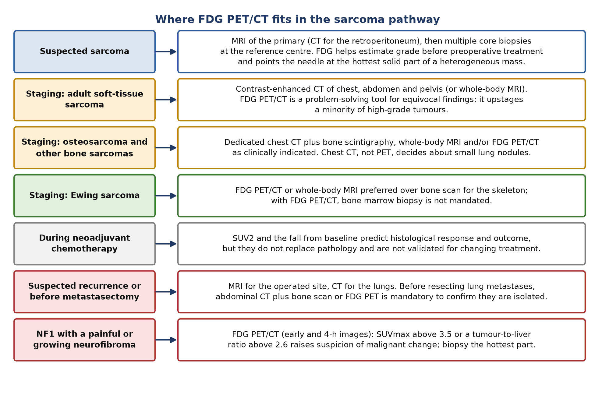
- CT stages adult soft-tissue sarcoma; PET solves problems. The ESMO–EURACAN–GENTURIS 2021 guideline stages with contrast-enhanced CT of chest, abdomen and pelvis and reserves FDG PET/CT for equivocal findings.
- Ewing sarcoma is the exception. FDG PET/CT or whole-body MRI is preferred over bone scan for the skeleton, and with PET/CT a bone marrow biopsy is not mandated.
- Chest CT, not PET, judges small lung nodules. In a prospective paediatric study PET found only 25% of lung metastases that CT found; PET was better for nodes and bone.
- NF1: know the numbers. An SUVmax above 3.5 on delayed images, or a tumour-to-liver ratio above 2.6, flags possible malignant change in a nerve sheath tumour.
- Response in bone sarcoma is prognostic, not yet actionable. After neoadjuvant chemotherapy an SUV2 below 2.5 was linked with better progression-free survival in Ewing sarcoma and osteosarcoma, but agreement with histological necrosis was only 58–69%.
- FAP-targeted imaging and therapy are investigational. ⁶⁸Ga-FAPI PET had a positive predictive value of 1.00 per patient in a prospective sarcoma cohort; ⁹⁰Y-FAPI-46 therapy gave disease control in 38% of heavily pretreated patients.
1. The clinical problem
Sarcomas are rare malignant tumours of mesenchymal tissue. They arise in soft tissue (soft-tissue sarcoma, STS) or bone (osteosarcoma, Ewing sarcoma, chondrosarcoma and rarer types), in adults and children, anywhere in the body. Their rarity and diversity are why European guidance insists that they are managed in sarcoma reference centres by a multidisciplinary team (MDT). The oncologist and surgeon ask imaging five questions: which part of the mass should be biopsied, how aggressive it is likely to be, has it spread, is preoperative treatment working, and has it come back? Magnetic resonance imaging (MRI) answers the local questions. ¹⁸F-fluorodeoxyglucose positron emission tomography/computed tomography (FDG PET/CT) contributes most to the first, third and last.
Classification and staging in force
- World Health Organization (WHO) 2020 classification of soft tissue and bone tumours: the current pathological standard named in the 2021 European Society for Medical Oncology (ESMO) guidelines. It separates undifferentiated round cell sarcomas (CIC-rearranged, BCOR-altered and EWSR1 non-ETS fused sarcomas) from Ewing sarcoma, and lists NTRK-rearranged tumours as an emerging entity.
- Grade: for STS, histological grade is usually assigned with the three-tier French Federation of Cancer Centres (FNCLCC) system. Grade is the main driver of FDG avidity, and of metastatic risk.
- American Joint Committee on Cancer (AJCC) eighth edition: staging is now site-specific. Bone sarcomas are staged separately for the appendicular skeleton, trunk, skull and face; the spine; and the pelvis, with a three-grade system. STS is staged separately for trunk and extremity, retroperitoneum, head and neck, and visceral sites.
- Guideline view of staging: ESMO–EURACAN–GENTURIS (with the European Reference Network for rare adult solid cancers and the Genetic Tumour Risk Syndromes network) notes that available staging classifications are of limited clinical value in STS and that risk is better estimated with validated nomograms.
| AJCC eighth edition change | What it means for the reader of the scan |
|---|---|
| STS of trunk and extremity: size categories ≤5 cm, >5–10 cm, >10–15 cm, >15 cm | Report the maximum dimension; the CT or MRI measurement sets T |
| Depth (superficial or deep to the fascia) removed from T | Depth still matters to the surgeon but no longer changes stage |
| Trunk and extremity STS: any T, N1, M0 is stage IV | A PET-positive regional node, if confirmed, upstages to stage IV; nodal spread is uncommon, so confirm it |
| Retroperitoneal STS: any T, N1, M0 remains stage IIIB | The same nodal finding has a different stage at this site |
| Bone sarcoma: three sites and three grades | Name the site group (appendicular, spine, pelvis) in the report |
Where nuclear medicine changes management
- Biopsy targeting and grade: ESMO states that when preoperative treatment is an option, imaging including FDG PET/CT may help estimate malignancy grade alongside pathology.
- Distant staging: for Ewing sarcoma and, as clinically indicated, other bone sarcomas; for STS when CT is equivocal, and before resection of apparently isolated lung metastases, when abdominal CT plus bone scan or FDG PET is mandatory.
- Malignant change in neurofibromatosis type 1 (NF1): FDG PET/CT is a highly sensitive test for malignant peripheral nerve sheath tumour (MPNST) and shows which part of a large tumour to biopsy.
- Response and recurrence: FDG predicts histological response and outcome and detects recurrence accurately, although neither use is written into guidelines as routine.
2. Tracers and why they work
FDG and tumour grade
FDG is transported into cells by glucose transporters and trapped after phosphorylation by hexokinase. In sarcoma, uptake rises with cellularity, proliferation and grade. The standardised uptake value (SUV), and its maximum in the tumour (SUVmax), are therefore surrogates of grade rather than of histological type. A meta-analysis of 15 studies (441 soft-tissue lesions) found that on visual reading FDG was positive in all intermediate- and high-grade sarcomas, 74% of low-grade sarcomas and 39% of benign lesions (including 11 of 12 inflammatory lesions). With an SUV cut-off of 2.0 the rates were 89%, 33% and 19%. FDG separates high-grade sarcoma from benign disease well; it does not separate low-grade sarcoma from benign disease.
| Usually intensely avid | Often low or variable uptake |
|---|---|
| Undifferentiated pleomorphic sarcoma, leiomyosarcoma | Grade 1 sarcomas of any type |
| Translocation sarcomas: Ewing, synovial, alveolar rhabdomyosarcoma | Atypical lipomatous tumour / well-differentiated liposarcoma |
| Dedifferentiated liposarcoma, pleomorphic liposarcoma | Myxoid liposarcoma |
| High-grade osteosarcoma | Alveolar soft part sarcoma, solitary fibrous tumour (intermediate-grade types with low SUVmax) |
| High-grade and dedifferentiated chondrosarcoma | Atypical cartilaginous tumour / grade 1 chondrosarcoma |
Pattern of FDG avidity by subtype, from a single-centre audit of 957 PET/CT scans in 493 patients (Macpherson 2018) and the subtype series cited below.
Lipomatous tumours
In 67 lipomatous tumours imaged before resection, mean SUVmax was 0.8 for lipoma, 2.3 for atypical lipomatous tumour, 3.0 for myxoid liposarcoma, 13.5 for pleomorphic liposarcoma and 16.3 for dedifferentiated liposarcoma; hibernoma, a benign brown-fat tumour, averaged 11.9. Low-grade and high-grade liposarcoma differed clearly (2.5 versus 12.8), but benign and malignant lesions did not, because hibernomas are hot and atypical lipomatous tumours and myxoid liposarcomas are cool. In an earlier series of 54 liposarcomas, an SUVmax above 3.6 predicted shorter disease-free survival (21 versus 44 months) better than grade or subtype. ESMO stresses that recognising the well-differentiated versus the dedifferentiated component of a retroperitoneal liposarcoma is critical to surgical planning: a focal FDG-avid solid nodule in a fatty mass is the classic target.
Cartilage tumours
Most conventional chondrosarcomas are low grade (atypical cartilaginous tumour / grade 1), which ESMO allows to be curetted or watched in the long bones, whereas grade 2–3 tumours and all pelvic or axial chondrosarcomas need wide excision. Biopsy under-grades these tumours, so FDG adds information. In 31 patients, mean SUV was 3.4 in grade 1, 5.4 in grade 2 and 7.1 in grade 3; an SUV above 4 predicted relapse (sensitivity 90%, specificity 76%). In 95 chondroid lesions, the best SUVmax cut-offs were 2.6 for enchondroma versus low-grade chondrosarcoma (sensitivity 68%, specificity 86%), 3.7 for low-grade versus grade 2–3 (83%, 84%) and 7.7 for grade 2–3 versus dedifferentiated chondrosarcoma (92%, 90%). These are single-centre thresholds and should be quoted with their source.
NF1 and malignant peripheral nerve sheath tumour
Plexiform neurofibromas in NF1 are common and usually mildly avid; MPNST is aggressive and usually much hotter. In 69 patients (85 lesions) imaged at 90 and 240 min, FDG PET/CT had a sensitivity of 97% and specificity of 87% for MPNST, and the authors recommended early plus 4-h imaging with an SUVmax cut-off of 3.5 on the delayed images. In 49 patients, normalising to the liver improved specificity: a tumour-to-liver ratio (TLR) above 2.6 and an SUVmax above 3.5 were both 100% sensitive, but specificity was 90.3% with the ratio versus 79.8% with the SUVmax cut-off. Uptake varies within a large plexiform tumour, so the whole lesion must be examined and the hottest region biopsied.
When FDG fails and what replaces it
- Low-grade tumours: atypical lipomatous tumour, myxoid liposarcoma and grade 1 chondrosarcoma can overlap with benign lesions; MRI and histology decide.
- Small lung metastases: partial-volume effects hide sub-centimetre nodules; the CT component, read in lung windows, and a dedicated chest CT decide.
- Benign avid lesions: giant cell tumour, chondroblastoma, schwannoma, hibernoma and inflammation mimic sarcoma; histology decides.
- Fibroblast activation protein (FAP) tracers: sarcomas express FAP on tumour cells and associated fibroblasts, so FAP inhibitor (FAPI) PET is being studied as a complement; it is investigational (section 8).
3. Indications by clinical scenario
| Scenario | Role of FDG PET/CT | Guideline position (ESMO–EURACAN–GENTURIS 2021 unless stated) |
|---|---|---|
| Suspected STS: diagnosis and biopsy | Choose the most metabolically active solid part of a heterogeneous mass; estimate grade | MRI is the main local modality; multiple core needle biopsies (14–16 G) at the reference centre. When preoperative treatment is an option, imaging including FDG PET/CT may help estimate grade |
| Staging adult STS | Problem-solving for equivocal CT findings | Contrast-enhanced CT of chest, abdomen and pelvis (whole-body MRI an alternative); FDG PET/CT is indicated as a problem-solving tool in equivocal cases |
| Staging osteosarcoma, chondrosarcoma and other bone sarcomas | Skeletal and nodal staging, skip lesions | Dedicated chest CT and bone scintigraphy, and/or whole-body MRI and/or FDG PET/CT or PET/MRI as clinically indicated; PET and whole-body MRI are increasingly used for bone and marrow metastases |
| Staging Ewing sarcoma | Whole-body skeletal and marrow staging | FDG PET/CT or whole-body MRI preferred over bone scan for skeletal imaging, if available; bone marrow biopsy and aspirate not mandated if FDG PET/CT is done |
| Rhabdomyosarcoma (mainly paediatric) | Nodal and distant staging | A Cochrane review found only two eligible studies (36 patients): insufficient evidence to define accuracy; follow paediatric protocols (see the separate paediatric article) |
| Apparently isolated lung metastases before metastasectomy | Exclude extrapulmonary disease | Abdominal CT plus a bone scan or FDG PET is mandatory before surgery for lung metastases |
| Response to neoadjuvant chemotherapy | Prognosis; research | No guideline recommendation; histological response of the resected tumour remains the reference in bone sarcoma |
| Radiotherapy planning | Not standard | No guideline role for FDG; MRI is the main local imaging modality. FDG may show where residual disease is most active, which is useful only if the MDT asks |
| Follow-up and suspected recurrence | Equivocal local or distant findings; whole-body restaging before salvage | High-grade bone sarcoma: clinical review, imaging of the primary site and chest X-ray or CT at set intervals; no routine PET |
| NF1 with a painful or growing nerve sheath tumour | Detect malignant change and target biopsy | Cohort evidence (sections 2 and 6): high sensitivity (97–100%) with moderate to high specificity |
- Adult STS staging: ESMO uses PET only for problem-solving, and a Canadian series of 109 limb and trunk sarcomas upstaged only 4.5% with PET. A UK audit of high-grade bone and soft-tissue sarcomas, however, found PET moved about 12% from M0 to M1. The difference reflects case mix (grade, histology, size); expect more benefit in large, high-grade and translocation sarcomas.
- Bone sarcoma staging: the ESMO bone guideline lists bone scintigraphy and chest CT as the general staging tests, yet for Ewing sarcoma prefers PET/CT or whole-body MRI over bone scan. In osteosarcoma the choice is left to the MDT.
- Response: FDG response predicts outcome in several series, but no guideline uses it to change treatment, and EURAMOS-1 showed that changing chemotherapy for poor histological responders did not help.
4. Evidence at a glance
| Study | Design and n | Key finding | What it changed |
|---|---|---|---|
| Ioannidis 2003 | Meta-analysis, 15 studies, 441 soft-tissue lesions | Visual reading: sensitivity 92%, specificity 73% for malignancy; all intermediate/high-grade sarcomas positive, 74% of low-grade, 39% of benign lesions | FDG grades but cannot clear a low-grade lesion |
| Roberge 2012 | Retrospective, 109 adult limb and trunk STS with chest CT | 98% of unresected primaries avid; PET upstaged 5 patients (4.5%); 3 false positives | Supports PET as problem-solver, not routine, in adult STS |
| Macpherson 2018 | Audit, 957 scans in 493 patients | SUVmax tracked grade; PET added value over CT/MRI in an estimated 21%, with 12% of high-grade cases upstaged to M1 | Case for PET in high-grade disease |
| Völker 2007 | Prospective multicentre, 46 paediatric sarcomas (Ewing, osteosarcoma, rhabdomyosarcoma) | Nodes: PET 95% vs conventional 25%; bone: 90% vs 57%; lung: CT 100% vs PET 25%; correct therapy decisions 91% with side-by-side reading vs 59% conventional | PET plus CT, never PET instead of chest CT |
| Seth 2022 | Meta-analysis, 31 studies, 735 patients with Ewing sarcoma | Sensitivity/specificity: bone metastases 83.9%/93.2%; lung metastases 76.1%/92.4%; recurrence 89.9%/92.6% | CT remains superior for lung metastases |
| Hawkins 2005 | Retrospective, 36 Ewing sarcoma family tumours | 4-year PFS 72% if SUV2 <2.5 vs 27% if ≥2.5; concordance with histology 68–69% | SUV2 as a prognostic marker |
| Hawkins 2009 | Retrospective, 40 extremity osteosarcomas | 4-year PFS 73% vs 39% (SUV2 <2.5 vs ≥2.5); concordance with histology 58% (SUV2) and 68% (ratio) | PET only partly mirrors necrosis |
| Evilevitch 2008 | Prospective, 42 high-grade STS before and after neoadjuvant therapy | Area under the curve 0.93 for FDG change vs 0.60 for size; ≥60% fall: sensitivity 100%, specificity 71% for ≥95% necrosis | Metabolic change beats size change in STS |
| EURAMOS-1 (Marina 2016) | RCT, 618 poor responders (≥10% viable tumour) of 2,260 registered | Adding ifosfamide and etoposide: event-free survival HR 0.98, more toxicity | Response-adapted intensification failed, so early PET response has no validated action yet |
| Warbey 2009 | Prospective, 69 NF1 patients, 85 lesions | Sensitivity 97%, specificity 87% for MPNST; SUVmax 3.5 on 4-h images recommended | Standard NF1 protocol |
| Salamon 2014 | 49 NF1 patients | TLR >2.6: sensitivity 100%, specificity 90.3% (vs 79.8% for SUVmax >3.5) | Liver normalisation improves specificity |
| Kessler 2022 | Prospective observational, 47 sarcoma patients, ⁶⁸Ga-FAPI | PPV 1.00 per patient; upstaging vs FDG in 18.6%; management changed in 30% | FAP imaging trials |
Abbreviations: PFS, progression-free survival; SUV1 and SUV2, SUVmax before and after neoadjuvant chemotherapy; RCT, randomised controlled trial; HR, hazard ratio; TLR, tumour-to-liver ratio; PPV, positive predictive value.
5. Patient preparation and acquisition
Preparation follows the European Association of Nuclear Medicine (EANM) FDG tumour guideline version 2.0: at least 4 h without food or sugary drinks, glucose checked before injection, and an uptake time of 60 min (acceptable range 55–75 min), kept identical on serial scans because SUV depends on it. What differs in sarcoma is below.
- Field of view: sarcomas start in the limbs, so extend the scan to include the whole limb carrying the primary, and usually vertex to toes. The EANM guideline defines whole-body imaging as the top of the head through the feet.
- Injection site: inject away from the primary and its draining nodes, and record the site, so that extravasation or reactive nodes are not mistaken for disease.
- Before biopsy when possible: a PET done to choose the biopsy target must precede the biopsy; afterwards, record the biopsy route and date, because the track and haematoma take up FDG.
- Young patients: brown fat uptake is more common in the young and in cold rooms. Keep the patient warm from 30–60 min before injection until the end of the scan, as EANM recommends. Expect symmetrical physeal uptake around the knees and elsewhere in adolescents.
- NF1: acquire early (about 90 min) and delayed (about 4 h) images, as in the largest prospective series; SUVmax differs between the two time points, and the published cut-off of 3.5 applies to the delayed images.
- Response studies: same scanner, reconstruction and uptake time as baseline; scan after the last cycle of neoadjuvant chemotherapy and before surgery, matching the protocol of the series whose thresholds you intend to use.
- History for the reader: dates of surgery, biopsy, radiotherapy and chemotherapy; growth factor support; any fracture or prosthesis; and, in NF1, which lesion is painful or growing.
6. Interpretation
What to measure
- SUVmax of the hottest solid part of the primary, with its location described so that the biopsy can target it; note necrotic or cystic regions that would give a non-diagnostic core.
- Heterogeneity: a focal hot nodule in a cool fatty or cartilaginous tumour suggests dedifferentiation; say so explicitly.
- In NF1: SUVmax on early and delayed images and the TLR for each symptomatic or growing lesion; list lesions above the thresholds.
- Distant disease: lungs (on the CT, in lung windows), bone and marrow, nodes (uncommon in most adult STS, so confirm an avid node before it upstages the patient), and soft tissue elsewhere, including the opposite limb.
Thresholds that have been published
| Question | Threshold | Performance and source |
|---|---|---|
| Enchondroma vs low-grade chondrosarcoma | SUVmax 2.6 | Sensitivity 68%, specificity 86% (Annovazzi, 95 lesions) |
| Low-grade vs grade 2–3 chondrosarcoma | SUVmax 3.7 | Sensitivity 83%, specificity 84% (Annovazzi) |
| Grade 2–3 vs dedifferentiated chondrosarcoma | SUVmax 7.7 | Sensitivity 92%, specificity 90% (Annovazzi) |
| Chondrosarcoma relapse risk | SUV 4 | Sensitivity 90%, specificity 76% for relapse (Brenner, 31 patients) |
| Liposarcoma outcome | SUVmax 3.6 | Above it, disease-free survival 21 vs 44 months (Brenner, 54 patients) |
| NF1: MPNST | SUVmax 3.5 on 4-h images | Sensitivity 97%, specificity 87% for the whole PET/CT reading (Warbey) |
| NF1: MPNST | Tumour-to-liver ratio 2.6 | Sensitivity 100%, specificity 90.3% (Salamon) |
| Osteosarcoma or Ewing sarcoma after induction | SUV2 <2.5 or SUV2/SUV1 ≤0.5 | Good response; SUV2 <2.5 linked with better 4-year PFS (Hawkins) |
| High-grade STS after neoadjuvant therapy | ≥60% fall in SUV | Sensitivity 100%, specificity 71% for ≥95% necrosis (Evilevitch) |
All are single-centre, retrospective or small prospective derivations; SUV depends on scanner, reconstruction and uptake time. Quote the source and use them to support, not replace, the visual read.
Pitfalls and mimics
| Pitfall | What it looks like | How to tell |
|---|---|---|
| Recent surgery, biopsy track, drain or seroma | Linear or rim uptake around a fluid collection or along the scar | Match to dates; a thin, even rim around fluid favours seroma; nodular, eccentric uptake needs MRI |
| Radiotherapy change | Diffuse uptake throughout the treated field, including muscle and skin | Geometric margins matching the field; compare with MRI |
| Brown fat | Symmetric neck, supraclavicular, axillary and paravertebral uptake in fat density | CT density; warming; common in young patients |
| Giant cell tumour and other giant-cell-rich or histiocytic bone lesions | Intense uptake in an epiphyseal lytic lesion | Giant cell tumours averaged SUV 4.6, higher than chondrosarcomas (2.2) and similar to osteosarcomas (3.1); chondroblastoma and Langerhans cell histiocytosis can be as hot |
| Fibrous dysplasia | Moderate uptake in an expanded ground-glass lesion | Mean SUV 2.1, no different from chondrosarcoma; CT pattern decides |
| Schwannoma | Well-defined nerve sheath mass, sometimes very avid | SUVmax 1.5–17.3 (median 3.7) in 22 resected schwannomas; higher with peritumoral lymphoid cuffs and in the gut |
| Hibernoma | Fat-containing mass with intense uptake | Mean SUVmax 11.9: brown fat tumour, benign |
| Desmoid-type fibromatosis, myositis ossificans, healing fracture | Moderate to intense uptake in a soft-tissue mass or along bone | Clinical history, CT pattern (peripheral mineralisation in myositis ossificans, fracture line) and MRI; biopsy if doubt remains |
| Inflammation and infection | Avid mass or collection | 11 of 12 inflammatory lesions were FDG-positive in the meta-analysis |
| Normal physes in adolescents | Symmetric transverse bands at the ends of long bones | Symmetry and position; compare left with right |
| FDG-negative sarcoma | Low-grade, myxoid or well-differentiated tumours with little uptake | A cool scan does not exclude sarcoma; MRI and biopsy decide |
| Small lung metastases | FDG-negative sub-centimetre nodules | Report them from the CT; dedicated chest CT |
Wording the conclusion
- Answer the question asked first: stage (with the AJCC site group), the biopsy target, or malignant change in NF1.
- Give the SUVmax of the hottest part and where it is, for example 'SUVmax 11.4 in the anteromedial solid component, 3 cm from the skin; central necrosis'.
- Separate definite metastases from indeterminate findings and say which test resolves each: 'three 4–6 mm lung nodules without uptake, below PET resolution; assess on dedicated chest CT'.
- In NF1, list each assessed lesion with early and delayed SUVmax and TLR, and state which exceed the thresholds.
7. Response assessment and follow-up
Bone sarcomas: FDG and histological response
In osteosarcoma and Ewing sarcoma the percentage of necrosis in the resected tumour after neoadjuvant chemotherapy is a strong prognostic factor; a good response is conventionally at least 90% necrosis, and EURAMOS-1 defined poor response as 10% or more viable tumour. Two series from one institution tested FDG against it. In 36 Ewing sarcoma family tumours, a good PET response was defined as SUV2 below 2.5 or SUV2/SUV1 of 0.5 or less; these agreed with histology in 68% and 69%, and 4-year PFS was 72% with SUV2 below 2.5 versus 27% above it. In 40 extremity osteosarcomas, agreement with histology was 58% for SUV2 and 68% for the ratio, and 4-year PFS was 73% versus 39%.
So FDG predicts outcome, but it is not a substitute for pathology. Nor is there yet a validated action: in EURAMOS-1, 618 poor histological responders were randomised to add ifosfamide and etoposide to postoperative MAP (cisplatin, doxorubicin, high-dose methotrexate); event-free survival did not improve (HR 0.98) and toxicity rose. An early metabolic response would need its own trial before it changes treatment.
Soft-tissue sarcoma
In 42 patients with high-grade STS scanned before and after neoadjuvant therapy, the fall in FDG uptake predicted histopathological response (at least 95% necrosis) far better than the change in size: area under the curve 0.93 versus 0.60. A decrease of 60% or more had a sensitivity of 100% and specificity of 71%, whereas size-based Response Evaluation Criteria in Solid Tumours (RECIST) had a sensitivity of only 25%. Many sarcomas do not shrink when they respond, and some swell with haemorrhage or necrosis, which is why metabolic response is attractive here. It remains a research tool.
Which criteria
No sarcoma-specific PET response criteria are recommended by guidelines. State what you measured (SUVmax or SULpeak, the same region, the same uptake time), the percentage change, and the threshold of the series you are applying. Do not call a response 'complete' because a necrotic tumour is cold centrally; describe residual rim uptake.
Follow-up and recurrence
- ESMO schedules: high-grade bone sarcoma is followed with examination, imaging of the primary site and chest X-ray or CT, roughly every 2–3 months for 2 years, 6-monthly to year 5 and 6–12-monthly to year 10. Intermediate- and high-grade STS may be seen every 3–4 months for 2–3 years, then twice yearly to year 5 and yearly after; low-grade STS 6-monthly for 5 years, then yearly. PET is not part of routine follow-up.
- PET versus MRI at the operated site: MRI is the local surveillance test. In the meta-analysis of soft-tissue lesions, FDG performed similarly for primary and recurrent lesions, and the limited head-to-head data showed no difference from MRI. PET helps when MRI is degraded by metal or confounded by postoperative and radiation change, and it restages the whole body before salvage surgery.
- Accuracy for recurrence: in Ewing sarcoma, pooled sensitivity was 89.9% and specificity 92.6%.
- Timing: early after surgery or radiotherapy, inflammatory uptake is expected; interpret against dates and prefer a later scan when the question allows.
8. Theranostics and emerging tracers
| Agent | Target | Evidence level (2026) |
|---|---|---|
| ¹⁸F-FDG | Glucose metabolism | Guideline-endorsed for Ewing sarcoma staging, as clinically indicated in other bone sarcomas, problem-solving in STS and before lung metastasectomy |
| ⁹⁹ᵐTc-diphosphonate bone scan | Bone turnover | Guideline-endorsed for bone sarcoma staging (ESMO); being replaced by PET/CT or whole-body MRI in Ewing sarcoma |
| ⁶⁸Ga-FAPI-46 PET | Fibroblast activation protein | Investigational. In 47 patients, uptake correlated with FAP expression; PPV 1.00 per patient and sensitivity 0.96 where histology was available. Detection rate 76.6% vs 81.4% for FDG, but FAPI upstaged 18.6% and changed management in 30% |
| ⁹⁰Y-FAPI-46 radioligand therapy | Fibroblast activation protein | Investigational. Of 119 patients screened, 21 had FAP uptake high enough (SUVmax ≥10 in more than half of the tumour), 16 with sarcoma. Disease control by RECIST in 8 of 21 (38%), one partial response; grade 3–4 thrombocytopenia or anaemia in 38% |
| ¹⁸F-sodium fluoride (NaF) PET | Bone turnover | Not part of sarcoma guidelines; no sarcoma-specific role established |
FAP is attractive in sarcoma because the tumour cells themselves, not only the stroma, can express it. The trials so far are small, uncontrolled and from few centres; FAP-directed therapy should be offered only within trials or formal compassionate-use programmes after multidisciplinary review.
9. Structured report example
- Clinical question: 62-year-old man; 11-cm deep mass of the left anterior thigh on MRI; core biopsy: pleomorphic sarcoma, grade not assessable on the small cores. Staging before planned preoperative treatment; any distant disease?
- Technique: ¹⁸F-FDG 250 MBq after a 6-h fast (glucose 5.1 mmol/l), right antecubital injection; imaging at 61 min, vertex to toes, low-dose CT. Diagnostic chest CT from the same day available.
- Findings: Left thigh mass 11.2 × 6.4 cm with a necrotic centre and peripheral uptake, SUVmax 13.5 in an anteromedial solid nodule. Two lung nodules (right lower lobe 12 mm, SUVmax 6.8; left upper lobe 9 mm, SUVmax 4.9) and four FDG-negative nodules of 3–5 mm. Focal uptake in the right ilium (SUVmax 8.0) and the T11 vertebral body (SUVmax 7.2) with lytic change on CT. No nodal disease.
- Conclusion: High-grade-pattern primary sarcoma of the left thigh with lung and bone metastases (M1; AJCC eighth edition stage IV, trunk and extremity). The anteromedial solid nodule is the most suitable biopsy target if more tissue is needed for grading. The small lung nodules are below PET resolution: characterise on chest CT.
- Recommendation: Discuss at the sarcoma MDT; consider biopsy of the iliac lesion if confirmation of metastatic disease would change management.
10. Take-home points
- Think grade: FDG is reliably positive in intermediate- and high-grade sarcoma, unreliable in low-grade tumours and positive in a substantial minority of benign lesions.
- Report the hottest solid part and where it is; it is the best biopsy target and flags dedifferentiation in fatty and cartilaginous tumours.
- Adult STS is staged with CT; PET solves problems. Ewing sarcoma is staged with PET/CT or whole-body MRI; bone sarcomas otherwise as clinically indicated.
- Never let a negative PET clear small lung nodules; the CT decides.
- In NF1, use delayed images, SUVmax 3.5 and the tumour-to-liver ratio 2.6, and send the biopsy needle to the hottest part.
- Metabolic response after neoadjuvant chemotherapy predicts outcome but has not been shown to guide treatment; histology remains the reference.
- Giant cell tumour, schwannoma, hibernoma, inflammation and postoperative change are hot; low-grade and myxoid sarcomas can be cool.
- FAP-targeted imaging and therapy are promising and investigational.
Test yourself
5 quick questions. Pick an answer to see the explanation.
References
- Sbaraglia M, Bellan E, Dei Tos AP. The 2020 WHO Classification of Soft Tissue Tumours: news and perspectives. Pathologica. 2021;113(2):70-84.
- Tanaka K, Ozaki T. New TNM classification (AJCC eighth edition) of bone and soft tissue sarcomas: JCOG Bone and Soft Tissue Tumor Study Group. Jpn J Clin Oncol. 2019;49(2):103-7.
- Gronchi A, Miah AB, Dei Tos AP, et al. Soft tissue and visceral sarcomas: ESMO-EURACAN-GENTURIS Clinical Practice Guidelines for diagnosis, treatment and follow-up. Ann Oncol. 2021;32(11):1348-65.
- Strauss SJ, Frezza AM, Abecassis N, et al. Bone sarcomas: ESMO-EURACAN-GENTURIS-ERN PaedCan Clinical Practice Guideline for diagnosis, treatment and follow-up. Ann Oncol. 2021;32(12):1520-36.
- Boellaard R, Delgado-Bolton R, Oyen WJG, et al. FDG PET/CT: EANM procedure guidelines for tumour imaging: version 2.0. Eur J Nucl Med Mol Imaging. 2015;42(2):328-54.
- Ioannidis JPA, Lau J. 18F-FDG PET for the diagnosis and grading of soft-tissue sarcoma: a meta-analysis. J Nucl Med. 2003;44(5):717-24.
- Macpherson RE, Pratap S, Tyrrell H, et al. Retrospective audit of 957 consecutive 18F-FDG PET-CT scans compared to CT and MRI in 493 patients with different histological subtypes of bone and soft tissue sarcoma. Clin Sarcoma Res. 2018;8:9.
- Roberge D, Vakilian S, Alabed YZ, et al. FDG PET/CT in initial staging of adult soft-tissue sarcoma. Sarcoma. 2012;2012:960194.
- Völker T, Denecke T, Steffen I, et al. Positron emission tomography for staging of pediatric sarcoma patients: results of a prospective multicenter trial. J Clin Oncol. 2007;25(34):5435-41.
- Seth N, Seth I, Bulloch G, et al. 18F-FDG PET and PET/CT as a diagnostic method for Ewing sarcoma: a systematic review and meta-analysis. Pediatr Blood Cancer. 2022;69(3):e29415.
- Vaarwerk B, Breunis WB, Haveman LM, et al. Fluorine-18-fluorodeoxyglucose (FDG) positron emission tomography (PET) computed tomography (CT) for the detection of bone, lung, and lymph node metastases in rhabdomyosarcoma. Cochrane Database Syst Rev. 2021;11(11):CD012325.
- Brenner W, Conrad EU, Eary JF. FDG PET imaging for grading and prediction of outcome in chondrosarcoma patients. Eur J Nucl Med Mol Imaging. 2004;31(2):189-95.
- Annovazzi A, Anelli V, Zoccali C, et al. 18F-FDG PET/CT in the evaluation of cartilaginous bone neoplasms: the added value of tumor grading. Ann Nucl Med. 2019;33(11):813-21.
- Brenner W, Eary JF, Hwang W, et al. Risk assessment in liposarcoma patients based on FDG PET imaging. Eur J Nucl Med Mol Imaging. 2006;33(11):1290-5.
- Baffour FI, Wenger DE, Broski SM. 18F-FDG PET/CT imaging features of lipomatous tumors. Am J Nucl Med Mol Imaging. 2020;10(1):74-82.
- Warbey VS, Ferner RE, Dunn JT, et al. [18F]FDG PET/CT in the diagnosis of malignant peripheral nerve sheath tumours in neurofibromatosis type-1. Eur J Nucl Med Mol Imaging. 2009;36(5):751-7.
- Salamon J, Veldhoen S, Apostolova I, et al. 18F-FDG PET/CT for detection of malignant peripheral nerve sheath tumours in neurofibromatosis type 1: tumour-to-liver ratio is superior to an SUVmax cut-off. Eur Radiol. 2014;24(2):405-12.
- Hawkins DS, Schuetze SM, Butrynski JE, et al. [18F]Fluorodeoxyglucose positron emission tomography predicts outcome for Ewing sarcoma family of tumors. J Clin Oncol. 2005;23(34):8828-34.
- Hawkins DS, Conrad EU, Butrynski JE, et al. [F-18]-fluorodeoxy-D-glucose-positron emission tomography response is associated with outcome for extremity osteosarcoma in children and young adults. Cancer. 2009;115(15):3519-25.
- Evilevitch V, Weber WA, Tap WD, et al. Reduction of glucose metabolic activity is more accurate than change in size at predicting histopathologic response to neoadjuvant therapy in high-grade soft-tissue sarcomas. Clin Cancer Res. 2008;14(3):715-20.
- Marina NM, Smeland S, Bielack SS, et al. Comparison of MAPIE versus MAP in patients with a poor response to preoperative chemotherapy for newly diagnosed high-grade osteosarcoma (EURAMOS-1): an open-label, international, randomised controlled trial. Lancet Oncol. 2016;17(10):1396-408.
- Aoki J, Watanabe H, Shinozaki T, et al. FDG PET of primary benign and malignant bone tumors: standardized uptake value in 52 lesions. Radiology. 2001;219(3):774-7.
- Miyake KK, Nakamoto Y, Kataoka TR, et al. Clinical, morphologic, and pathologic features associated with increased FDG uptake in schwannoma. AJR Am J Roentgenol. 2016;207(6):1288-96.
- Kessler L, Ferdinandus J, Hirmas N, et al. 68Ga-FAPI as a diagnostic tool in sarcoma: data from the 68Ga-FAPI PET prospective observational trial. J Nucl Med. 2022;63(1):89-95.
- Fendler WP, Pabst KM, Kessler L, et al. Safety and efficacy of 90Y-FAPI-46 radioligand therapy in patients with advanced sarcoma and other cancer entities. Clin Cancer Res. 2022;28(19):4346-53.