PET/CT in Mesothelioma and Thymic Epithelial Tumours
- PET does not make the diagnosis. For malignant versus benign pleural disease, FDG PET pooled a sensitivity of 95% and specificity of 82%, but semiquantitative PET/CT reading reached only 81% and 74%. Thoracoscopic or image-guided biopsy decides; PET chooses the site.
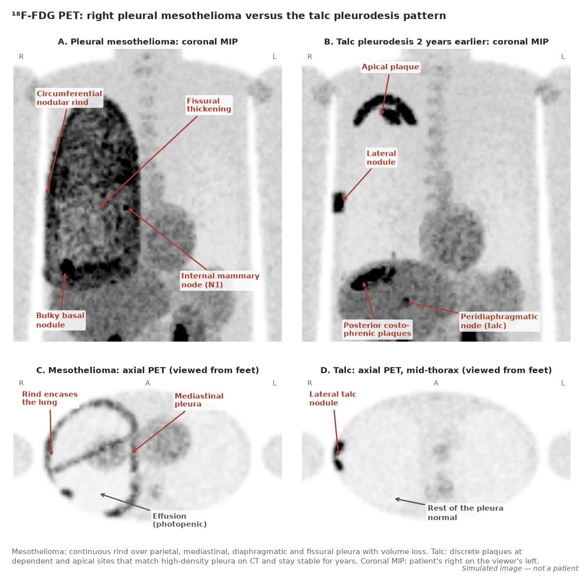
- Scan before talc. Talc pleurodesis causes intense, dense (about 140–380 HU) pleural uptake that persists for years, and can make the draining nodes avid. A pre-pleurodesis PET/CT is the reference for everything that follows.
- Ninth-edition cT is measured. Pleural mesothelioma cT1–cT3 now depend on Psum, the sum of the maximum pleural thickness at three levels (12 mm and 30 mm cut-offs), plus fissural thickness over 5 mm; cT4 is still defined by invasion. N and M are unchanged.
- PET's staging job is N and M. It finds occult nodal or distant disease in 29–38% of surgical candidates. Local invasion is better judged on CT and MRI, and avid nodes still need sampling.
- Fewer patients go to radical surgery. In MARS 2, extended pleurectomy decortication added to chemotherapy gave shorter survival (median 19.3 versus 24.8 months). Most patients now receive immunotherapy or chemotherapy, and response is read on CT with modified RECIST 1.1.
- Volume matters more than SUVmax. Baseline SUVmax and total lesion glycolysis are prognostic, and an early fall in total glycolytic volume predicted survival where SUVmax and CT did not.
- Thymic tumours: uptake follows grade. SUVmax rises from low-risk thymoma (A, AB, B1) to high-risk thymoma (B2, B3) to thymic carcinoma. PET separates carcinoma from thymoma with pooled sensitivity 89% and specificity 77%, but it is not a routine test for a thymic mass.
- Know the thymic mimics. Rebound hyperplasia after chemotherapy keeps the gland's shape and loses signal on chemical-shift MRI. Somatostatin receptor imaging selects advanced thymoma for octreotide. FAPI and mesothelin tracers remain investigational.
1. The clinical problem
Pleural mesothelioma and thymic epithelial tumours are rare thoracic cancers, but they reach every PET centre. Both spread along surfaces rather than as a single measurable mass, and both have benign lookalikes that take up ¹⁸F-fluorodeoxyglucose (FDG). The nuclear medicine physician therefore has three tasks. First, help find the right place to biopsy without claiming a diagnosis. Second, stage the patients whose treatment depends on it. Third, recognise the mimics before they change management.
Pleural mesothelioma
Pleural mesothelioma (the qualifier 'malignant' has been dropped in current terminology) is usually caused by asbestos and presents with a unilateral effusion or pleural thickening. There are three histological subtypes: epithelioid (the large majority), biphasic and sarcomatoid. Performance status, histological subtype and tumour volume are the main prognostic factors in routine practice (ERS/ESTS/EACTS/ESTRO 2020). For the tumour board the questions are: where is the best biopsy target; is the disease confined to one hemithorax and its nodes; is there a reason not to enrol a fit patient in a radical-treatment trial; and, later, is it responding?
Staging system in force: the ninth edition for pleural mesothelioma
The International Association for the Study of Lung Cancer (IASLC) built the ninth-edition proposals from 3,598 eligible cases diagnosed between 2013 and 2022. The main change is in clinical T. Earlier editions used qualitative CT descriptors such as endothoracic fascia, lung parenchymal and non-transmural diaphragmatic invasion, which CT cannot show reliably. These were removed. Instead, the maximum pleural thickness is measured perpendicular to the chest wall or mediastinum in the upper, middle and lower thirds of the hemithorax (divided at the top of the aortic arch and the top of the left atrium). The sum is Psum. Fissural thickness (Fmax) is measured on sagittal images. Median overall survival was 49.8 months for cT1, 27.5 months for cT2, 21.1 months for cT3 and 12.6 months for cT4.
| Category | Ninth-edition clinical definition | What the PET/CT report can add |
|---|---|---|
| cT1 | Ipsilateral pleura only; Psum 12 mm or less; no fissural involvement (Fmax 5 mm or less) | Confirm the thickening is avid and continuous; state whether the fissures are involved |
| cT2 | Psum over 12 mm and up to 30 mm without fissural involvement; or Psum up to 30 mm with fissural involvement (Fmax over 5 mm), mediastinal fat invasion or a solitary focus of chest-wall soft-tissue invasion | Fissural and mediastinal fat uptake; a single chest-wall focus |
| cT3 | Psum over 30 mm, with or without fissural involvement, mediastinal fat invasion or solitary chest-wall soft-tissue invasion | Psum comes from CT; PET shows where the bulk is |
| cT4 | Any Psum with rib invasion, invasion of mediastinal organs (heart, spine, oesophagus, trachea, great vessels), diffuse chest-wall, transdiaphragmatic or transpericardial invasion | Suspected transdiaphragmatic or diffuse chest-wall disease: confirm with MRI or laparoscopy |
| N1 | Ipsilateral intrathoracic nodes (including internal mammary, peridiaphragmatic and intercostal) | Name every avid station; many lie outside endoscopic reach |
| N2 | Contralateral intrathoracic or any supraclavicular nodes | The node that removes surgical options: sample it |
| M1 | Distant metastasis (unchanged) | Occult distant disease is PET's main staging contribution |
Stage groups were revised to match: stage I is T1N0; stage II is T1N1 or T2N0; stage IIIA is T1N2, T2N1, T2N2 or any T3; stage IIIB is any T4; stage IV is any M1. Pathological T keeps the eighth-edition descriptors except that fissural involvement becomes pT2.
Thymic epithelial tumours
The fifth-edition (2021) World Health Organization (WHO) classification groups thymomas (types A, AB, B1, B2 and B3, plus rare variants), thymic carcinomas (squamous cell carcinoma is the commonest) and thymic neuroendocrine tumours (typical and atypical carcinoid, large-cell and small-cell carcinoma). For imaging purposes thymomas are split into low-risk (A, AB, B1) and high-risk (B2, B3) groups. Thymoma is associated with autoimmune disease, most often myasthenia gravis.
Two staging systems run in parallel. The Masaoka-Koga system is surgical and still guides adjuvant radiotherapy: stage I, completely encapsulated; IIA, microscopic transcapsular invasion; IIB, macroscopic invasion of thymic or surrounding fat, or adherence to but not through the mediastinal pleura or pericardium; III, macroscopic invasion of neighbouring organs; IVA, pleural or pericardial dissemination; IVB, lymphatic or haematogenous metastasis. The TNM system (introduced in the eighth edition) is applicable to all thymic epithelial tumours. The ninth-edition IASLC proposals, built on 11,347 patients, split T1 by size into T1a (5 cm or less) and T1b (over 5 cm), drop the mediastinal pleura as a descriptor and move lung or phrenic nerve invasion from T3 to T2. N (N1 anterior perithymic nodes; N2 deep intrathoracic or cervical nodes), M (M1a separate pleural or pericardial nodules; M1b lung parenchymal or distant organ metastasis) and the stage groups are unchanged.
Where nuclear medicine changes management
- Choosing the biopsy site: the most avid, accessible pleural or mediastinal focus gives the best yield.
- Selecting for radical treatment: occult N2 or M1 disease removes a patient from surgical trials.
- Recognising mimics: talc, empyema and rebound thymic hyperplasia avoid a false upstaging.
- Response and prognosis: metabolic volume adds information where CT measurement of a rind is imprecise.
- Receptor imaging: somatostatin receptor (SSTR) imaging selects thymic tumours for somatostatin analogue therapy.
2. Tracers and why they work
FDG enters tumour cells through glucose transporters, is phosphorylated by hexokinase and is trapped. Mesothelioma is usually FDG-avid, and the rind-like growth pattern produces a continuous band of uptake around the lung. Inflammatory cells use the same pathway, which is why pleural infection, talc granulomas and recent surgery are the recurrent false positives. In thymic epithelial tumours uptake tracks biological aggressiveness: it rises with WHO risk group and is highest in thymic carcinoma.
| Tumour | Usual FDG avidity | Practical point |
|---|---|---|
| Epithelioid pleural mesothelioma | Moderate to high | Early, thin disease can be faint; a normal PET does not exclude it |
| Biphasic and sarcomatoid mesothelioma | Usually avid | Worse prognosis; histological subtype is one of the main prognostic factors |
| Peritoneal mesothelioma | Variable; lower in epithelioid | Small-volume peritoneal disease is under-staged |
| Low-risk thymoma (A, AB, B1) | Low to moderate | Uptake just above blood pool; smooth encapsulated mass |
| High-risk thymoma (B2, B3) | Moderate | Overlaps with both neighbours; B3 can be intense |
| Thymic carcinoma | High | Nodal and distant disease far more likely than in thymoma |
| Thymic neuroendocrine tumours | Variable | Pair FDG with SSTR PET |
When FDG fails and what replaces it
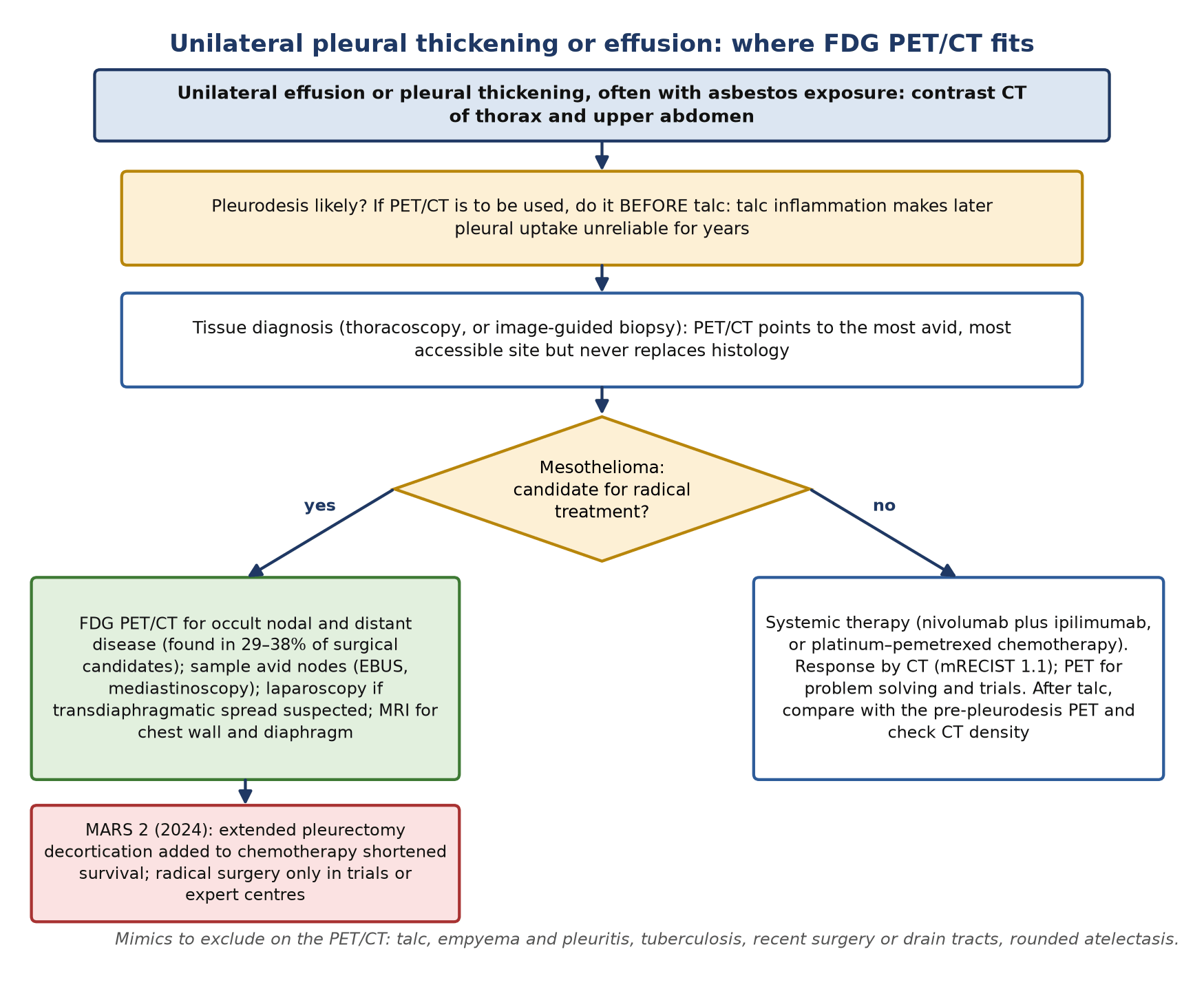
- After talc pleurodesis: the pleura is no longer readable by FDG; compare with a pre-pleurodesis scan and use CT density and MRI.
- Local invasion (chest wall, diaphragm, pericardium): contrast-enhanced MRI (ASCO 2018).
- Nodal staging: sampling by endobronchial ultrasound (EBUS) or mediastinoscopy, accepting that internal mammary and peridiaphragmatic nodes are out of reach.
- Thymic hyperplasia versus tumour: chemical-shift MRI.
- Somatostatin receptor status: ¹¹¹In-pentetreotide scintigraphy or ⁶⁸Ga-DOTATATE/DOTATOC PET.
- Research: ⁶⁸Ga-FAPI (fibroblast activation protein inhibitor) PET and radiolabelled anti-mesothelin antibodies.
3. Indications by clinical scenario
| Scenario | Role of nuclear medicine | Guideline position (strength where given) |
|---|---|---|
| Suspected pleural malignancy | FDG PET/CT can guide the biopsy to the most avid site; it does not replace tissue | ASCO 2018: PET/CT is a valuable adjunct to distinguish benign from malignant pleural disease, with tissue confirmation. Meta-analysis: PET/CT with semiquantitative reading is not accurate enough to be recommended routinely for this purpose |
| Before talc pleurodesis | Acquire PET/CT first if it will be needed at all | ASCO 2018: talc inflammation renders later PET unreliable for pleural disease |
| Initial staging of pleural mesothelioma | Whole-body FDG PET/CT for nodal and distant disease | ASCO 2018: PET/CT should usually be obtained for initial staging; may be omitted when definitive surgery is not being considered (evidence based, intermediate quality, strong). ESMO 2022: contrast CT of thorax and upper abdomen is the standard, with more extensive staging for radical-treatment candidates |
| Candidate for radical multimodality treatment | PET/CT plus sampling of avid nodes; MRI for local invasion | ASCO 2018: mediastinoscopy or EBUS for enlarged or PET-avid mediastinal nodes (strong); laparoscopy if transdiaphragmatic disease suspected. ERS/ESTS/EACTS/ESTRO 2020: treat such patients within trials in expert centres |
| Response to systemic therapy | CT with modified RECIST 1.1 is the standard; PET volumetric response is investigational | ASCO 2018: response from the sum of up to six pleural measurements (strong); tumour volumetry not yet recommended |
| Peritoneal mesothelioma | PET/CT as an adjunct to CT for extent and extraperitoneal disease | No PET-specific recommendation; small peritoneal deposits are under-staged |
| Thymic mass, likely thymoma | Not routine | SEOM-GECP-GETTHI 2021: contrast CT is standard [IV, A]; MRI for suspected hyperplasia or cyst [IV, B]; PET not generally recommended to assess thymic masses [IV, C] |
| Thymic carcinoma, aggressive histology or advanced stage | FDG PET/CT to complete staging and characterise suspected recurrence | SEOM-GECP-GETTHI 2021: PET can be considered in these settings |
| Advanced or refractory thymoma considered for somatostatin analogues | SSTR imaging (octreotide scintigraphy or SSTR PET) | Octreotide-scan positivity was the entry criterion of the ECOG phase II trial of octreotide with or without prednisone |
The guidelines differ in emphasis rather than direction. ASCO recommends PET/CT for most patients at initial staging, while ESMO treats CT as the baseline test for all and reserves fuller staging for those considered for radical treatment. Since MARS 2, that group has become small, so in practice PET/CT is requested mainly for trial candidates, for problem-solving and to choose a biopsy site.
4. Evidence at a glance
| Study | Design and size | Key finding | What it changed |
|---|---|---|---|
| Treglia 2014 | Meta-analysis, 11 studies (16 reviewed, 745 patients) | Malignant versus benign pleural lesions: sensitivity 95%, specificity 82%, AUC 0.95; no gain from PET/CT over PET | PET is accurate but not specific; mimics remain |
| Porcel 2015 | Meta-analysis, 14 studies, 407 malignant and 232 benign cases | Semiquantitative reading less sensitive than visual (82% versus 91%); PET/CT with SUV thresholds: sensitivity 81%, specificity 74% | No SUV cut-off replaces tissue |
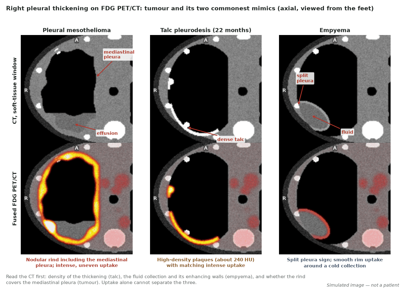
| Kwek 2004 | 9 patients after talc pleurodesis | Pleural SUV mean 5.4 (range 2.0–16.3) about 22 months after talc; matching dense thickening, mean 230 HU (140–380); unchanged on serial imaging | Correlate every pleural focus with CT density |
| IASLC ninth edition, T (Gill 2024) | International database, 3,598 eligible; 1,790 with Psum | Psum cut-offs at 12 and 30 mm and Fmax over 5 mm separate survival; median OS 49.8, 27.5, 21.1 and 12.6 months for cT1–cT4 | Measured clinical T |
| MARS 2 (Lim 2024) | Phase 3 randomised, 335 patients | Extended pleurectomy decortication plus chemotherapy versus chemotherapy: median survival 19.3 versus 24.8 months; restricted mean survival at 2 years 1.9 months shorter; serious adverse events 318 versus 169 | Radical surgery restricted to trials and expert centres |
| CheckMate 743 (Baas 2021) | Phase 3 randomised, 605 patients | Nivolumab plus ipilimumab versus platinum–pemetrexed: median OS 18.1 versus 14.1 months, HR 0.74 | First-line immunotherapy; new response-assessment questions |
| Francis 2007 | Prospective, 23 patients, PET before and after one cycle of chemotherapy | Fall in total glycolytic volume predicted survival (p=0.015); SUVmax (p=0.097) and CT (p=0.131) did not | Volumetric PET response |
| Wang 2022 | Meta-analysis, 19 studies, 1,819 patients | Pooled HR for overall survival: SUVmax 1.29, TLG 1.47 (univariate); SUVmax 1.20 and TLG 1.13 in multivariate analysis; MTV not independent | Baseline uptake and glycolytic burden are prognostic |
| Kessler 2024 | Prospective, 41 patients, ⁶⁸Ga-FAPI-46 and FDG | Similar sensitivity (per patient 100% versus 97.3%); per-region specificity 81.1% versus 36.8% | FAPI a candidate for problem-solving; still investigational |
| Kim 2022 | Meta-analysis of FDG PET in thymic epithelial tumours | Thymic carcinoma versus thymoma: sensitivity 89%, specificity 77%; high- versus low-risk tumour: 90% and 81% | FDG characterises a proven thymic tumour |
| Loehrer 2004 (ECOG) | Phase II, 38 assessable octreotide-scan-positive patients | 2 complete and 10 partial responses to octreotide with or without prednisone; none of 6 without pure thymoma responded | Receptor imaging to select somatostatin analogue therapy |
5. Patient preparation and acquisition
Standard oncological FDG preparation applies (fasting for at least 4 hours, blood glucose recorded, uptake time about 60 minutes, identical protocol for any scan that will be compared). The points below are specific to pleural and thymic disease.
- Timing against pleural procedures: whenever possible, scan before talc pleurodesis. After thoracoscopy, biopsy or drain insertion, record the date and site: track and biopsy uptake is common and can mimic chest-wall invasion.
- History to record: asbestos exposure, pleurodesis (agent and date), indwelling pleural catheter, recent infection or empyema, prior radiotherapy, chemotherapy or immunotherapy dates, myasthenia gravis and steroid use.
- Coverage: skull base to mid-thigh. Include the whole abdomen and pelvis in peritoneal mesothelioma and whenever transdiaphragmatic spread is suspected.
- Respiration: acquire the low-dose CT in shallow breathing so that basal pleural and diaphragmatic disease registers with PET.
- Quantitative follow-up: if metabolic volume or TLG will be tracked, keep scanner, reconstruction, uptake time and segmentation method constant; record the threshold used.
- Thymic imaging in young adults: note age and time since chemotherapy; plan chemical-shift MRI rather than repeat PET when rebound hyperplasia is the question.
- SSTR imaging: follow the neuroendocrine tumour protocol and record somatostatin analogue treatment.
6. Interpretation
The pleura
Describe the distribution (focal, multifocal, circumferential rind), whether the mediastinal, diaphragmatic and fissural pleura are involved, the intensity against the mediastinal blood pool and liver, and the CT correlate (thickness, nodularity, density, calcification, fluid). Circumferential or nodular thickening and involvement of the mediastinal pleura point towards malignancy on CT, and intense uptake in such thickening supports it. Uptake alone is not specific: in the meta-analyses, specificity was only about 74–82%, and semiquantitative thresholds performed worse than visual reading. Do not quote a single SUV cut-off as if it separated benign from malignant disease.
Nodes and distant disease
Mesothelioma drains to nodes that lung cancer rarely uses: internal mammary, peridiaphragmatic (cardiophrenic), intercostal and paravertebral, as well as the mediastinum. List every avid station and say which are ipsilateral (N1) and which contralateral or supraclavicular (N2). Nodes adjacent to avid pleura are hard to separate from it; say so rather than guessing. Up to half of involved mediastinal nodes lie outside the reach of mediastinoscopy and endosonography (ASCO 2018), so an avid internal mammary node matters even if EBUS is negative. For M, look at the contralateral pleura, peritoneum, liver, bone and lung. Brain imaging is not routine in the absence of symptoms.
Thymic masses
Report size (for T1a versus T1b), margins, fat invasion, contact with the pericardium, great vessels and lung, pleural or pericardial implants (M1a) and nodes (N1 perithymic; N2 deep intrathoracic or cervical). Give SUVmax with a comment on the risk group it suggests, but not a histological diagnosis: pooled SUVmax differences between low-risk thymoma, high-risk thymoma and carcinoma are real, yet individual values overlap. In the meta-analysis of Treglia and colleagues, carcinoma exceeded low-risk thymoma by a weighted mean of 4.8 SUV units and high-risk thymoma by 3.5; high-risk exceeded low-risk thymoma by only 1.2.
Normal thymus and rebound hyperplasia
Thymic FDG uptake is common in the young and after treatment. In 559 PET/CT studies of patients aged 3–40 years, thymic uptake was present in 25% of studies: in 73% of untreated patients up to 13 years of age but only 8% of those aged 31–40. It was seen in 17% of baseline scans and 27–40% of follow-up scans after chemotherapy, most often as an inverted-V shape, with a mean SUVmax of 3.73 (SD 1.22). Features favouring hyperplasia are youth, recent chemotherapy, preserved bilobed shape, smooth margins and mild homogeneous uptake. When doubt remains, dual-echo chemical-shift MRI settles it: hyperplastic thymus contains microscopic fat and loses signal on opposed-phase images, tumour does not. In 92 adults, a signal intensity index cut-off of 8.92% separated hyperplasia from tumour with 100% sensitivity and specificity, whereas the chemical-shift ratio overlapped in some young adults.
Pitfalls and mimics
| Finding | Why it misleads | How to avoid the error |
|---|---|---|
| Talc pleurodesis | Intense nodular or plaque-like pleural uptake, commonly posterior costophrenic and apical, persisting for years | High-density (calcific-range) pleura on CT at the same site; comparison with pre-pleurodesis PET; stability |
| Talc in draining nodes | Avid anterior peridiaphragmatic, paracardiac or internal mammary nodes | High attenuation within the node on the non-contrast CT |
| Empyema and parapneumonic effusion | Smooth rim uptake around a collection; pleural thickening | Split pleura sign, lenticular collection, clinical infection; aspirate |
| Tuberculous or rheumatoid pleuritis | Diffuse avid pleural thickening | Clinical context; biopsy |
| Recent thoracoscopy, biopsy or drain | Linear uptake along the track and chest wall | Record procedure dates; track shape |
| Benign asbestos-related pleural plaques and rounded atelectasis | Pleural lesions in an exposed patient | Calcified plaques and the comet-tail of rounded atelectasis usually show little uptake; new avid thickening still needs tissue |
| Rebound thymic hyperplasia | Anterior mediastinal uptake after chemotherapy mimics relapse or a thymic tumour | Age, timing, shape; chemical-shift MRI |
| Thymic cyst | Anterior mediastinal mass on CT | Fluid density, no uptake; MRI confirms fluid |
| Lymphoma and germ cell tumour | Intensely avid anterior mediastinal mass mimicking thymic carcinoma | Nodal disease elsewhere, B symptoms, raised lactate dehydrogenase; tumour markers (alpha-fetoprotein, beta-hCG) in young men; biopsy |
| Brown fat and vaccination nodes | Supraclavicular and mediastinal uptake | Fat density on CT; vaccination history |
Wording the conclusion
- For a pleural question without histology: describe the pattern, give the best biopsy target, and state what cannot be excluded (for example, FDG-avid circumferential right pleural thickening with mediastinal pleural involvement, suspicious for malignancy; most avid accessible site: posterolateral lower zone.)
- After talc: say explicitly that pleural uptake is uninterpretable where it matches dense talc, and report only new, non-dense or extrapleural findings.
- For staging: give cT from the CT measurements where available (Psum and Fmax), list nodal stations as N1 or N2, and flag any lesion that would remove the patient from radical treatment for confirmation.
- For a thymic mass: suggest a risk group, not a histology, and name any finding that changes stage (fat invasion, implants, nodes).
Peritoneal mesothelioma
Peritoneal mesothelioma is much rarer than pleural disease and is treated, when possible, by cytoreductive surgery with hyperthermic intraperitoneal chemotherapy. FDG PET/CT helps to show extent and extraperitoneal spread but under-stages small-volume peritoneal disease. In 42 patients, SUVmax and metabolic tumour volume were lower in epithelioid than in non-epithelioid tumours; among 18 patients explored surgically, PET/CT staged T correctly in 72.2% and overall TNM stage in 94.4%. A negative PET does not exclude peritoneal deposits, and the peritoneal cancer index still comes from cross-sectional imaging and surgical exploration.
7. Response assessment and follow-up
Modified RECIST 1.1 on CT
The rind cannot be measured with standard RECIST. Modified RECIST measures tumour thickness perpendicular to the chest wall or mediastinum and sums up to six measurements, each at least 1 cm thick, with no more than two sites on each of three CT sections separated by at least 1 cm (ASCO 2018, strong recommendation). The 2018 revision (modified RECIST 1.1) aligns the method with RECIST 1.1 and defines minimally measurable disease, measurable lesions, acceptable measurement sites, nodes, non-pleural lesions and bilateral disease. Measure at the same sites on every scan. ASCO considers CT tumour volumetry promising but still investigational.
Metabolic response
No PET response criteria have been validated specifically for mesothelioma, and PET is not a routine response test outside trials. It helps when CT is ambiguous, for example when effusion, collapse or fibrosis hide the rind. Volume-based measures work better than SUVmax: in 23 patients scanned before and after one cycle of chemotherapy, a fall in total glycolytic volume predicted survival while falls in SUVmax and CT measurements did not. Under nivolumab in 30 patients with recurrent disease, the European Organisation for Research and Treatment of Cancer (EORTC) criteria, PERCIST and immunotherapy-modified PERCIST (imPERCIST) each classified 16.7% of patients as complete metabolic responders, whereas CT showed no complete response. All three PET criteria predicted progression-free survival better than CT, and imPERCIST was the most accurate.
- Talc changes the baseline: pleurodesis uptake is intense and stays on serial imaging, so a post-talc pleural SUV cannot be compared with a pre-talc one.
- Under checkpoint inhibitors, new or increased uptake early in treatment may be inflammation. In a stable patient, correlate with CT and consider a confirmatory scan before calling progression.
- Use the same segmentation method and threshold for metabolic volume on every scan, and report the method.
Prognosis
Baseline uptake carries prognostic information. In a meta-analysis of 19 studies and 1,819 patients, higher SUVmax (pooled hazard ratio for death 1.29) and total lesion glycolysis (TLG, 1.47) predicted shorter survival; both remained significant in multivariate analyses (1.20 and 1.13), whereas metabolic tumour volume did not. These are group-level findings; there is no validated cut-off for individual decisions.
Follow-up
Surveillance of both diseases is CT-based. For thymic epithelial tumours, PET/CT can be used to characterise lesions suspicious for recurrence (SEOM-GECP-GETTHI 2021). After pleurodesis or pleural surgery, a new avid focus should be judged against the post-procedure baseline and the CT density before it is called recurrence.
8. Theranostics and emerging tracers
| Tracer | Target | Evidence level (2026) | What it may add |
|---|---|---|---|
| ¹¹¹In-pentetreotide or ⁶⁸Ga-DOTATATE/DOTATOC | Somatostatin receptor | In clinical use to select thymic tumours for somatostatin analogues | In the ECOG phase II trial, octreotide-scan-positive thymoma responded to octreotide, with more responses after prednisone was added |
| ⁶⁸Ga-FAPI-46 | Fibroblast activation protein | Investigational (prospective single-centre, 41 patients) | Uptake correlated with FAP expression; per-region specificity 81.1% versus 36.8% for FDG with similar sensitivity; may help separate tumour from inflammation |
| ¹¹¹In-amatuximab | Mesothelin | Investigational (first-in-human, 6 patients) | Tumour uptake in mesothelioma and pancreatic cancer, higher in mesothelioma; tumour-to-background ratios 1.2–62 |
| Radioligand therapy | FAP, mesothelin or SSTR | No approved radioligand therapy for mesothelioma or thymic epithelial tumours | Receptor imaging would select patients for future trials |
FAPI is attractive in mesothelioma because the tumour and its stroma express FAP strongly, and the higher specificity may matter most after pleurodesis or infection. The evidence is still single-centre and observational, so FAPI findings should not change management outside a study. Mesothelin is a therapeutic target of antibodies and antibody-drug conjugates; imaging it is at an early, first-in-human stage.
9. Structured report example
- Indication: 71-year-old former shipyard worker; epithelioid pleural mesothelioma on thoracoscopic biopsy (right); being assessed for a radical-treatment trial. No pleurodesis; indwelling catheter since 2 weeks.
- Technique: ¹⁸F-FDG 280 MBq, uptake 62 min, glucose 6.1 mmol/L; skull base to mid-thigh; low-dose CT in shallow breathing.
- Pleura: circumferential nodular right pleural thickening including mediastinal and diaphragmatic pleura and the major fissure (Fmax 7 mm), SUVmax 9.8; Psum on the diagnostic CT 34 mm. Catheter track uptake in the right lateral chest wall, consistent with the procedure. No rib destruction or transdiaphragmatic extension.
- Nodes: right internal mammary node 8 mm, SUVmax 5.2, and right cardiophrenic node 7 mm, SUVmax 4.1 (N1). No contralateral or supraclavicular uptake.
- Distant: no extrathoracic avid disease; no peritoneal uptake.
- Conclusion: cT3 (Psum over 30 mm) cN1 cM0, ninth-edition stage IIIA. Chest-wall uptake follows the catheter track and is not called invasion.
- Recommendation: contrast MRI if chest-wall or diaphragmatic invasion needs clarifying; sample nodes only if the result would change eligibility, recognising that internal mammary nodes are not reachable by EBUS.
10. Take-home points
- FDG PET/CT is sensitive but not specific for pleural malignancy; it guides biopsy and never replaces it.
- Scan before talc pleurodesis. Afterwards, dense avid pleura and dense avid nodes on the drainage pathway are talc until proven otherwise.
- Ninth-edition cT for pleural mesothelioma is measured: Psum cut-offs of 12 and 30 mm and fissural thickness over 5 mm; cT4 remains invasion-based.
- PET's main staging value is occult nodal and distant disease in patients considered for radical treatment, now a small group after MARS 2.
- Response is read on CT with modified RECIST 1.1; metabolic volume and TLG add prognostic information but are not yet validated for decisions.
- In thymic epithelial tumours, uptake rises with WHO risk group; use it to suggest grade and complete staging, not to name the histology.
- Rebound thymic hyperplasia keeps the gland's shape; chemical-shift MRI resolves doubt. SSTR imaging selects thymoma for somatostatin analogues.
- FAPI and mesothelin imaging are promising but investigational.
Test yourself
5 quick questions. Pick an answer to see the explanation.
References
- Gill RR, Nowak AK, Giroux DJ, et al. The International Association for the Study of Lung Cancer Mesothelioma Staging Project: proposals for revisions of the "T" descriptors in the forthcoming ninth edition of the TNM classification for pleural mesothelioma. J Thorac Oncol. 2024;19(9):1310-25.
- Murphy DJ, Nowak AK, Frauenfelder T, et al. Radiologist's guide to the ninth edition TNM staging system for quantitative and qualitative assessment of pleural mesothelioma. Radiology. 2025;316(3):e250531.
- Scherpereel A, Opitz I, Berghmans T, et al. ERS/ESTS/EACTS/ESTRO guidelines for the management of malignant pleural mesothelioma. Eur Respir J. 2020;55(6):1900953.
- Popat S, Baas P, Faivre-Finn C, et al. Malignant pleural mesothelioma: ESMO Clinical Practice Guidelines for diagnosis, treatment and follow-up. Ann Oncol. 2022;33(2):129-42.
- Kindler HL, Ismaila N, Armato SG, et al. Treatment of malignant pleural mesothelioma: American Society of Clinical Oncology clinical practice guideline. J Clin Oncol. 2018;36(13):1343-73.
- Treglia G, Sadeghi R, Annunziata S, et al. Diagnostic accuracy of 18F-FDG-PET and PET/CT in the differential diagnosis between malignant and benign pleural lesions: a systematic review and meta-analysis. Acad Radiol. 2014;21(1):11-20.
- Porcel JM, Hernández P, Martínez-Alonso M, et al. Accuracy of fluorodeoxyglucose-PET imaging for differentiating benign from malignant pleural effusions: a meta-analysis. Chest. 2015;147(2):502-12.
- Kwek BH, Aquino SL, Fischman AJ. Fluorodeoxyglucose positron emission tomography and CT after talc pleurodesis. Chest. 2004;125(6):2356-60.
- Lim E, Waller D, Lau K, et al. Extended pleurectomy decortication and chemotherapy versus chemotherapy alone for pleural mesothelioma (MARS 2): a phase 3 randomised controlled trial. Lancet Respir Med. 2024;12(6):457-66.
- Baas P, Scherpereel A, Nowak AK, et al. First-line nivolumab plus ipilimumab in unresectable malignant pleural mesothelioma (CheckMate 743): a multicentre, randomised, open-label, phase 3 trial. Lancet. 2021;397(10272):375-86.
- Armato SG, Nowak AK. Revised modified Response Evaluation Criteria in Solid Tumors for assessment of response in malignant pleural mesothelioma (version 1.1). J Thorac Oncol. 2018;13(7):1012-21.
- Francis RJ, Byrne MJ, van der Schaaf AA, et al. Early prediction of response to chemotherapy and survival in malignant pleural mesothelioma using a novel semiautomated 3-dimensional volume-based analysis of serial 18F-FDG PET scans. J Nucl Med. 2007;48(9):1449-58.
- Wang Y, Xu Y, Kan Y, et al. Prognostic value of 18F-FDG PET/CT in malignant pleural mesothelioma: a meta-analysis. Acta Radiol. 2023;64(2):552-62.
- Kitajima K, Maruyama M, Yokoyama H, et al. Response to immune checkpoint inhibitor therapy in patients with unresectable recurrent malignant pleural mesothelioma shown by FDG-PET and CT. Cancers (Basel). 2021;13(5):1098.
- Kessler L, Schwaning F, Metzenmacher M, et al. Fibroblast activation protein-directed imaging outperforms 18F-FDG PET/CT in malignant mesothelioma: a prospective, single-center, observational trial. J Nucl Med. 2024;65(8):1188-93.
- Lindenberg L, Thomas A, Adler S, et al. Safety and biodistribution of 111In-amatuximab in patients with mesothelin expressing cancers using single photon emission computed tomography-computed tomography (SPECT-CT) imaging. Oncotarget. 2015;6(6):4496-504.
- Zhang H, Pang L, Hou J, et al. Significance of 18F-fluorodeoxyglucose PET/computed tomography in the initial staging of malignant peritoneal mesothelioma. Nucl Med Commun. 2025;46(8):746-52.
- Marx A, Chan JKC, Chalabreysse L, et al. The 2021 WHO classification of tumors of the thymus and mediastinum: what is new in thymic epithelial, germ cell, and mesenchymal tumors? J Thorac Oncol. 2022;17(2):200-13.
- Ruffini E, Huang J, Cilento V, et al. The International Association for the Study of Lung Cancer Thymic Epithelial Tumors Staging Project: proposal for a stage classification for the forthcoming (ninth) edition of the TNM classification of malignant tumors. J Thorac Oncol. 2023;18(12):1655-71.
- Treglia G, Sadeghi R, Giovanella L, et al. Is (18)F-FDG PET useful in predicting the WHO grade of malignancy in thymic epithelial tumors? A meta-analysis. Lung Cancer. 2014;86(1):5-13.
- Kim K, Jeong JH, Kim SJ. Diagnostic test accuracy of 18F-FDG PET or PET/CT for characterization of histologic type of thymic epithelial tumor: a meta-analysis. Clin Nucl Med. 2022;47(1):36-42.
- Loehrer PJ, Wang W, Johnson DH, et al. Octreotide alone or with prednisone in patients with advanced thymoma and thymic carcinoma: an Eastern Cooperative Oncology Group phase II trial. J Clin Oncol. 2004;22(2):293-9.
- Priola AM, Priola SM, Ciccone G, et al. Differentiation of rebound and lymphoid thymic hyperplasia from anterior mediastinal tumors with dual-echo chemical-shift MR imaging in adulthood: reliability of the chemical-shift ratio and signal intensity index. Radiology. 2015;274(1):238-49.
- Jerushalmi J, Frenkel A, Bar-Shalom R, et al. Physiologic thymic uptake of 18F-FDG in children and young adults: a PET/CT evaluation of incidence, patterns, and relationship to treatment. J Nucl Med. 2009;50(6):849-53.
- Remon J, Bernabé R, Diz P, et al. SEOM-GECP-GETTHI clinical guidelines for the treatment of patients with thymic epithelial tumours (2021). Clin Transl Oncol. 2022;24(4):635-45.