Nuclear Medicine in Breast Cancer
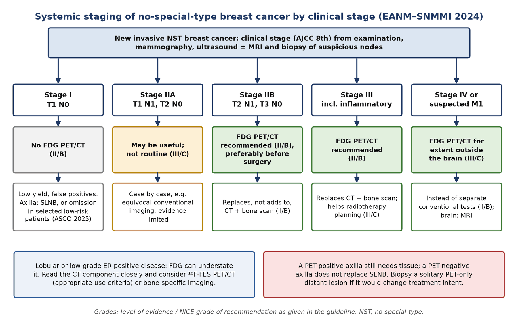
- Stage decides the test. The joint EANM–SNMMI guideline (2024) recommends FDG PET/CT for systemic staging of no-special-type cancer from clinical stage IIB (including T3N0) through stage III and inflammatory disease, in place of CT plus bone scan. It is not recommended in stage I and is optional in stage IIA.
- Randomised evidence. In PETABC (369 patients, stage IIB T3N0 or III), PET/CT upstaged 23% to stage IV versus 11% with CT and bone scan, and fewer patients went on to curative-intent combined-modality treatment.
- Biology matters. Triple-negative, HER2-positive and grade 3 tumours are FDG-avid; lobular and low-grade oestrogen-receptor-positive tumours often are not. Read the CT component, and consider ¹⁸F-FES.
- FES shows the receptor. ¹⁸F-fluoroestradiol PET (US approval 2020) maps functional oestrogen receptor at every site. Tamoxifen and fulvestrant block it for up to 8 and 28 weeks; aromatase inhibitors do not.
- The axilla is surgical. PET does not replace sentinel node biopsy. Lymphoscintigraphy with a deep injection also maps internal mammary drainage; SPECT/CT localises unusual nodes.
- De-escalation is here. SOUND and INSEMA showed that omitting axillary surgery was non-inferior in selected small, clinically node-negative cancers; ASCO (2025) now advises against routine SLNB in a defined low-risk group.
- Response and recurrence. Early FDG response is useful in triple-negative and HER2-positive disease during neoadjuvant therapy; FDG PET/CT is the test of choice for suspected recurrence when conventional imaging is equivocal. Report response with PERCIST or EORTC criteria.
1. The clinical problem
Breast cancer is the most commonly diagnosed cancer in women worldwide. About 75–80% of invasive cancers are of no special type (NST), formerly called invasive ductal carcinoma, and 10–15% are invasive lobular carcinoma (ILC). Treatment depends as much on biology as on anatomy: oestrogen and progesterone receptors (ER, PR), human epidermal growth factor receptor 2 (HER2) and proliferation define the subtypes that decide systemic therapy.
What the oncologist needs from imaging
- Local extent: mammography, ultrasound and, selectively, MRI; nuclear medicine plays a minor role (molecular breast imaging, section 9).
- Axilla: ultrasound with needle biopsy of suspicious nodes, then sentinel lymph node biopsy (SLNB) in the clinically node-negative patient. Lymphoscintigraphy is a nuclear medicine procedure at the centre of this step.
- Extra-axillary nodes and distant disease: the question that decides curative versus palliative intent in higher-stage disease. This is where FDG PET/CT changes management.
- Receptor status across sites: metastases can differ from the primary; ¹⁸F-FES PET shows ER expression at every site at once.
- Response: early response during neoadjuvant therapy, and response of bone-predominant metastatic disease that CT cannot measure.
Staging system in force: AJCC 8th edition
The American Joint Committee on Cancer (AJCC) 8th edition remains current for breast cancer; breast has not yet been released in AJCC Version 9. It has two parts. The anatomic stage uses tumour (T), node (N) and metastasis (M) categories. The prognostic stage adds grade, ER, PR, HER2 and, for T1–2 N0 ER-positive HER2-negative cancers, a multigene assay result (a low Oncotype DX recurrence score assigns prognostic stage IA). Imaging defines the clinical anatomic stage, and guideline recommendations for PET use the clinical anatomic stage.
| Stage | TNM groups | Imaging consequence |
|---|---|---|
| I | T1 N0 | No systemic staging unless symptoms; SLNB (or omission in selected patients) |
| IIA | T0–1 N1; T2 N0 | Systemic staging optional; FDG PET/CT case by case |
| IIB | T2 N1; T3 N0 | FDG PET/CT recommended |
| IIIA | T0–3 N2; T3 N1 | FDG PET/CT recommended (locally advanced) |
| IIIB | T4 N0–2 (includes inflammatory T4d) | FDG PET/CT recommended |
| IIIC | Any T N3 | FDG PET/CT recommended |
| IV | Any T, any N, M1 | Whole-body extent; biopsy one site for receptors |
Clinical anatomic stage groups (AJCC 8th) and what they mean for nuclear medicine.
- Nodal categories that PET changes: N2b is internal mammary nodes without axillary nodes; N3a is infraclavicular (level III); N3b is internal mammary plus axillary; N3c is ipsilateral supraclavicular.
- What is metastatic, not nodal: contralateral axillary, internal mammary or supraclavicular nodes and any cervical node are M1.
Subtypes and why they matter for tracer choice
| Subtype | Markers | FDG avidity | Imaging implication |
|---|---|---|---|
| Luminal A-like | HR+ HER2−, low grade and proliferation | Often low | FDG may understate disease; bone metastases often sclerotic; FES useful |
| Luminal B-like | HR+ HER2−, high grade or proliferation | Intermediate | FDG usually adequate; FES if equivocal |
| HER2-positive | HER2 amplified or overexpressed (HR+ or −) | Usually high | FDG for staging and early response; visceral and brain spread |
| Triple-negative | ER−, PR−, HER2− | High | FDG for staging and early response; visceral (especially lung) metastases |
| Invasive lobular | Almost always ER+ (about 95%) | Often low | Metastases to peritoneum, gut, serosa and bone (sometimes miliary, FDG-negative); consider FES |
HR, hormone receptor. Luminal-like tumours are about 65% of cases, HER2-positive 15–20% and triple-negative 10–15%.
2. Tracers and why they work
- ¹⁸F-FDG: uptake reflects glucose transporter 1 (GLUT1) and hexokinase activity, viable tumour cell density and hypoxia-driven glycolysis. NST is more avid than lobular cancer, triple-negative more than ER-positive, and grade 3 more than lower grades. High uptake carries a worse prognosis.
- ¹⁸F-FES (fluoroestradiol): a radiolabelled oestrogen that binds functional ER. Uptake correlates with ER immunohistochemistry. It does not image PR or HER2.
- Bone tracers (⁹⁹ᵐTc-MDP/HDP, ¹⁸F-NaF): show osteoblastic reaction, not tumour. NaF PET has higher sensitivity and resolution than planar bone scintigraphy.
- Radiocolloids for sentinel nodes: ⁹⁹ᵐTc-nanocolloidal albumin (5–100 nm, preferred in most of Europe), filtered ⁹⁹ᵐTc-sulphur colloid (mostly 100–220 nm; unfiltered mean about 305–340 nm), ⁹⁹ᵐTc-antimony trisulphide (3–30 nm) and the receptor-targeted ⁹⁹ᵐTc-tilmanocept (about 7 nm), which binds mannose receptors on macrophages.
When FDG fails and what replaces it
- Small lesions: lesions of 10 mm or less (about 4–5 mm with digital PET) and micrometastatic nodes are missed. This is why PET cannot replace SLNB.
- Low-avidity histology: lobular, tubular, mucinous, low-grade ER-positive and in situ disease. In stage III, PET/CT revealed unsuspected distant metastases in 22% of NST patients but 11% of ILC patients; sclerotic, sometimes miliary, lobular bone metastases may be visible only on the CT component.
- Sclerotic bone metastases: may be faint on FDG; read the CT windows and consider bone-specific imaging.
- Replacement: ¹⁸F-FES for ER-positive disease (approved in the USA and France); bone scan or NaF PET for purely sclerotic disease; fluciclovine and fibroblast activation protein inhibitor (FAPI) PET are investigational (section 9).
3. Indications by clinical scenario
The joint EANM–SNMMI guideline on FDG PET/CT in NST breast cancer (Vaz et al., 2024; endorsed by the ACR, ESSO, ESTRO, EUSOBI/ESR and EUSOMA) graded its recommendations by level of evidence (I–IV) and NICE grade (A–D). It applies to NST cancer; the panel explicitly did not extend it to lobular cancer.
| Scenario | EANM–SNMMI 2024 recommendation (evidence/grade) | Comment and other guidelines |
|---|---|---|
| Stage I (T1N0) | Not recommended (II/B) | In 325 operable cancers PET suggested metastases in 13; only 3 (0.9%) were confirmed and 10 (3.0%) were false positives |
| Stage IIA (T1N1, T2N0) | May be useful; not enough data for routine use (III/C) | Distant disease in 0.8–11% across series; NCCN also allows it in selected IIA cases |
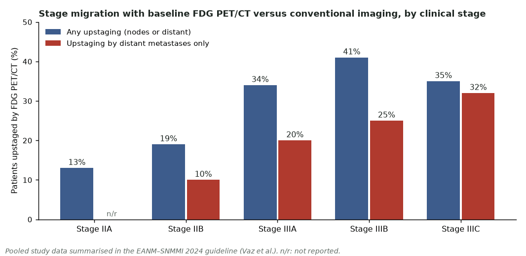
| Stage IIB (T2N1, T3N0) | Recommended, preferably before surgery (II/B) | Upstaging 19% overall, 10% by distant disease alone |
| Stage III, including inflammatory | Recommended (II/B); instead of, not in addition to, conventional imaging (II/B) | PETABC randomised trial; ESMO and ABC also accept PET/CT in place of CT and bone scan in high-risk disease |
| Radiotherapy planning (IIB–III) | Recommended in treatment planning; may improve radiotherapy planning (III/C) | Changed regional radiotherapy in about 20% of node-positive patients |
| Stage IV extent | Useful outside the brain (III/C); instead of separate conventional tests (II/B) | Brain requires MRI |
| Early response, neoadjuvant therapy | May be used, particularly triple-negative and HER2-positive (II/B) | NCCN and ESMO do not recommend PET in this setting |
| Response, metastatic disease | May play a role (III/C); especially bone metastases (III/C) | ESMO: may give earlier guidance in bone-predominant disease; NCCN prefers RECIST on CT |
| Suspected recurrence | Useful when conventional imaging is equivocal (I/A); with symptoms (I/A); rising tumour markers (II/B); to guide biopsy (IV/D); radiotherapy planning (III/C) | All guidelines agree: no PET for surveillance of asymptomatic patients |
| Bone metastases | Can substitute for CT and/or bone scan (II/B) | NCCN still asks for concordant PET and CT, or a bone scan or NaF PET |
| Reporting | PERCIST or EORTC (III/C); quantitative SUV, MTV and TLG are prognostic (I/A) | Use EARL- or ACR/IAC-accredited scanners for response |
Sources: Vaz et al. 2024; comparison with NCCN, ESMO and ABC guidelines from Groheux et al. 2024.
Where the guidelines disagree
- The EANM–SNMMI guideline recommends FDG PET/CT more widely than NCCN, ESMO or the Advanced Breast Cancer (ABC) consensus. NCCN and ESMO mainly use it to resolve equivocal conventional imaging, although ESMO and ABC accept it instead of CT and bone scan in high-risk and metastatic disease.
- For neoadjuvant response, EANM–SNMMI supports early FDG assessment in triple-negative and HER2-positive disease; NCCN and ESMO give PET no role there.
- For bone, EANM–SNMMI accepts FDG PET/CT alone; NCCN still asks for concordant PET and CT findings or a separate bone scan or NaF PET/CT.
The axilla: sentinel node biopsy and its de-escalation
- Clinically node-negative (cN0): SLNB is the staging standard. FDG PET/CT cannot exclude small-volume nodal disease, and an FDG-avid axillary node still needs tissue.
- One or two positive sentinel nodes: ACOSOG Z0011 (891 women, T1–T2, breast-conserving surgery with whole-breast radiotherapy) showed no survival loss without axillary dissection: 10-year overall survival 86.3% versus 83.6%. AMAROS (1,425 randomised) showed axillary radiotherapy is an alternative to dissection with less lymphoedema (11.9% versus 24.5% at 5 years) and 10-year axillary recurrence of 1.82% versus 0.93%.
- Omitting SLNB altogether: SOUND (1,405 analysed; tumours up to 2 cm with a negative axillary ultrasound) found 5-year distant disease-free survival of 98.0% without axillary surgery versus 97.7% with SLNB. INSEMA (5,502 randomised 1:4; T1–T2 up to 5 cm, clinically node-negative, breast conservation) found 5-year invasive disease-free survival of 91.9% versus 91.7% (hazard ratio 0.91), with axillary recurrence 1.0% versus 0.3%.
- ASCO 2025: clinicians should not recommend routine SLNB in postmenopausal patients aged 50 or over with a negative axillary ultrasound and a grade 1–2, 2 cm or smaller, HR-positive HER2-negative cancer treated with breast-conserving therapy. SLNB may be offered in clinically node-negative cT3–T4c or multicentric tumours, in pregnancy, obesity and men, and after previous breast or axillary surgery.
- After neoadjuvant therapy in node-positive disease: SLNB alone is less reliable. In ACOSOG Z1071 (cN1 disease, two or more nodes removed) the false-negative rate was 12.6%. Removing the biopsy-proven, clipped node as well (targeted axillary dissection, localised with an ¹²⁵I seed in the original series) cut the false-negative rate to 2.0%.
Internal mammary drainage
Deep (peritumoral or intratumoral) injection is needed to map internal mammary nodes (IMNs). They are seen in about one-third of patients; 63–92% can be harvested, and 11–27% of harvested IMNs contain metastases. IMN involvement worsens prognosis, and mapping causes stage migration and changes radiotherapy and systemic treatment, but survival benefit from IMN biopsy is unproven, so practice varies. The nuclear medicine task is to report the drainage accurately and localise it.
4. Evidence at a glance
| Study | Design and size | Key finding | What it changed |
|---|---|---|---|
| PETABC (Dayes 2023) | Randomised, 369 with stage III or IIB (T3N0), six Ontario centres | Upstaged to stage IV: 23% with PET/CT vs 11% with CT and bone scan (difference 12.3%); curative-intent combined-modality therapy 81% vs 89% | First randomised evidence; underpins the EANM–SNMMI stage IIB–III recommendation |
| Han and Choi 2021 | Meta-analysis, 29 studies of FDG PET, PET/CT or PET/MRI at initial staging | Pooled change in stage 25%; change in management 18% | Quantified management impact |
| Hildebrandt 2016 | Prospective, 100 with suspected recurrence; FDG PET/CT vs contrast CT vs CT plus bone scan | Area under the curve for distant recurrence 0.99 vs 0.84 vs 0.86 | Level I/A recommendation for suspected recurrence |
| AVATAXHER (Coudert 2014) | Randomised phase 2, 142 HER2-positive, PET after cycle 1 | pCR 53.6% in PET responders; in poor responders 43.8% with added bevacizumab vs 24.0% without | Proof that early PET can steer therapy; no disease-free survival gain at 5 years |
| PHERGain (Pérez-García 2024) | Randomised phase 2, 356 HER2-positive stage I–IIIA; PET after 2 cycles of trastuzumab–pertuzumab | 3-year invasive disease-free survival 94.8% in the PET-adapted arm; about a third of patients avoided chemotherapy | PET-guided chemotherapy de-escalation is feasible; not yet standard |
| ACOSOG Z0011 (Giuliano 2017) | Randomised, 891 with 1–2 positive SLNs, breast conservation | 10-year overall survival 86.3% (SLNB alone) vs 83.6% (dissection) | No axillary dissection for limited SLN disease |
| AMAROS (Bartels 2023) | Randomised, 1,425 SLN-positive cT1–2 | Axillary recurrence 1.82% (radiotherapy) vs 0.93% (dissection); lymphoedema 11.9% vs 24.5% | Axillary radiotherapy preferred to dissection |
| SOUND (Gentilini 2023) | Randomised, 1,405 with tumours up to 2 cm and negative ultrasound | 5-year distant disease-free survival 98.0% (no surgery) vs 97.7% (SLNB) | Omission of axillary surgery in selected patients |
| INSEMA (Reimer 2025) | Randomised 1:4, 5,502 cT1–2 cN0, breast conservation | 5-year invasive disease-free survival 91.9% vs 91.7%; axillary recurrence 1.0% vs 0.3% | Supports ASCO 2025 omission criteria |
| ACOSOG Z1071 (Boughey 2013); TAD (Caudle 2016) | Prospective; 649 cN1 after chemotherapy; 208 with clipped nodes | SLNB false-negative rate 12.6%; with the clipped node (TAD) 2.0% | Targeted axillary dissection after neoadjuvant therapy |
| Linden 2006 | Prospective, 47 with ER-positive metastatic disease starting endocrine therapy | No response in 15 patients with FES SUV below 1.5; 11 of 32 (34%) responded above it | FES predicts lack of endocrine benefit |
pCR, pathological complete response; SLN, sentinel lymph node; TAD, targeted axillary dissection.
5. Patient preparation and acquisition
FDG PET/CT: what differs in breast cancer
- Standard preparation: fast at least 4 h, hydrate, blood glucose below 200 mg/dL (11.1 mmol/L), rest warm for an uptake time of 60 ± 5 min, arms up, skull base to mid-thigh.
- Timing after therapy: metabolic flare occurs 7–10 days after starting endocrine therapy (tamoxifen, fulvestrant or aromatase inhibitors). The panel suggests scanning at least 10 days (15 if possible) after the last dose of chemotherapy or endocrine therapy; this is expert opinion. Do not interrupt targeted therapy for a scan.
- After radiotherapy: wait at least 3 months before looking for recurrence in the radiotherapy field.
- Record: biopsy and surgery dates, radiotherapy fields, growth factor injections, corticosteroids and recent vaccinations (with the arm used).
- Options: contrast-enhanced CT helps in metastatic disease and makes later CT comparison easier; prone hanging-breast images and a flat table top help breast assessment and radiotherapy planning.
- Response studies: baseline, interim and final scans on the same accredited scanner.
¹⁸F-FES PET/CT
- Activity and timing: the US label recommends 222 MBq (range 111–222 MBq) with imaging 80 min after injection (range 20–80 min); the SNMMI/EANM standard describes 111–280 MBq with about 60 min uptake. Effective dose is about 4.9 mSv for 222 MBq, plus the CT.
- Drug interactions: tamoxifen and other ER modulators, and fulvestrant, block FES binding for up to 8 and 28 weeks respectively. Image before these drugs start; do not delay indicated therapy for a scan. Aromatase inhibitors do not block the receptor and were allowed in the registration studies.
- Other preparation: the label asks for good hydration and frequent voiding, a pregnancy check, and interruption of breastfeeding for 4 hours.
Breast lymphoscintigraphy (EANM–SNMMI 2013)
| Step | Recommendation |
|---|---|
| Activity | 5–30 MBq for surgery the same day; up to 150 MBq if injected the afternoon before (two-day protocol) |
| Volume | 0.05–0.5 mL for superficial injections; 0.5–1.0 mL for peritumoral injections |
| Injection site | Superficial (periareolar, subareolar, intradermal, subdermal) shows axillary nodes best; deep (peritumoral, intratumoral) is needed for internal mammary and other extra-axillary nodes; dual injection lowers false negatives. Avoid injecting into a seroma or prosthesis |
| Technique | Mix the syringe gently before injection; massage can speed drainage; lidocaine with the tracer reduces pain |
| Imaging | Planar at 15–30 min, 1 h and 2–4 h (up to 18–24 h if needed); anterior, 45° anterior oblique and lateral, 3–5 min each; low-energy high-resolution collimator; mark the skin in the surgical position |
| SPECT/CT | For non-visualisation, obesity, extra-axillary or unusual drainage (internal mammary, intramammary, contralateral), previous breast surgery, or confusing planar images |
| Non-visualisation | Consider a second injection, perhaps at a different site; most patients still have a node found at surgery, and mapping fails altogether in about 1–2% |
| Pregnancy | Low-activity lymphoscintigraphy is justified (fetal dose negligible below 10 MBq); avoid blue dye |
Tracer uptake in a node does not predict whether it is the sentinel node, so no quantification is needed; report location and number.
6. Interpretation and reporting
Reading the staging FDG study
- Nodes by station: name axillary level I–II, level III (infraclavicular), internal mammary and supraclavicular nodes, because each changes the N category (N1 to N3c). Contralateral nodes are M1.
- Bone: FDG is sensitive for lytic and marrow disease; sclerotic lesions can be faint. In 102 patients with newly diagnosed metastatic disease, FDG PET found 87% of bone lesions, bone scan 26% and contrast CT 39%, and FDG changed management in 16%.
- Lung: PET is less sensitive than dedicated CT for small nodules; read the CT lung windows.
- Brain: not assessed reliably by FDG; MRI if symptoms or in high-risk subtypes.
- Incidental focal breast uptake: found in 0.61% of women having PET/CT for other reasons, and 38.7% of these are malignant. Recommend diagnostic mammography and ultrasound.
- Quantify: report SUVmax, and where possible metabolic tumour volume (MTV) and total lesion glycolysis (TLG). They are prognostic, but no validated cut-offs exist.
Reading FES
- Uptake reflects ER density and function. Positivity is judged against background outside high-uptake organs; quantitative SUV helps when visual reading is equivocal (an SUV of 1.5 separated likely non-responders in one prospective study), but scanner calibration matters.
- Physiological uptake: liver, gallbladder and bowel (hepatobiliary excretion), kidneys and bladder, uterus and ovaries. Liver metastases are hard to assess.
- A negative FES scan in a patient on tamoxifen or fulvestrant, or within 8 or 28 weeks of stopping, is not evidence of ER loss.
- FES-positive lesions predict endocrine sensitivity only in part; FES-negative lesions in a patient with ER-positive biopsy suggest heterogeneity, and pathology should take precedence over a discordant negative scan.
Normal variants, pitfalls and mimics
| Finding | Cause | How to tell |
|---|---|---|
| Bilateral supraclavicular, axillary and paravertebral uptake | Brown fat | Symmetrical, fat density on CT; prevent with warmth |
| Diffuse bilateral breast uptake | Lactation | Symmetrical, glandular; ask about breastfeeding |
| Uptake at the biopsy, lumpectomy or drain site | Post-procedural inflammation, seroma wall | Rim pattern around fluid; know the dates |
| Chest wall, skin or lung uptake in the radiotherapy field | Radiation inflammation, pneumonitis | Geographic to the field; wait 3 months |
| Focal breast uptake with fat or calcification on CT | Fat necrosis | Fat-containing lesion; correlate with mammography |
| Uptake around an implant, axillary or chest wall nodes | Silicone granuloma, implant rupture | High-density silicone on CT |
| Axillary nodes ipsilateral to a recent vaccine | Reactive nodes (COVID-19 and other vaccines) | Record the arm and date; small, oval nodes with fatty hila; contralateral to the cancer they must not be called M1 without tissue |
| Diffuse marrow and splenic uptake | Growth factors, marrow recovery after chemotherapy | Homogeneous; history of G-CSF |
| New or brighter bone lesions early in treatment | Flare (bone scan or FDG) during healing | Rising sclerosis on CT, falling markers; bone scan increases within 6 months may be flare |
| FDG-negative known cancer | Lobular or low-grade histology, lesion 10 mm or smaller, hyperglycaemia | Read the CT; consider FES |
| Benign avid breast lesions | Fibroadenoma, papilloma, mastitis, granulomatous disease | Correlate with breast imaging and biopsy |
G-CSF, granulocyte colony-stimulating factor.
Reading the lymphoscintigram
- A sentinel node is one on a direct lymphatic drainage pathway from the tumour, not simply the hottest node; nodes that appear later along the same channel are second-echelon nodes.
- Report each node basin (axilla, internal mammary with interspace, intramammary, infraclavicular, supraclavicular, contralateral) and mark the skin.
- Watch for contamination and for a node hidden by the injection site in upper outer quadrant tumours; an oblique view or SPECT/CT resolves both.
Wording the conclusion
- State the stage consequence: for example, "ipsilateral supraclavicular node, cN3c" or "FDG-avid bone lesions in keeping with M1; biopsy of the iliac lesion is advised if it will change treatment intent".
- Say when a negative result is weak: "low FDG avidity of the primary (lobular histology) limits the sensitivity of this study".
- Separate treatment effects from disease: "uptake in the right chest wall corresponds to the radiotherapy field (completed 6 weeks ago)".
7. Response assessment and follow-up
| Category | PERCIST 1.0 | EORTC 1999 |
|---|---|---|
| Measurement | SULpeak of the hottest lesion (up to 5 lesions, 2 per organ); measurable if at least 1.5 × liver mean SUL + 2 SD | SUV in a region defined on the baseline scan |
| Complete metabolic response | Uptake in the target lesion below liver mean and indistinguishable from blood pool | Complete resolution to background |
| Partial metabolic response | Fall of at least 30% and at least 0.8 SUL units | Fall of 15–25% after one cycle; more than 25% after more cycles |
| Stable metabolic disease | Neither response nor progression | Increase of less than 25% or fall of less than 15% |
| Progressive metabolic disease | Rise of more than 30% and 0.8 SUL units, a visible increase in extent, or new lesions | Rise of more than 25%, visible increase in extent (20% in longest dimension) or new lesions |
Keep uptake time within 15 min of baseline and use the same scanner. The EANM–SNMMI guideline accepts either system; use immunotherapy-specific criteria when checkpoint inhibitors are given.
- Neoadjuvant therapy: in four meta-analyses, early FDG response predicted pathological complete response with sensitivity 80–85% and specificity 66–79%. Cut-offs vary with subtype and regimen, so use the trial protocol. Luminal tumours rarely reach complete response and early PET adds little. At the end of treatment PET underestimates residual tumour: MRI is better for the breast, and no imaging test reliably identifies complete axillary response.
- Metastatic disease: FDG detected progression first in half of 87 patients in the MESTAR study, a mean of 6 months earlier than contrast CT, and PET response predicted survival. Use the same modality at baseline and follow-up.
- Bone-only or bone-predominant disease: RECIST cannot measure most bone lesions. A fall in FDG uptake with increasing sclerosis on CT indicates response. Early FDG at 4 weeks of endocrine therapy predicted outcome in a prospective study of 23 women, and is being tested in the ECOG-ACRIN EA1183 (FEATURE) trial.
- Flare: on bone scan, more or brighter lesions within the first months of effective therapy can represent healing; on FDG, a transient rise 7–10 days after starting endocrine therapy is a flare, not progression.
- Surveillance: no guideline recommends PET, bone scan or CT in asymptomatic patients after curative treatment; annual mammography and clinical review are standard.
8. Theranostics and emerging tracers
| Tracer or technique | What it shows | Evidence level (2026) |
|---|---|---|
| ¹⁸F-FES PET | Functional ER | Approved (USA 2020, France); SNMMI appropriate use criteria and SNMMI/EANM procedure standard |
| ¹⁸F-NaF PET | Osteoblastic activity | Accepted alternative to bone scan in NCCN; comparative outcome data limited |
| ⁸⁹Zr-trastuzumab and other HER2 PET | HER2 expression across lesions, including HER2-low disease | Investigational; heterogeneity shown in prospective studies |
| FAPI PET | Cancer-associated fibroblasts | Investigational; small retrospective series suggest it may outperform FDG in some patients; its role in lobular cancer is being studied |
| ¹⁸F-fluciclovine | Amino acid transport | Investigational; preliminary data in lobular cancer |
| Molecular breast imaging (⁹⁹ᵐTc-sestamibi) | Tumour perfusion and mitochondrial uptake in dense breasts | Single-centre prospective screening data; not part of the EANM–SNMMI staging recommendations |
| Positron emission mammography and dedicated breast PET | High-resolution (about 1.6 mm) breast FDG imaging | Research and specialist use; misses lesions near the chest wall |
No radioligand therapy is approved for breast cancer.
- FES in practice: the SNMMI appropriate use criteria rate FES as most appropriate to assess ER function when endocrine therapy is considered at first diagnosis of metastatic disease or after progression on endocrine therapy, to assess lesions that are difficult or dangerous to biopsy, and when other tests are inconclusive. Staging of lobular and low-grade ER-positive cancer may also be appropriate. In seven patients with metastatic lobular cancer imaged with both tracers, FES showed 254 lesions and FDG 111, and FES found more metastases in five of seven.
- Molecular breast imaging: in 1,585 women with dense breasts, adding sestamibi imaging (300 MBq, effective dose 2.4 mSv) to mammography raised cancer detection from 3.2 to 12.0 per 1,000, at the cost of more recalls (11.0% to 17.6%) and biopsies (1.3% to 4.2%). There are no outcome or randomised data.
- HER2 imaging: HER2-targeted PET can show HER2-positive metastases in patients with HER2-negative primaries and may help select patients for HER2-directed drugs, a question sharpened by treatments for HER2-low disease. It remains a research tool.
9. Structured report example
- Clinical question: 52-year-old woman, right breast NST carcinoma, grade 3, ER-negative, PR-negative, HER2-negative; clinical T3 N1 on ultrasound. Staging before neoadjuvant chemotherapy.
- Technique: 3.5 MBq/kg ¹⁸F-FDG, glucose 5.4 mmol/L, uptake 62 min; skull base to mid-thigh with low-dose CT, arms up. Core biopsy 9 days ago.
- Findings, breast: 5.5 cm right upper outer quadrant mass, SUVmax 14.2; post-biopsy change in its lateral margin.
- Findings, nodes: right axillary level I–II nodes (SUVmax 9.1), a level III (infraclavicular) node, a 9 mm right supraclavicular node (SUVmax 6.3) and a right internal mammary node in the second interspace (SUVmax 5.8). No contralateral or cervical nodes.
- Findings, distant: FDG-avid lytic lesions in T8 (SUVmax 8.0) and the right iliac wing (SUVmax 7.2). Liver, lungs and adrenals clear on this study.
- Conclusion: Right breast primary with N3c nodal disease (infraclavicular, supraclavicular and internal mammary nodes) and two bone metastases (M1): stage IV. Biopsy of the iliac lesion is recommended to confirm metastatic disease and receptor status before treatment intent is changed.
10. Take-home points
- Use clinical anatomic stage (AJCC 8th): FDG PET/CT from stage IIB (including T3N0) through III and inflammatory disease, instead of CT plus bone scan; not in stage I.
- PETABC doubled detection of stage IV disease in locally advanced cancer; biopsy a solitary PET-only lesion before changing treatment intent.
- Know the N categories: internal mammary and ipsilateral supraclavicular nodes are regional (N2b–N3c); contralateral nodes are M1.
- Lobular and low-grade ER-positive cancers are often FDG-poor: read the CT and consider FES.
- FES is blocked by tamoxifen (up to 8 weeks) and fulvestrant (up to 28 weeks), not by aromatase inhibitors; liver assessment is poor.
- Lymphoscintigraphy: superficial injection for the axilla, deep injection for internal mammary nodes; SPECT/CT for unusual drainage. PET never replaces SLNB, and SLNB itself may now be omitted in selected low-risk patients.
- Report response with PERCIST or EORTC criteria, and expect flare early in endocrine therapy and in healing bone.
Test yourself
5 quick questions. Pick an answer to see the explanation.
References
- Vaz SC, Woll JPP, Cardoso F, et al. Joint EANM-SNMMI guideline on the role of 2-[18F]FDG PET/CT in no special type breast cancer (endorsed by the ACR, ESSO, ESTRO, EUSOBI/ESR, and EUSOMA). Eur J Nucl Med Mol Imaging. 2024;51(9):2706-32.
- Groheux D, Vaz SC, Poortmans P, et al. Role of [18F]FDG PET/CT in patients with invasive breast carcinoma of no special type: literature review and comparison between guidelines. Breast. 2024;78:103806.
- Giuliano AE, Connolly JL, Edge SB, et al. Breast cancer: major changes in the American Joint Committee on Cancer eighth edition cancer staging manual. CA Cancer J Clin. 2017;67(4):290-303.
- Dayes IS, Metser U, Hodgson N, et al. Impact of 18F-labeled fluorodeoxyglucose positron emission tomography-computed tomography versus conventional staging in patients with locally advanced breast cancer. J Clin Oncol. 2023;41(23):3909-16.
- Han S, Choi JY. Impact of 18F-FDG PET, PET/CT, and PET/MRI on staging and management as an initial staging modality in breast cancer: a systematic review and meta-analysis. Clin Nucl Med. 2021;46(4):271-82.
- Hildebrandt MG, Gerke O, Baun C, et al. [18F]Fluorodeoxyglucose (FDG)-positron emission tomography (PET)/computed tomography (CT) in suspected recurrent breast cancer: a prospective comparative study of dual-time-point FDG-PET/CT, contrast-enhanced CT, and bone scintigraphy. J Clin Oncol. 2016;34(16):1889-97.
- Coudert B, Pierga JY, Mouret-Reynier MA, et al. Use of [18F]-FDG PET to predict response to neoadjuvant trastuzumab and docetaxel in patients with HER2-positive breast cancer, and addition of bevacizumab to neoadjuvant trastuzumab and docetaxel in [18F]-FDG PET-predicted non-responders (AVATAXHER): an open-label, randomised phase 2 trial. Lancet Oncol. 2014;15(13):1493-502.
- Pérez-García JM, Cortés J, Ruiz-Borrego M, et al. 3-year invasive disease-free survival with chemotherapy de-escalation using an 18F-FDG-PET-based, pathological complete response-adapted strategy in HER2-positive early breast cancer (PHERGain): a randomised, open-label, phase 2 trial. Lancet. 2024;403(10437):1649-59.
- Ulaner GA, Mankoff DA, Clark AS, et al. Summary: appropriate use criteria for estrogen receptor-targeted PET imaging with 16α-18F-fluoro-17β-fluoroestradiol. J Nucl Med. 2023;64(3):351-4.
- Mankoff D, Balogová S, Dunnwald L, et al. Summary: SNMMI procedure standard/EANM practice guideline for estrogen receptor imaging of patients with breast cancer using 16α-[18F]fluoro-17β-estradiol PET. J Nucl Med. 2024;65(2):221-3.
- Cerianna (fluoroestradiol F 18) injection, for intravenous use. Prescribing information (initial US approval 2020; revised 05/2020). US Food and Drug Administration, application 212155.
- Ulaner GA, Jhaveri K, Chandarlapaty S, et al. Head-to-head evaluation of 18F-FES and 18F-FDG PET/CT in metastatic invasive lobular breast cancer. J Nucl Med. 2021;62(3):326-31.
- Linden HM, Stekhova SA, Link JM, et al. Quantitative fluoroestradiol positron emission tomography imaging predicts response to endocrine treatment in breast cancer. J Clin Oncol. 2006;24(18):2793-9.
- Giammarile F, Alazraki N, Aarsvold JN, et al. The EANM and SNMMI practice guideline for lymphoscintigraphy and sentinel node localization in breast cancer. Eur J Nucl Med Mol Imaging. 2013;40(12):1932-47.
- Giuliano AE, Ballman KV, McCall L, et al. Effect of axillary dissection vs no axillary dissection on 10-year overall survival among women with invasive breast cancer and sentinel node metastasis: the ACOSOG Z0011 (Alliance) randomized clinical trial. JAMA. 2017;318(10):918-26.
- Bartels SAL, Donker M, Poncet C, et al. Radiotherapy or surgery of the axilla after a positive sentinel node in breast cancer: 10-year results of the randomized controlled EORTC 10981-22023 AMAROS trial. J Clin Oncol. 2023;41(12):2159-65.
- Gentilini OD, Botteri E, Sangalli C, et al. Sentinel lymph node biopsy vs no axillary surgery in patients with small breast cancer and negative results on ultrasonography of axillary lymph nodes: the SOUND randomized clinical trial. JAMA Oncol. 2023;9(11):1557-64.
- Reimer T, Stachs A, Veselinovic K, et al. Axillary surgery in breast cancer: primary results of the INSEMA trial. N Engl J Med. 2025;392(11):1051-64.
- Park KU, Somerfield MR, Anne N, et al. Sentinel lymph node biopsy in early-stage breast cancer: ASCO guideline update. J Clin Oncol. 2025;43(14):1720-41.
- Boughey JC, Suman VJ, Mittendorf EA, et al. Sentinel lymph node surgery after neoadjuvant chemotherapy in patients with node-positive breast cancer: the ACOSOG Z1071 (Alliance) clinical trial. JAMA. 2013;310(14):1455-61.
- Caudle AS, Yang WT, Krishnamurthy S, et al. Improved axillary evaluation following neoadjuvant therapy for patients with node-positive breast cancer using selective evaluation of clipped nodes: implementation of targeted axillary dissection. J Clin Oncol. 2016;34(10):1072-8.
- Wahl RL, Jacene H, Kasamon Y, Lodge MA. From RECIST to PERCIST: evolving considerations for PET response criteria in solid tumors. J Nucl Med. 2009;50 Suppl 1:122S-50S.
- Young H, Baum R, Cremerius U, et al. Measurement of clinical and subclinical tumour response using [18F]-fluorodeoxyglucose and positron emission tomography: review and 1999 EORTC recommendations. Eur J Cancer. 1999;35(13):1773-82.
- Van den Wyngaert T, Strobel K, Kampen WU, et al. The EANM practice guidelines for bone scintigraphy. Eur J Nucl Med Mol Imaging. 2016;43(9):1723-38.
- Rhodes DJ, Hruska CB, Conners AL, et al. Molecular breast imaging at reduced radiation dose for supplemental screening in mammographically dense breasts. AJR Am J Roentgenol. 2015;204(2):241-51.
- New bone foci after starting endocrine therapy · Intermediate