PET/CT in Multiple Myeloma and Plasma-Cell Disorders
- Lysis, not uptake, defines bone disease. Under the 2014 International Myeloma Working Group (IMWG) criteria, one or more osteolytic lesions of at least 5 mm on CT or PET/CT meet the bone criterion; FDG uptake without lysis on the CT does not.
- MRI focal lesions count too. More than one focal lesion of at least 5 mm on MRI is a myeloma-defining event, which is why whole-body MRI is the preferred test in smouldering myeloma.
- Pick the test for the question. Whole-body low-dose CT (WB-LDCT) is the first-line bone survey; ¹⁸F-FDG PET/CT is an alternative at diagnosis and the preferred test for response; bone scintigraphy has no place in staging because myeloma lesions are purely lytic.
- Baseline PET is prognostic. More than 3 FDG-avid focal lesions, a focal-lesion SUVmax above 4.2, and paramedullary or extramedullary disease all predict shorter survival.
- Report with IMPeTUs. Record marrow, focal lesions, lytic lesions, fractures, paramedullary and extramedullary disease, each with a Deauville score where applicable.
- Complete metabolic response means everything at or below liver. Marrow and every previously involved site at Deauville 1–3 after therapy predicts longer survival, and adds to marrow minimal residual disease (MRD) negativity.
- About 1 in 10 myelomas is FDG-negative. Low hexokinase-2 expression explains most; a negative baseline scan cannot be used to follow the disease.
- Target-based tracers are coming, not here. CXCR4 PET, CD38 immuno-PET and CXCR4-directed radioligand therapy remain investigational.
1. The clinical problem
Plasma-cell disorders run from monoclonal gammopathy of undetermined significance (MGUS), through smouldering myeloma, to active multiple myeloma, with solitary plasmacytoma, POEMS syndrome and light-chain (AL) amyloidosis as related entities. Imaging answers four questions: is there a myeloma-defining event that means treatment should start; is an apparently solitary plasmacytoma really solitary; how much disease is there and where (including outside the bone); and has treatment eradicated it?
The IMWG 2014 definitions
Multiple myeloma needs clonal bone marrow plasma cells of 10% or more, or a biopsy-proven bony or extramedullary plasmacytoma, plus at least one myeloma-defining event (Rajkumar 2014). The events are the CRAB features of end-organ damage and three biomarkers of malignancy (SLiM): Sixty per cent or more clonal marrow plasma cells, an involved-to-uninvolved serum free Light-chain ratio of 100 or more (involved light chain at least 100 mg/L), and more than one focal lesion on MRI.
| Entity | Defining features (IMWG 2014) | What imaging contributes |
|---|---|---|
| Non-IgM MGUS | Serum M-protein < 30 g/L, clonal marrow plasma cells < 10%, no CRAB features or amyloidosis; progression about 1% per year | Excludes bone disease when there are symptoms or high-risk features |
| Smouldering myeloma | Serum IgG or IgA M-protein ≥ 30 g/L or urinary M-protein ≥ 500 mg/24 h, and/or clonal marrow plasma cells 10–60%; no myeloma-defining event or amyloidosis | Whole-body imaging is mandatory: a lytic lesion on CT or more than one MRI focal lesion reclassifies the patient as having myeloma |
| Multiple myeloma | Clonal plasma cells ≥ 10% or biopsy-proven plasmacytoma, plus ≥ 1 CRAB feature or SLiM biomarker | Bone criterion (B of CRAB) and MRI criterion (M of SLiM); baseline for response |
| Solitary plasmacytoma | Single biopsy-proven bone or soft-tissue lesion, no clonal marrow plasma cells, no other lesion on imaging, no CRAB features; about 10% progress within 3 years | Must show that the lesion is solitary; defines the radiotherapy target |
| Solitary plasmacytoma with minimal marrow involvement | As above but clonal marrow plasma cells < 10%; progression 60% (bone) or 20% (soft tissue) within 3 years | As above; with 10% or more clonal cells it is myeloma |
Selected IMWG 2014 definitions (Rajkumar 2014).
The imaging rules inside the criteria
- CRAB bone lesion: one or more osteolytic lesions on skeletal radiography, CT or PET/CT. Clear osteolysis of 5 mm or more on CT, including WB-LDCT or the CT of a PET/CT, qualifies even if radiographs are normal.
- Uptake alone is not enough: IMWG states that increased uptake on PET/CT without underlying osteolytic destruction on the CT is not adequate for the diagnosis.
- Fewer than 10% clonal marrow cells: more than one bone lesion is needed, to separate myeloma from a solitary plasmacytoma with minimal marrow involvement.
- MRI biomarker: more than one focal lesion, each 5 mm or more. In 149 patients with smouldering myeloma, 70% of those with more than one MRI focal lesion progressed within 2 years. Small or equivocal lesions need CT or PET/CT, or a repeat scan in 3–6 months, before myeloma is diagnosed.
- Not bone disease: osteoporosis or vertebral compression fractures without lytic lesions, and diffuse MRI marrow infiltration alone, do not meet the criteria.
Where nuclear medicine changes management
- Finding lytic lesions that radiographs missed, which starts treatment in a patient labelled smouldering.
- Upstaging an apparent solitary plasmacytoma to myeloma, which changes treatment from radiotherapy alone to systemic therapy.
- Detecting paramedullary and extramedullary disease, which carry a poor prognosis and which marrow sampling cannot see.
- Defining imaging MRD negativity after treatment, alongside marrow MRD.
Staging systems for prognosis (the International Staging System and its revisions, R-ISS and R2-ISS) use laboratory and cytogenetic markers; they do not include imaging. PET findings add prognostic information on top of them.
2. Tracers and imaging modalities
Why FDG works, and why it sometimes fails
Malignant plasma cells in focal lesions and extramedullary sites are usually glycolytic, so ¹⁸F-FDG shows active disease and separates it from inactive, burnt-out lytic holes. Uptake is heterogeneous, from barely above marrow to very intense. In 227 newly diagnosed patients imaged with both FDG PET and diffusion-weighted MRI, 11% had disease on MRI but a false-negative PET; these cases expressed markedly less hexokinase-2, the enzyme that phosphorylates and traps FDG (Rasche 2017). The EANM guideline puts the FDG-negative fraction at 10–20%. Such patients cannot be followed with FDG.
Why the bone scan fails
Myeloma activates osteoclasts and suppresses osteoblasts, so lesions are purely lytic with little new bone formation for a bone-seeking tracer to bind. The IMWG imaging statement concluded that bone scintigraphy has no place in the routine staging of myeloma (Dimopoulos 2009). A 'normal bone scan' should never be used to exclude myeloma bone disease. POEMS syndrome, with sclerotic or mixed lesions, is the exception in the plasma-cell family (section 6).
The three whole-body tests
| Test | Strengths | Limitations |
|---|---|---|
| WB-LDCT (vertex to knees, no contrast) | Detects lysis far better than radiographs; quick (< 1 min); widely available; effective dose about 3.2–4.8 mSv; shows fractures at risk | Cannot show marrow infiltration before bone is destroyed; cannot separate active from healed lesions |
| Whole-body MRI with diffusion-weighted imaging (DWI) | Most sensitive for focal and diffuse marrow infiltration before lysis; best for cord compression; no radiation | 30–50 min; claustrophobia, implants; limited availability; lesions normalise slowly after treatment |
| ¹⁸F-FDG PET/CT | Whole-body lysis (CT) plus activity (PET) in one test; shows paramedullary and extramedullary disease; prognostic; normalises early with response | 10–20% FDG-negative disease; less sensitive than MRI for diffuse infiltration; false positives from fractures, inflammation and reactive marrow |
Whole-body imaging options (EANM 2024 summary of IMWG and other guidelines).
Head-to-head data: in the prospective IMAJEM study, MRI of spine and pelvis was positive at diagnosis in 95% and PET/CT in 91% (not significantly different), but after therapy only PET normalisation predicted outcome (Moreau 2017). A retrospective series summarised by EANM found higher per-patient sensitivity for whole-body MRI than PET/CT (91.3% vs 69.6%) with similar treatment decisions, and DWI detects more focal lesions than PET, especially in smouldering myeloma. PET/CT, in turn, sees lesions outside a spine-and-pelvis MRI field (about 10% of disease lies outside it).
Other PET tracers
¹¹C-methionine detected focal lesions in 76% of 78 patients against 60% for FDG in the largest retrospective comparison, and a systematic review found a patient-level sensitivity advantage of more than 10% in five studies (194 patients). ¹¹C-acetate showed marrow involvement in 94% of 64 patients against 66% for FDG. Short half-life and cyclotron access limit both to specialist centres; the EANM guideline suggests methionine in selected FDG-negative cases. CXCR4 and CD38 tracers are covered in section 8.
3. Indications by clinical scenario
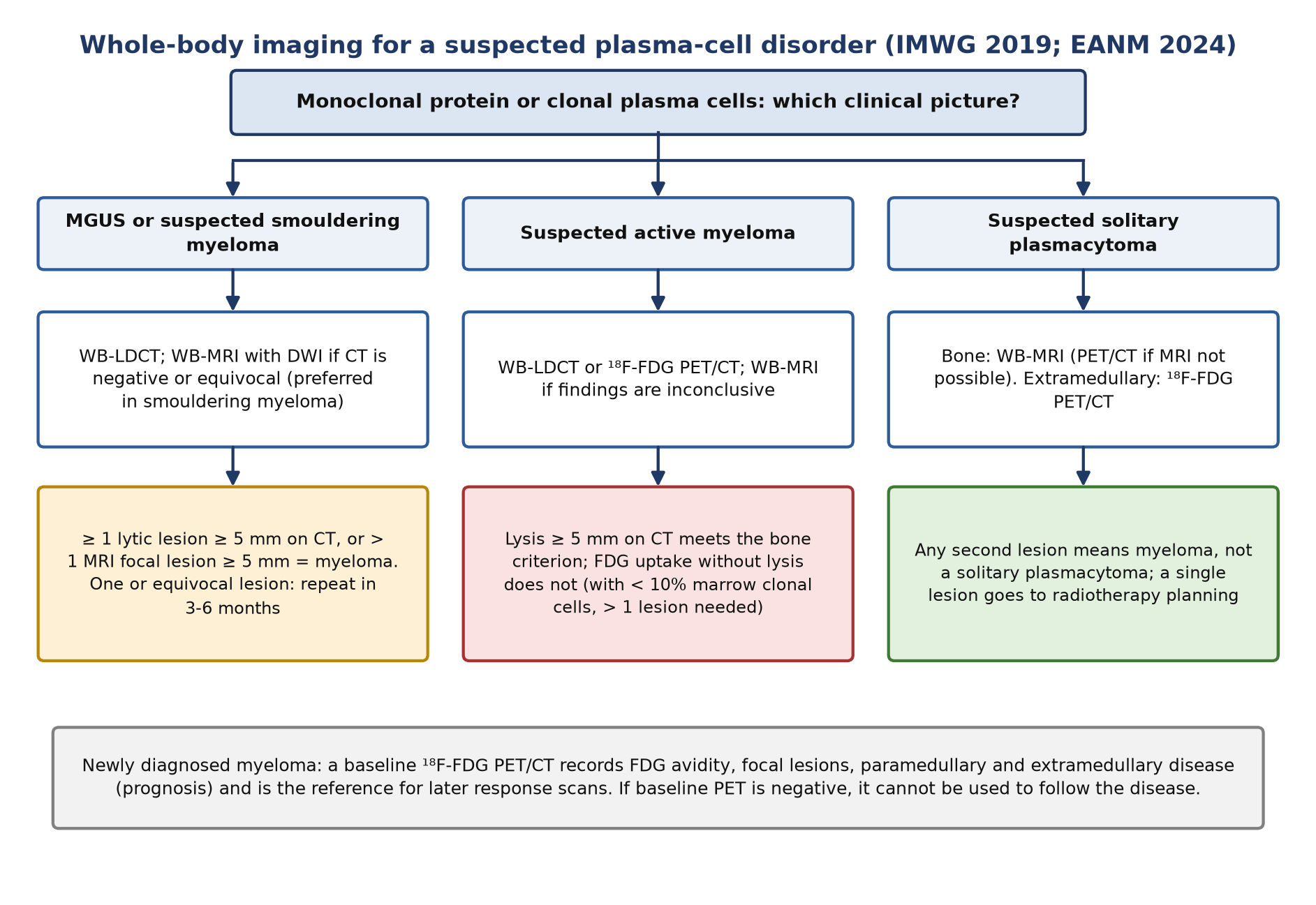
| Scenario | Recommended imaging | Source and strength |
|---|---|---|
| MGUS | WB-LDCT when imaging is indicated; no prospective data on PET/CT (in 338 patients, 30 had myeloma bone lesions, mainly with a higher M-protein) | IMWG 2019; EANM 2024 (consensus) |
| Suspected smouldering myeloma | WB-LDCT, then whole-body MRI (preferred); FDG PET/CT to separate smouldering from active disease if MRI is unavailable | IMWG 2014, 2017 and 2019 (consensus) |
| Suspected multiple myeloma | WB-LDCT or FDG PET/CT; whole-body MRI if inconclusive | IMWG 2019; ESMO and European Myeloma Network: WB-LDCT first; NICE: whole-body MRI first |
| Newly diagnosed myeloma, baseline | FDG PET/CT to record avidity, focal lesions, paramedullary and extramedullary disease and to serve as the response baseline | IMWG 2017; EANM 2024 |
| Suspected solitary bone plasmacytoma | Whole-body MRI; FDG PET/CT is mandatory if whole-body MRI cannot be done | IMWG 2017 and 2019 |
| Suspected extramedullary plasmacytoma | FDG PET/CT | IMWG 2019 |
| Non-secretory myeloma | Whole-body MRI or FDG PET/CT to follow disease that cannot be measured in blood | NICE (as summarised by EANM 2024) |
| Response assessment | FDG PET/CT is the preferred technique; pre-maintenance scan has the strongest data | IMWG 2017; EHA–ESMO 2021 and EANM 2024 |
| Suspected relapse | WB-LDCT (or targeted CT); MRI if negative; FDG PET/CT optional, useful for active versus healed lesions and extramedullary disease | IMWG 2019; EHA–ESMO 2021 (per EANM 2024) |
| Follow-up | Yearly FDG PET/CT if the post-treatment scan was positive; yearly PET/CT in marrow-MRD-negative patients to confirm extramedullary negativity; same technique each time | IMWG; EHA–ESMO 2021 (per EANM 2024) |
IMWG and EANM statements are consensus recommendations without formal grades. Guidelines disagree on the first test at diagnosis: IMWG, ESMO and the European Myeloma Network start with WB-LDCT (PET/CT acceptable), NICE starts with whole-body MRI.
4. Evidence at a glance
| Study | Design, n | Key finding | What it changed |
|---|---|---|---|
| Bartel 2009 (Total Therapy 3) | Prospective, 239 newly diagnosed | More than 3 FDG-avid focal lesions was the leading independent adverse factor for overall and event-free survival; complete FDG suppression before first transplant conferred better outcome | Established focal-lesion count and FDG suppression as prognostic |
| Zamagni 2011 | Prospective, 192 transplant-eligible | At baseline, ≥ 3 focal lesions (44%), SUVmax > 4.2 (46%) and extramedullary disease (6%) predicted shorter PFS (4-year 50%, 43% and 28%); PET negative 3 months after transplant in 65%, with better PFS and OS | SUVmax > 4.2 and extramedullary disease as baseline risk markers |
| Rasche 2017 | 227 newly diagnosed, FDG PET and DWI-MRI | 11% FDG false-negative; hexokinase-2 expression 5.3-fold lower in these cases | Explains FDG-negative myeloma |
| Siontis 2015 | Retrospective, 122 smouldering patients observed | Progression within 2 years: 75% if PET/CT positive vs 30% if negative; 87% with osteolysis, 61% with uptake but no osteolysis | Supported adding CT/PET-CT lysis to the IMWG criteria |
| Zamagni 2016 | Prospective, 120 smouldering patients | PET/CT positive in 16%; 2-year progression 58% vs 33%; median time to progression 1.1 vs 4.5 years; lesions had no osteolysis | PET-positive smouldering myeloma is high risk, but uptake without lysis is still not a defining event |
| IMAJEM (Moreau 2017) | Prospective, 134, within IFM/DFCI 2009 | MRI positive 95% vs PET/CT 91% at diagnosis; PET normalisation after 3 cycles (32%) gave 30-month PFS 78.7% vs 56.8%; normalisation before maintenance (62%) predicted PFS and OS; MRI normalisation did not | PET/CT, not MRI, for response |
| Zamagni 2021 (IFM 2009 + EMN02/HO95) | Pooled, 228 transplant-eligible | Focal-lesion and marrow uptake below liver (Deauville < 4) before maintenance independently predicted longer PFS and OS | Standard definition of complete metabolic response |
| Nanni 2018 (IMPeTUs) | 86 patients, 211 scans, 5 blinded readers | Agreement above 75% at all time points; Deauville score 4 most reproducible | Reproducible reporting for trials |
| FORTE imaging (Zamagni 2023) | 109 transplant-eligible, paired PET and flow MRD | Complete metabolic response before maintenance in 63%; PFS HR 0.40 (univariate); PET CMR plus flow-MRD negativity: PFS HR 0.41 (multivariate) | Validated Deauville criteria; PET complements marrow MRD |
| CASSIOPET (Kraeber-Bodéré 2025) | 225 with pre-maintenance PET, daratumumab era | 92% of 175 baseline-positive patients became PET-negative; PET negativity predicted PFS; double PET and flow-MRD negativity gave the best PFS | PET response keeps its value with anti-CD38 therapy |
5. Patient preparation and acquisition
Fasting, glucose checks and uptake conditions follow the EANM tumour-imaging procedure. The myeloma-specific points come from the 2024 EANM plasma-cell guideline (Nanni 2024).
- Field of view: vertex to toes, or at least to the knees, because myeloma involves the skull and appendicular skeleton. Acquire the PET from the feet upwards so the bladder is scanned soon after voiding.
- Arms down: arms alongside the body (humeri are common disease sites), on foam supports to limit beam-hardening across the spine; avoid CT truncation, using an extended field of view if available.
- CT component: WB-LDCT without contrast is the standard; the IMWG Bone Working Group WB-LDCT parameters apply to the CT of the PET/CT so that the lysis criterion can be read from it. If contrast is needed, check renal function and hydrate: dehydration promotes light-chain precipitation and acute kidney injury.
- History to record: myeloma type, known extramedullary sites, date and type of last treatment, corticosteroids and growth factors (erythropoietin, G-CSF), fractures, vertebroplasty and orthopaedic hardware, infection, and earlier imaging for comparison.
- Positioning and pain: bone pain is common; give analgesia and position carefully, as fractures and cord compression are real risks.
- Reconstruction: IMPeTUs and the Deauville response criteria were developed on OSEM reconstructions; EANM advises not applying them to time-of-flight or other noise-optimised algorithms. Keep the reconstruction constant between baseline and follow-up.
- Reference regions: liver region of at least 3 cm radius in the central right lobe away from the edges; mediastinal blood pool inside the aortic arch lumen, avoiding the wall and calcification.
- Check before the patient leaves: urgent findings such as impending fracture or spinal cord compression need same-day communication.
6. Interpretation
What counts as disease
| Finding | Definition (EANM 2024, after IMPeTUs) |
|---|---|
| Focal lesion (FL) | Visually detectable focal uptake above the surrounding marrow, in the skeleton, on 2 or more adjacent slices, with or without osteolysis on CT |
| Diffuse marrow involvement | Diffuse uptake in the axial skeleton above liver, homogeneous or heterogeneous, which may extend to the limbs |
| Paramedullary disease (PMD) | Soft tissue contiguous with involved bone (a lesion breaking out through the cortex) |
| Extramedullary disease (EMD) | Abnormal soft tissue without contiguous bone involvement (nodes, skin, liver, pleura and others) |
| Lytic lesion | Osteolysis on the CT; 5 mm or more meets the IMWG bone criterion |
The Deauville scale as used in myeloma
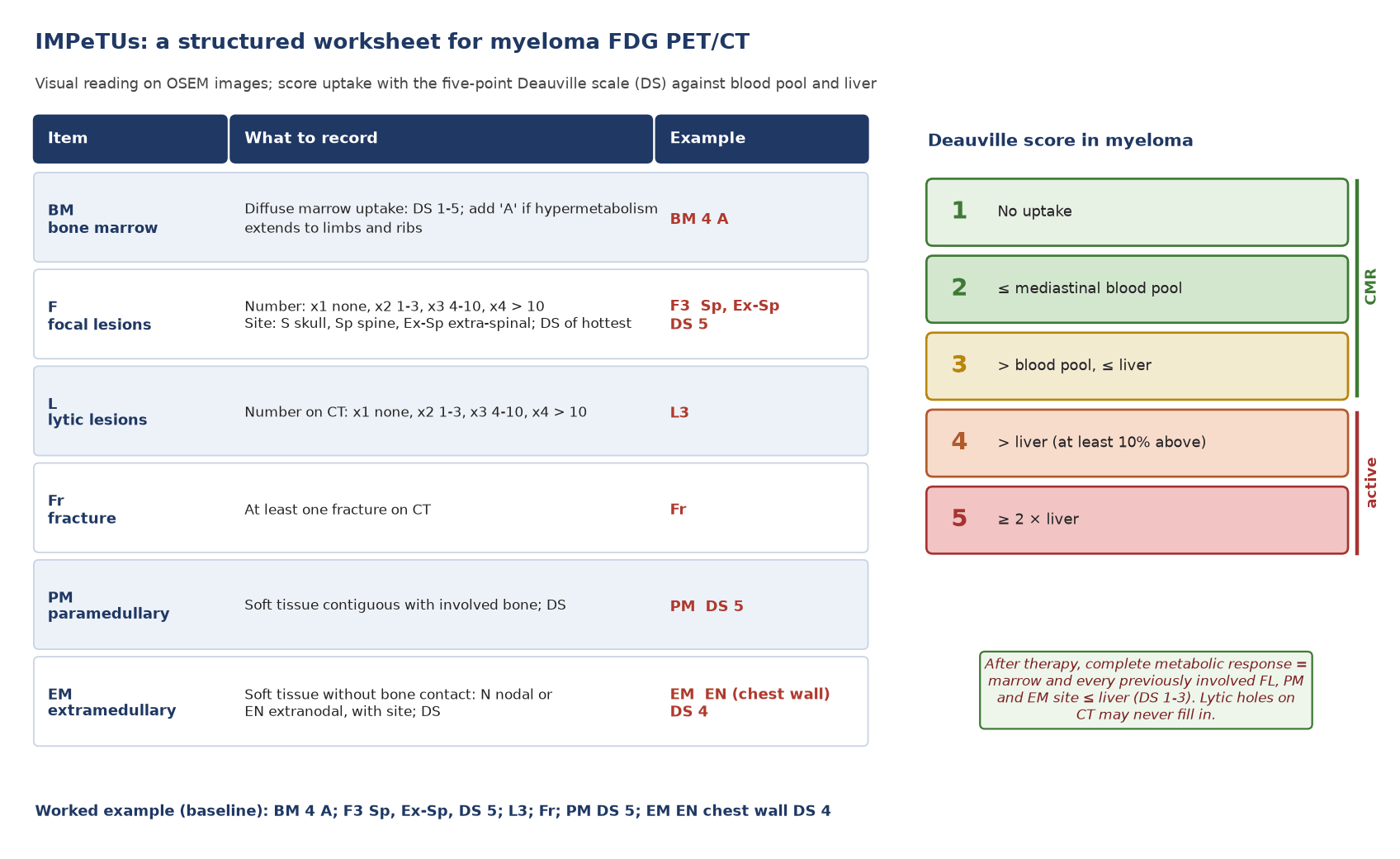
The EANM guideline defines the five points for myeloma as: 1 no uptake; 2 at or below the mediastinal blood pool; 3 above blood pool but at or below liver; 4 above liver (at least 10% more); 5 markedly above liver (at least twice). Note that score 5 is anchored at twice the liver, which differs slightly from lymphoma practice.
IMPeTUs
The Italian Myeloma criteria for PET Use (IMPeTUs) were agreed within the EMN02 trial as a purely visual, descriptive system (Nanni 2016) and proved reproducible in 86 patients and 211 scans read by five blinded reviewers (Nanni 2018). Lesion-to-background SUV ratios may support, but do not replace, the visual score.
| Item | What is recorded | Grading |
|---|---|---|
| BM (bone marrow) | Diffuse marrow uptake; add 'A' if hypermetabolism extends to limbs and ribs | Deauville 1–5 |
| F (focal lesions) | Number: x1 none, x2 1–3, x3 4–10, x4 more than 10; site: S skull, Sp spine, Ex-Sp extra-spinal | Deauville score of the hottest lesion |
| L (lytic lesions) | Number on CT: x1 none, x2 1–3, x3 4–10, x4 more than 10 | None |
| Fr (fracture) | At least one fracture on CT | None |
| PM (paramedullary) | At least one | Deauville 1–5 |
| EM (extramedullary) | At least one; N nodal or EN extranodal, with site | Deauville 1–5 |
IMPeTUs items (Nanni 2016; EANM 2024 Table 1).
Baseline findings that carry prognosis
- Number of focal lesions: more than 3 FDG-avid lesions (Bartel 2009) or 3 or more (Zamagni 2011) predicted shorter survival.
- Intensity: a focal-lesion SUVmax above 4.2 predicted shorter PFS and OS, and its persistence after induction predicted early progression (Zamagni 2011). A pooled analysis cited by EANM found bone SUVmax to be the only independent PET factor, with an OS threshold of 7.1; thresholds are cohort-dependent.
- Extramedullary disease: present in 5–11% at diagnosis and more than 20% at relapse; independently predicted PFS and OS in IMAJEM.
- Paramedullary disease: reported in 18% of the CASSIOPET baseline population and associated with shorter PFS (EANM summary of CASSIOPET).
- A normal baseline PET: better prognosis than a positive one, but it also means FDG cannot be used for response.
- Volumetric measures: total metabolic tumour volume and total lesion glycolysis are prognostic in some series, but cut-offs differ widely and segmentation is not standardised; not for routine reports.
Smouldering myeloma and MGUS
A positive PET in smouldering myeloma predicts progression: 2-year rates of 75% vs 30% (Siontis 2015) and 58% vs 33% (Zamagni 2016). Focal uptake with underlying lysis of 5 mm or more is a myeloma-defining event. Focal uptake without lysis is not, however high the risk: report it clearly, recommend whole-body MRI (where more than one focal lesion of 5 mm or more would define myeloma) and close follow-up, and let the haematologist decide.
Solitary plasmacytoma and radiotherapy planning
Before radiotherapy, whole-body imaging must exclude other lesions: whole-body MRI for a bony lesion and FDG PET/CT for an extramedullary one, with PET/CT required whenever whole-body MRI cannot be done (Cavo 2017). PET/CT finds lesions outside a spine-and-pelvis MRI field. Two or more focal lesions on PET/CT at diagnosis predicted a shorter time to myeloma. For planning, the International Lymphoma Radiation Oncology Group recommends 35–40 Gy for solitary bone plasmacytomas under 5 cm and 40–50 Gy for extramedullary plasmacytomas (Tsang 2018); PET/CT and MRI help define the gross tumour volume, particularly soft-tissue extension. Follow-up is yearly for 5 years with the same technique used at diagnosis.
POEMS syndrome and AL amyloidosis
- POEMS: polyneuropathy and a monoclonal plasma-cell disorder (almost always lambda) plus a major criterion, one of which is sclerotic bone lesions (IMWG 2014). In 90 patients, FDG PET/CT showed 140 avid bone lesions, mostly mixed lytic-sclerotic and often in the pelvis, with hepatosplenomegaly in 65 and serous effusions in 46; lesion metabolism fell 3 months after therapy (Pan 2015). Read sclerotic lesions on the CT carefully: they are part of the diagnosis, not an incidental finding.
- AL amyloidosis: cardiac uptake of bone-seeking tracers (⁹⁹ᵐTc-DPD, -PYP, -HMDP) is over 99% sensitive for transthyretin (ATTR) amyloid, but false positives come almost entirely from AL amyloid. Grade 2–3 uptake allows a non-biopsy diagnosis of ATTR only when serum and urine show no monoclonal protein (Gillmore 2016). In a patient with a plasma-cell disorder, a positive scan needs tissue typing. See the article on nuclear medicine in cardiac amyloidosis.
Pitfalls and mimics
| Pitfall | Typical pattern | How to resolve |
|---|---|---|
| Anaemia, erythropoietin, G-CSF | Diffuse, homogeneous marrow uptake that lowers lesion contrast and mimics diffuse infiltration | Record growth factors and haemoglobin; marrow Deauville 4 or more with limb involvement makes infiltration more likely |
| Recent fracture | Linear or band-like uptake along a fracture line; may take at least a month to settle after trauma | Use the CT; pathological fractures sit in lytic bone |
| Vertebroplasty, kyphoplasty and metalwork | Uptake at the cement or implant interface; CT streak artefact | Check non-attenuation-corrected images; healing or infection can both cause uptake |
| Degenerative change | Uptake at osteophytes, facet joints, tendons and recent Schmorl nodes (sclerotic rim, contact with the disc) | Read shape and site on CT; common in a population with median age near 70 |
| Infection and inflammation | Soft-tissue or nodal uptake that mimics extramedullary disease; patients are immunosuppressed | Correlate clinically; biopsy or targeted imaging when management depends on it |
| Medication-related osteonecrosis of the jaw | Moderately increased FDG uptake with medullary sclerosis in the mandible or maxilla after bisphosphonates or denosumab | Dental history; do not call a jaw focus myeloma without lysis or soft tissue |
| FDG-negative myeloma | Lytic lesions on CT or marrow disease without uptake (10–20%) | State that the scan cannot be used for response; use MRI or WB-LDCT for follow-up |
| Healed lytic lesions | Persistent holes on CT without uptake after therapy | Not active disease; lytic lesions rarely fill in, so CT size alone cannot judge response |
| Immunotherapy effects | Immune-related uptake with bispecific antibodies or CAR-T | Interpret early changes with caution (EANM) |
Sources: EANM 2024; Nanni 2020 (anaemia, erythropoietin, vertebroplasty, metalwork); Reinert 2024 (osteonecrosis of the jaw).
Wording the conclusion
- Give the IMPeTUs string, then translate it: 'more than 10 FDG-avid focal lesions, the hottest Deauville 5 (SUVmax 9.8) at L3; paramedullary extension at T11; no extramedullary disease'.
- State whether there is lysis of 5 mm or more on CT, because that, not the uptake, answers the diagnostic question.
- Name the baseline risk features that matter (more than 3 focal lesions, SUVmax above 4.2, paramedullary or extramedullary disease) and flag urgent findings (fracture risk, cord or root compression).
- If the scan is FDG-negative, say so explicitly and recommend another test for future response assessment.
7. Response assessment and follow-up
Two definitions of PET negativity
- IMWG 2016 (imaging MRD): MRD negativity in the marrow plus disappearance of every area of increased uptake found at baseline or a preceding PET/CT, or a decrease to less than the mediastinal blood pool SUV, or to less than surrounding normal tissue (Kumar 2016).
- Deauville-based complete metabolic response (Zamagni 2021; EANM 2024): uptake at or below liver (Deauville 1–3) in the marrow and in all previously involved focal lesions, paramedullary and extramedullary sites. This threshold was chosen a posteriori from outcome data in 228 patients and validated in FORTE and CASSIOPET; EANM recommends it, while noting it still needs further external validation before formal inclusion in the IMWG criteria.
The two are not the same: a lesion between blood pool and liver (Deauville 3) is residual uptake under the IMWG 2016 wording but a complete metabolic response under the Deauville criteria. Say which definition you applied.
| Category | Definition (IMPeTUs-based, EANM 2024) |
|---|---|
| Complete metabolic response | Uptake ≤ liver (Deauville 1–3) in marrow and all previously involved focal, paramedullary and extramedullary sites |
| Partial metabolic response | Fewer lesions and/or lower uptake, but at least one lesion still above liver (Deauville 4–5) |
| Stable metabolic disease | No significant change in marrow or previously involved sites compared with baseline |
| Progressive metabolic disease | New focal, paramedullary or extramedullary lesions consistent with myeloma |
PET and marrow MRD are complementary
Marrow MRD samples one site; myeloma is patchy and can persist in focal lesions or outside the bone. In FORTE, patients with both a complete metabolic response and flow-cytometry MRD negativity before maintenance had the longest PFS (HR 0.41). In CASSIOPET, double negativity again gave the best PFS in the daratumumab era. MRD-negative but PET-positive patients are few (about 5–7%) but relapse earlier, and in the PETHEMA/GEM2012 trial six patients progressed with extramedullary plasmacytomas on PET despite undetectable marrow MRD and no M-protein (EANM 2024).
Timing and consequences
- There is no consensus on scan timing; the strongest data are for a scan after induction and transplant, before maintenance.
- Scan against a baseline FDG PET/CT with the same reconstruction; response cannot be judged if the baseline was FDG-negative (IMWG then advises a WB-LDCT at the end of treatment as the new reference).
- IMWG: unless there is progression, treatment should not be changed on post-treatment imaging alone.
- Progression on CT (IMWG 2016): new osteolytic lesions; a 50% or greater increase from nadir in the sum of the products of diameters of more than one lesion; or a 50% or greater increase in the longest diameter of a previous lesion over 1 cm in short axis.
Relapse and surveillance
At suspected relapse, WB-LDCT is the standard, with MRI if it is negative; FDG PET/CT is optional but separates active from healed lesions and shows extramedullary disease, which is more common at relapse. For follow-up, IMWG advises yearly PET/CT when the post-treatment scan was positive, and EHA–ESMO advises yearly PET/CT in marrow-MRD-negative patients to confirm extramedullary negativity. Smouldering myeloma is followed with yearly spine-and-pelvis or whole-body MRI for at least 5 years, with PET/CT if MRI is not feasible.
8. Theranostics and emerging tracers
| Agent | Target and data | Evidence level (2026) |
|---|---|---|
| ⁶⁸Ga-pentixafor PET | CXCR4 chemokine receptor, overexpressed on myeloma cells. Prospective newly diagnosed cohort: positive in 28/30 (93%) vs 16/30 (53%) with FDG; retrospective 34 patients: more disease than FDG in 68%, stage changed in 41% | Investigational |
| ¹⁷⁷Lu- or ⁹⁰Y-pentixather radioligand therapy | CXCR4-directed therapy given with chemotherapy as conditioning before autologous stem-cell rescue, because CXCR4 is also on haematopoietic cells. In 8 heavily pretreated patients: 1 complete and 5 partial remissions, median PFS 54 days, one death from sepsis and one from tumour lysis | Investigational (compassionate use, small series) |
| ⁸⁹Zr-DFO-daratumumab immuno-PET | CD38 (the daratumumab target). Phase I, 10 patients, 74 MBq, four scans over 8 days: safe, with uptake at osseous myeloma sites | Investigational |
| ¹¹C-methionine, ¹¹C-acetate | Amino acid and lipid metabolism; more lesions than FDG in comparative series | Specialist use in FDG-negative disease (EANM); not widely available |
| ⁶⁸Ga-FAPI | Fibroblast activation protein in the tumour stroma. Retrospective 14 patients: extra lesions in some but no overall superiority over FDG | Investigational |
No radiopharmaceutical is approved for imaging or treating myeloma in 2026. CXCR4-directed therapy is marrow-ablative by design, so it belongs in transplant-centre protocols.
9. Structured report example
- Clinical question: baseline before induction and transplant; lytic disease and extramedullary spread?
- Technique: ¹⁸F-FDG 250 MBq, uptake 60 min, glucose 5.4 mmol/L; vertex to feet, arms down, low-dose CT; OSEM reconstruction. No growth factors; haemoglobin 98 g/L.
- Reference regions: mediastinal blood pool SUVmax 1.8; liver SUVmax 2.6.
- Findings: more than 10 FDG-avid focal lesions in skull, spine, ribs, pelvis and both femora, hottest at L2 (SUVmax 8.9, Deauville 5), most with lysis of 5 mm or more on CT. Soft tissue extends from T11 into the left paravertebral space (SUVmax 9.4). A 3 cm right chest-wall soft-tissue mass without bone contact (SUVmax 6.2). Diffuse marrow uptake just above liver, not in the limbs. Old healed fracture of the left 8th rib. No cord compression.
- IMPeTUs: BM 4; F4 S, Sp, Ex-Sp, DS 5; L4; Fr; PM DS 5; EM EN (chest wall) DS 5.
- Conclusion: FDG-avid myeloma with multiple lytic lesions (meets the IMWG bone criterion), paramedullary disease at T11 and an extramedullary chest-wall deposit: adverse PET features (more than 3 focal lesions, SUVmax above 4.2, paramedullary and extramedullary disease). Suitable baseline for response assessment; the chest-wall mass is the most accessible site if histology is needed.
10. Take-home points
- Lysis of 5 mm or more on CT meets the IMWG bone criterion; FDG uptake without lysis does not, however suspicious.
- WB-LDCT is the first bone survey, whole-body MRI is best for smouldering myeloma and bony plasmacytoma, and FDG PET/CT is the test for response and extramedullary disease. Do not use a bone scan.
- Report with IMPeTUs and the myeloma Deauville scale; name the hottest site, the paramedullary and extramedullary disease, and fractures.
- More than 3 focal lesions, SUVmax above 4.2 and paramedullary or extramedullary disease are adverse at baseline.
- Complete metabolic response means marrow and all previous sites at or below liver; combine it with marrow MRD.
- Check baseline avidity: 10–20% of myelomas are FDG-negative and need another test for follow-up.
- In a patient with a monoclonal protein, cardiac uptake on a bone scan does not diagnose ATTR amyloid.
Test yourself
5 quick questions. Pick an answer to see the explanation.
References
- Rajkumar SV, Dimopoulos MA, Palumbo A, et al. International Myeloma Working Group updated criteria for the diagnosis of multiple myeloma. Lancet Oncol. 2014;15(12):e538-48.
- Cavo M, Terpos E, Nanni C, et al. Role of 18F-FDG PET/CT in the diagnosis and management of multiple myeloma and other plasma cell disorders: a consensus statement by the International Myeloma Working Group. Lancet Oncol. 2017;18(4):e206-17.
- Hillengass J, Usmani S, Rajkumar SV, et al. International myeloma working group consensus recommendations on imaging in monoclonal plasma cell disorders. Lancet Oncol. 2019;20(6):e302-12.
- Kumar S, Paiva B, Anderson KC, et al. International Myeloma Working Group consensus criteria for response and minimal residual disease assessment in multiple myeloma. Lancet Oncol. 2016;17(8):e328-46.
- Nanni C, Deroose CM, Balogova S, et al. EANM guidelines on the use of [18F]FDG PET/CT in diagnosis, staging, prognostication, therapy assessment, and restaging of plasma cell disorders. Eur J Nucl Med Mol Imaging. 2024;52(1):171-92.
- Nanni C, Zamagni E, Versari A, et al. Image interpretation criteria for FDG PET/CT in multiple myeloma: a new proposal from an Italian expert panel. IMPeTUs (Italian Myeloma criteria for PET USe). Eur J Nucl Med Mol Imaging. 2016;43(3):414-21.
- Nanni C, Versari A, Chauvie S, et al. Interpretation criteria for FDG PET/CT in multiple myeloma (IMPeTUs): final results. IMPeTUs (Italian myeloma criteria for PET USe). Eur J Nucl Med Mol Imaging. 2018;45(5):712-9.
- Nanni C. PET-FDG: Impetus. Cancers (Basel). 2020;12(4):1030.
- Zamagni E, Nanni C, Dozza L, et al. Standardization of 18F-FDG-PET/CT according to Deauville criteria for metabolic complete response definition in newly diagnosed multiple myeloma. J Clin Oncol. 2021;39(2):116-25.
- Bartel TB, Haessler J, Brown TL, et al. F18-fluorodeoxyglucose positron emission tomography in the context of other imaging techniques and prognostic factors in multiple myeloma. Blood. 2009;114(10):2068-76.
- Zamagni E, Patriarca F, Nanni C, et al. Prognostic relevance of 18-F FDG PET/CT in newly diagnosed multiple myeloma patients treated with up-front autologous transplantation. Blood. 2011;118(23):5989-95.
- Moreau P, Attal M, Caillot D, et al. Prospective evaluation of magnetic resonance imaging and [18F]fluorodeoxyglucose positron emission tomography-computed tomography at diagnosis and before maintenance therapy in symptomatic patients with multiple myeloma included in the IFM/DFCI 2009 trial: results of the IMAJEM study. J Clin Oncol. 2017;35(25):2911-8.
- Zamagni E, Oliva S, Gay F, et al. Impact of minimal residual disease standardised assessment by FDG-PET/CT in transplant-eligible patients with newly diagnosed multiple myeloma enrolled in the imaging sub-study of the FORTE trial. EClinicalMedicine. 2023;60:102017.
- Kraeber-Bodéré F, Jamet B, Zweegman S, et al. Prognostic value of premaintenance FDG PET/CT response in patients with newly diagnosed myeloma from the CASSIOPEIA trial. Blood. 2025;146(25):3050-8.
- Rasche L, Angtuaco E, McDonald JE, et al. Low expression of hexokinase-2 is associated with false-negative FDG-positron emission tomography in multiple myeloma. Blood. 2017;130(1):30-4.
- Siontis B, Kumar S, Dispenzieri A, et al. Positron emission tomography-computed tomography in the diagnostic evaluation of smoldering multiple myeloma: identification of patients needing therapy. Blood Cancer J. 2015;5(10):e364.
- Zamagni E, Nanni C, Gay F, et al. 18F-FDG PET/CT focal, but not osteolytic, lesions predict the progression of smoldering myeloma to active disease. Leukemia. 2016;30(2):417-22.
- Dimopoulos M, Terpos E, Comenzo RL, et al. International myeloma working group consensus statement and guidelines regarding the current role of imaging techniques in the diagnosis and monitoring of multiple myeloma. Leukemia. 2009;23(9):1545-56.
- Tsang RW, Campbell BA, Goda JS, et al. Radiation therapy for solitary plasmacytoma and multiple myeloma: guidelines from the International Lymphoma Radiation Oncology Group. Int J Radiat Oncol Biol Phys. 2018;101(4):794-808.
- Pan Q, Li J, Li F, et al. Characterizing POEMS syndrome with 18F-FDG PET/CT. J Nucl Med. 2015;56(9):1334-7.
- Gillmore JD, Maurer MS, Falk RH, et al. Nonbiopsy diagnosis of cardiac transthyretin amyloidosis. Circulation. 2016;133(24):2404-12.
- Lapa C, Herrmann K, Schirbel A, et al. CXCR4-directed endoradiotherapy induces high response rates in extramedullary relapsed multiple myeloma. Theranostics. 2017;7(6):1589-97.
- Ulaner GA, Sobol NB, O'Donoghue JA, et al. CD38-targeted immuno-PET of multiple myeloma: from xenograft models to first-in-human imaging. Radiology. 2020;295(3):606-15.
- Elboga U, Sahin E, Cayirli YB, et al. Comparison of [68Ga]-FAPI PET/CT and [18F]-FDG PET/CT in multiple myeloma: clinical experience. Tomography. 2022;8(1):293-302.
- Reinert CP, Pfannenberg C, Gückel B, et al. Preoperative assessment of medication-related osteonecrosis of the jaw using [18F]fluoride positron emission tomography (PET)/CT and [18F]fluorodeoxyglucose PET/MRI in correlation with histomorphometry and micro-CT: a prospective comparative study. Diagnostics (Basel). 2024;14(4):428.
- Paraprotein and low back pain in a 62-year-old · Intermediate