Nuclear Medicine in Paediatric Oncology
- Children are not small adults. Tumour types, normal variants (thymus, physes, brown fat, Waldeyer's ring) and radiation risk all differ; every scan must be justified and optimised.
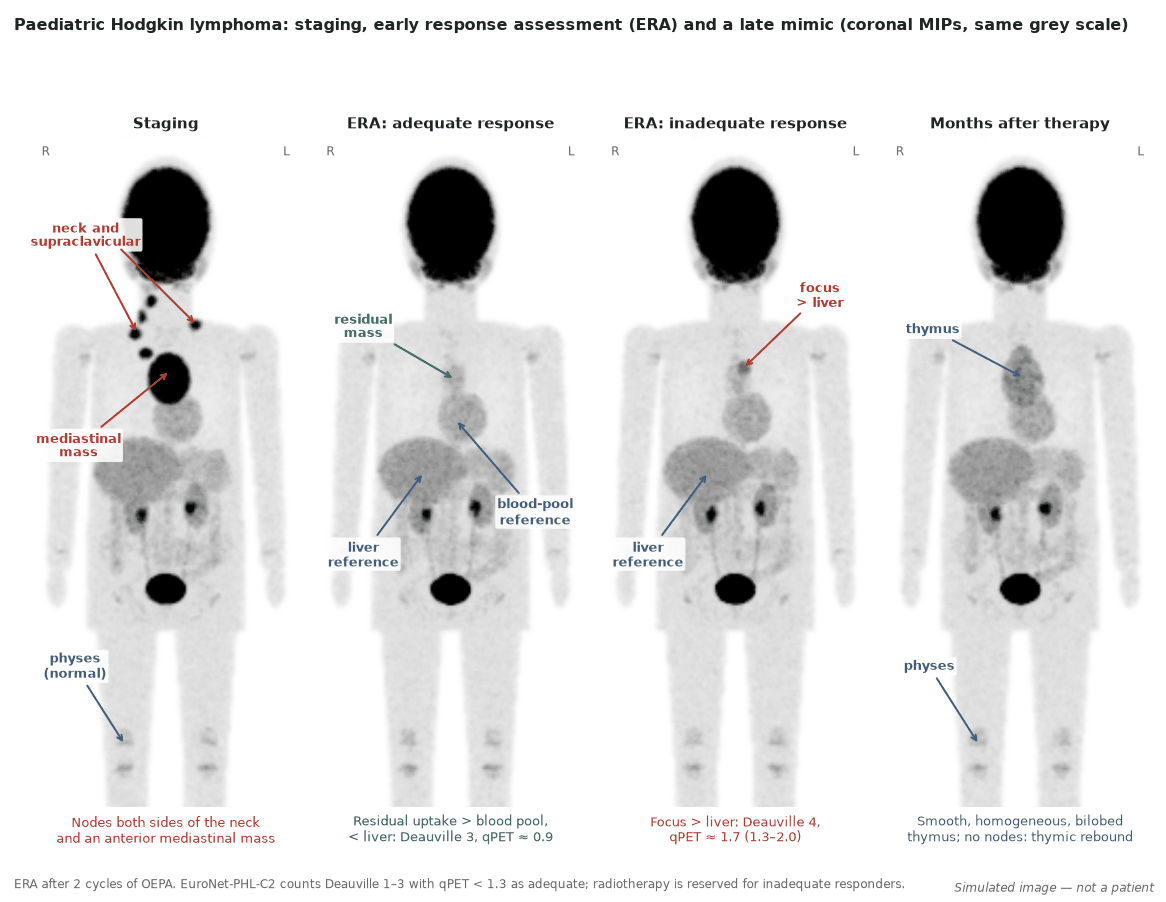
- Hodgkin lymphoma: PET decides radiotherapy. In EuroNet-PHL-C1, 40% of intermediate- and advanced-stage patients with an adequate early response were spared radiotherapy with 5-year event-free survival of 90.1%; EuroNet-PHL-C2 made Deauville 1–3 with qPET below 1.3 the definition of adequate response.
- qPET adds to Deauville. qPET (residual lesion SUVpeak divided by mean liver SUV) splits Deauville 5 into a large group with good outcome and a small group above 3 with 5-year event-free survival of only 47.6%.
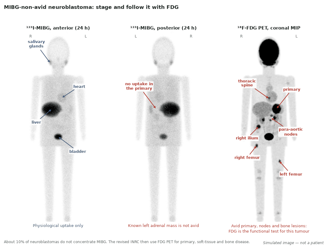
- Neuroblastoma: MIBG first, FDG when MIBG fails. ¹²³I-MIBG with SIOPEN or Curie scoring stages and follows about 90% of tumours; FDG replaces it for MIBG-non-avid disease under the revised response criteria.
- Scores carry prognosis. After induction, a SIOPEN score above 3 or a Curie score above 2 marks very poor event-free survival.
- Sarcoma and LCH. FDG PET/CT is recommended for distant and nodal staging of rhabdomyosarcoma; in Langerhans cell histiocytosis it is the most sensitive functional test, although guidelines still stage bone with the skeletal survey.
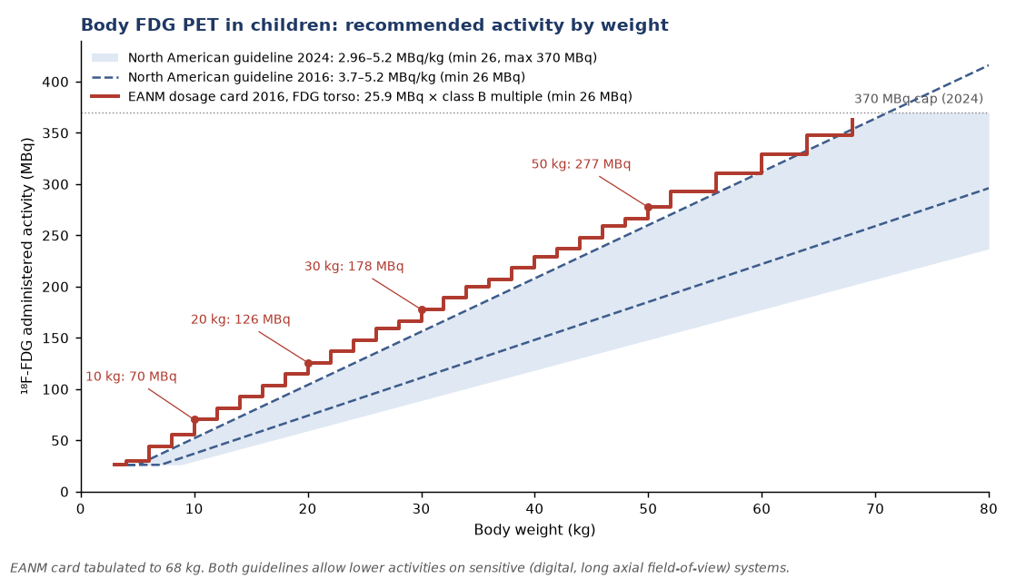
- Dose by weight, not by habit. The 2024 North American guideline gives body FDG 2.96–5.2 MBq/kg (minimum 26, maximum 370 MBq); the EANM card uses 25.9 MBq × a class B weight multiple. Match the CT to the question, and consider PET/MRI.
1. The clinical problem
Childhood cancer is rare, biologically distinct from adult cancer and mostly curable. Survival now depends less on finding one more lesion than on giving each child just enough treatment: enough to cure, and no more, because survivors carry decades of risk from radiotherapy, anthracyclines and alkylating agents. Imaging therefore has three jobs: stage accurately enough to assign a risk group, show early whether treatment is working so that it can be reduced or intensified, and do so with the lowest radiation burden that answers the question.
Staging systems in force
| Tumour | System | What nuclear medicine contributes |
|---|---|---|
| Hodgkin lymphoma (HL) | Ann Arbor stage plus risk factors; EuroNet groups early, intermediate and advanced disease into treatment groups or levels (TG/TL 1–3) | FDG PET/CT staging and early response assessment (ERA) after 2 cycles of OEPA |
| Neuroblastoma | International Neuroblastoma Risk Group Staging System (INRGSS, 2009): L1, L2, M, MS | ¹²³I-MIBG (or FDG if non-avid) defines stage M and scores skeletal disease |
| Rhabdomyosarcoma | European (EpSSG/CWS) risk grouping, in which nodal and distant metastases change the group | FDG PET/CT with chest CT for nodal and distant disease |
| Ewing sarcoma, osteosarcoma | Localised versus metastatic; site of metastases | FDG PET/CT for bone and nodal metastases; chest CT remains the test for lung nodules |
| Langerhans cell histiocytosis (LCH) | Single-system versus multisystem; risk organs (haematopoietic system, liver, spleen); CNS-risk lesions | FDG PET/CT maps active lesions and early response |
INRGSS after Monclair 2009; LCH categories after Haupt 2013.
INRGSS in one paragraph
The INRG staging system is applied before any treatment. Locoregional tumours are L1 if none of the 20 image-defined risk factors (IDRFs) is present and L2 if one or more is present; IDRFs are features on CT or MRI, such as vessel encasement or extension into the spinal canal, that make upfront resection hazardous. Distant metastases define M, except MS: metastases confined to skin, liver and/or bone marrow in children younger than 18 months. In the INRG validation cohort, 5-year event-free survival was 90% for L1 and 78% for L2. MIBG and FDG decide between locoregional and metastatic disease; IDRFs themselves are radiological (CT or MRI) findings.
Where nuclear medicine changes management
- In paediatric HL, the ERA PET after two cycles decides whether a child receives radiotherapy.
- In neuroblastoma, MIBG (or FDG) establishes stage M and the post-induction score predicts outcome and triggers alternative strategies.
- In rhabdomyosarcoma and Ewing sarcoma, FDG PET/CT upstages occult nodal and bone disease that changes radiotherapy fields and risk group.
- In LCH, FDG shows extent (single- versus multisystem) and early response, which decides whether systemic therapy continues.
2. Tracers and why they work
| Tracer | Mechanism | Main paediatric uses | Limits |
|---|---|---|---|
| ¹⁸F-FDG | Glucose transport and hexokinase trapping | HL staging and response; sarcomas; LCH; MIBG-non-avid neuroblastoma | Brown fat, thymus, physes, marrow after G-CSF, brain background |
| ¹²³I-MIBG | Norepinephrine transporter (NET) uptake into neural-crest cells | Neuroblastoma staging, response, selection for ¹³¹I-MIBG therapy | About 10% of neuroblastomas do not express NET; planar/SPECT resolution |
| ¹⁸F-MFBG | NET substrate labelled for PET | Neuroblastoma, in trials | Investigational; not yet a validated scoring basis |
| ⁶⁸Ga-DOTATATE | Somatostatin receptor type 2 | Neuroblastoma selected for PRRT; neuroendocrine tumours | Investigational in neuroblastoma |
| ¹⁸F-DOPA | L-amino-acid transport and decarboxylation | Neuroblastoma (research); paediatric gliomas | Striatal and pancreatic background; limited availability |
| ¹⁸F-FET, ¹¹C-MET, ¹⁸F-DOPA | Amino-acid transport | Paediatric gliomas: biopsy target, recurrence versus treatment effect | Needs co-registered MRI |
Avidity by tumour
- Hodgkin lymphoma: the inflammatory background around Hodgkin and Reed–Sternberg cells is intensely avid; FDG is the reference functional test.
- Neuroblastoma: about 90% of tumours express NET, so MIBG is both the imaging test and the theranostic partner of ¹³¹I-MIBG. FDG shows more of the disease in stage 1–2 tumours and in the chest, abdomen and pelvis, whereas MIBG detects more skeletal and marrow disease in stage 4 (Sharp 2009: 44 of 85 stage 4 scans showed more sites on MIBG, 11 on FDG).
- Sarcomas: Ewing sarcoma and rhabdomyosarcoma are usually avid; FDG is less sensitive than CT for small lung nodules.
- LCH: active lesions are avid, and uptake falls before radiographs change, which is why FDG detects early response.
- Wilms tumour and hepatoblastoma: FDG is not part of standard staging. Wilms tumour appears among the less common indications with some evidence in the SNMMI/EANM guideline; in AFP-secreting hepatoblastoma, serum alpha-fetoprotein detected every relapse before imaging and was more specific than CT or PET/CT, so PET is reserved for localising a confirmed biochemical relapse.
- Germ cell tumours: FDG may help staging and recurrence (SNMMI/EANM lists them among less common indications); tumour markers and cross-sectional imaging lead.
- CNS tumours: amino-acid PET plays a more pertinent role than FDG because normal brain takes up little amino acid; interpret only with co-registered MRI (joint EANM/SIOPE/RAPNO/SNMMI guideline 2022).
3. Indications by clinical scenario
| Tumour and scenario | Test | Position of guideline or protocol |
|---|---|---|
| HL: staging | FDG PET/CT, vertex to toes at baseline | SNMMI/EANM paediatric FDG guideline 2021 (consensus, no grades) |
| HL: early response after 2 cycles | FDG PET/CT scored with Deauville and qPET | EuroNet-PHL-C2 protocol: adequate response = Deauville 1–3 and qPET < 1.3; no radiotherapy |
| HL: surveillance in remission | Not indicated | Relapse is investigated when suspected; thymic regrowth alone is not relapse |
| Neuroblastoma: staging and response | ¹²³I-MIBG whole body, scored (SIOPEN or Curie) | EANM guideline 2018; revised INRC 2017: MIBG replaces bone scintigraphy for osteomedullary disease |
| Neuroblastoma: MIBG-non-avid | FDG PET/CT | Revised INRC 2017; SNMMI/EANM 2021 |
| Neuroblastoma: before ¹³¹I-MIBG therapy | ¹²³I-MIBG to confirm avidity | Phase II data; treatment within specialist centres |
| Rhabdomyosarcoma: staging | FDG PET/CT or PET/MRI plus chest CT; MRI for the primary | European (EpSSG/CWS/ESPR) imaging guideline 2021 recommends it |
| Ewing sarcoma and osteosarcoma: staging and response | FDG PET/CT, whole body | SNMMI/EANM 2021 (total-body coverage); response prognostic in cohorts |
| LCH: extent and response | FDG PET/CT or PET/MRI | Histiocyte Society 2013: skeletal survey remains mandatory; PET 'most sensitive' but not an alternative |
| Paediatric gliomas | Amino-acid PET (FET, DOPA, MET) with MRI | Joint EANM/SIOPE/RAPNO/SNMMI 2022 (consensus) |
| Wilms tumour, hepatoblastoma, germ cell tumours | FDG only for problem-solving | Not in standard staging; markers and MRI/CT lead |
Paediatric guidelines are consensus documents without graded recommendations; the paediatric FDG guideline states that its recommendations represent expert opinion.
4. Evidence at a glance
| Study | Design, n | Key finding | What it changed |
|---|---|---|---|
| EuroNet-PHL-C1, TG2/3 (Mauz-Körholz 2022) | Titration study with embedded randomisation; 1,287 per protocol | 514 (40%) with adequate response after 2 OEPA had no radiotherapy: 5-year EFS 90.1%. COPDAC vs COPP 86.1% vs 89.9% | Radiotherapy omitted after adequate ERA; dacarbazine replaced gonadotoxic procarbazine |
| EuroNet-PHL-C1, TG1 (Mauz-Körholz 2023) | Titration study; 713 early-stage | Adequate responders without radiotherapy: 5-year EFS 86.5%, below the 90% target; inadequate responders with 19.8 Gy: 88.6% | Omission acceptable overall, but children with risk factors may need more treatment |
| qPET in C1 (Kurch 2025) | Retrospective analysis of ERA scans, 1,447 | 5-year EFS about 89% if qPET < 1.3; 84.3% for 1.3–2; 83.1% for 2–3; 47.6% above 3 | Deauville 5 split into a large favourable group and a small high-risk group |
| EuroNet-PHL-C2 (Körholz, conference abstract) | Randomised, 2,249 evaluable TL2/3 | Adequate responders: EFS 96.0% with DECOPDAC-21 vs 91.2% with COPDAC-28; radiotherapy given to 12.8% vs 35.6% | Intensified consolidation reduces radiotherapy further; full publication awaited |
| Sharp 2009 | Retrospective, 113 paired MIBG/FDG scans | FDG better in stage 1–2; MIBG showed more sites in stage 4 (44 of 85 scans vs 11) | MIBG first; FDG for low stage, weakly avid disease and key decision points |
| Ladenstein 2018 (SIOPEN/HR-NBL1 and COG-A3973) | Validation, 341 + 216 | Post-induction SIOPEN ≤ 3: 5-year EFS 36% vs 14% (SIOPEN) and 43% vs 16% (COG) | SIOPEN > 3 after induction flags patients for alternative strategies |
| Yanik 2013 (COG-A3973) | Retrospective, 280 | Post-induction Curie > 2: 3-year EFS 15.4% vs 44.9% | Curie > 2 after induction is a poor-risk marker |
| Wang 2023 | Prospective, 40 children | ¹⁸F-MFBG found 784 lesions vs 532 on ¹²³I-MIBG SPECT/CT; 4 of 34 MIBG-negative patients were MFBG-positive | Supports PET-based NET imaging; not yet standard |
| Hawkins 2005 and 2009 | Retrospective, 36 Ewing and 40 osteosarcoma | SUV after neoadjuvant chemotherapy < 2.5: 4-year PFS 72% vs 27% (Ewing), 73% vs 39% (osteosarcoma) | Post-induction SUV is prognostic; concordance with histological response only 58–69% |
| Vaarwerk 2021 (Cochrane) | Systematic review, 2 studies, 36 children with rhabdomyosarcoma | Sensitivity 100% for bone and nodal metastases in both small studies; too few data for pooled estimates | FDG is recommended, but the evidence base is thin |
5. Patient preparation and acquisition
What follows is what differs in children; otherwise standard FDG tumour practice applies (SNMMI/EANM paediatric FDG guideline 2021).
- Explain and prepare the family: verbal and written information; measure height and weight on the day; place the cannula in advance with anaesthetic cream; use distraction.
- Fasting: 4–6 h before FDG, continued during uptake; plain water is encouraged unless the child is nil by mouth for anaesthesia. Glucose-containing fluids and parenteral nutrition are replaced by saline. Glucose should be below 11.1 mmol/L (200 mg/dL), ideally below 7.8 mmol/L.
- Sedation and anaesthesia: avoid where possible. When general anaesthesia is needed, the child is nil by mouth for 6–8 h and anaesthesia is given just before acquisition, not during uptake. Feed-and-wrap (feeding, then swaddling so the infant sleeps) suits studies that need no fast; it cannot be used unchanged for FDG, which needs the 4–6 h fast, so plan infant FDG scans with the anaesthetic team.
- Brown fat: commoner in children. Keep the uptake room at least 24 °C with a warm blanket and avoid cold for 1–2 days. Premedication options: oral propranolol 1 mg/kg (usually 20–40 mg, maximum 40 mg) 60–90 min before injection in children over 10 years; intravenous fentanyl under sedation monitoring; or oral diazepam for anxious children.
- Quiet uptake: no talking, playing, chewing or crying if avoidable; a pacifier is allowed but causes oral muscle uptake, so note it.
- Bladder: void just before acquisition; change the nappy; catheterisation is not routine, but catheter or furosemide 0.5–1 mg/kg (maximum 40 mg) helps with pelvic tumours.
- Timing after treatment: at least 10 days after chemotherapy and 2–3 months after radiotherapy are suggested; allow 3 weeks after long-acting G-CSF.
- Coverage: vertex to toes for baseline lymphoma, sarcoma, neuroblastoma and LCH; skull base to mid-thigh can suffice for lymphoma follow-up without bone or limb disease.
- Uptake time: 60 min, and the same at every follow-up scan so that SUVs and qPET remain comparable.
Administered activity
| Guideline | Body FDG | Brain FDG | Other paediatric PET tracers |
|---|---|---|---|
| North American consensus, 2016 (as quoted in the SNMMI/EANM 2021 guideline) | 3.7–5.2 MBq/kg, minimum 26 MBq | 3.7 MBq/kg, minimum 14 MBq | — |
| North American consensus, 2024 update | 2.96–5.2 MBq/kg, minimum 26 MBq, maximum 370 MBq | 1.85–3.7 MBq/kg, minimum 14, maximum 148 MBq | ¹⁸F-DOPA 2.96–5.92 MBq/kg (29.6–222 MBq); ⁶⁸Ga-DOTATATE 2.0 MBq/kg (14–200 MBq); ¹²³I-MIBG 5.2 MBq/kg (37–370 MBq) |
| EANM paediatric dosage card (version 5.7.2016) | Class B, baseline 25.9 MBq × weight multiple (e.g. 2.71 at 10 kg, 10.71 at 50 kg), minimum 26 MBq | Class B, baseline 14.0 MBq, minimum 14 MBq | ⁶⁸Ga peptides: class B, 12.8 MBq baseline, minimum 14; ¹²³I-MIBG: class B, 28.0 MBq, minimum 37 |
The 2024 North American update advises the low end for smaller patients and lower activities on digital or long axial field-of-view systems.
Radiation dose and ALARA
- PET component: effective dose per MBq rises as children get smaller: 0.095 mSv/MBq at 1 year, 0.050 at 5 years and 0.025 at 15 years (SNMMI/EANM 2021); adult 0.019. Weight-based dosing therefore gives roughly similar totals across ages: about 3.9 mSv at 1 year, 4.3 at 5, 5.1 at 10 and 5.9 mSv at 15 years for mid-range activities.
- CT component: 1 to more than 10 mSv depending on purpose. Decide per region whether the CT is for attenuation correction only, attenuation plus localisation, or diagnosis; use weight- or size-based paediatric protocols with dose modulation, never age alone. Suggested CTDIvol for chest-abdomen-pelvis attenuation correction ranges from 0.6–0.8 mGy under 1 year to 1.6–2.6 mGy over 15 years (32 cm phantom).
- Follow-up scans: low-dose CT, adding a limited diagnostic CT only where needed.
- PET/MRI: in 18 children with solid tumours, whole-body PET/MRI detected 61 foci against 62 on PET/CT, with a 73% reduction in radiation dose.
6. Interpretation
Hodgkin lymphoma: Deauville and qPET
- Deauville score: the most intense residual site compared with mediastinal blood pool (score 2 if not above it) and liver (score 3 if above blood pool but not above liver; 4 if moderately above liver; 5 if markedly above liver or new lesions). Score 1 is no uptake above background.
- qPET: the peak SUV of the hottest residual lesion divided by the mean liver SUV, measured on the same scan. The published correspondences are qPET below 1.3 for Deauville 3 or less, 1.3–2 for Deauville 4 and above 2 for Deauville 5; above 3 identifies the small high-risk group (5-year EFS 47.6% in C1).
- Which threshold? EuroNet-PHL-C1 was designed in 2005 and counted residual uptake above blood pool (approximately Deauville 3 or more) as positive. EuroNet-PHL-C2 counts Deauville 1–3 with qPET below 1.3 as an adequate response. Other paediatric groups use different time points and thresholds, so name the protocol and threshold in the report.
- Liver as reference in children: keep uptake time and reconstruction identical to baseline, because qPET is a ratio that drifts with uptake time and noise.
Neuroblastoma: MIBG scores
- SIOPEN score: 12 skeletal segments, each scored 0–6 by extent (1–3 = number of discrete lesions; 4 = more than three lesions or diffuse involvement of less than 50%; 5 = diffuse 50–95%; 6 = whole segment); maximum 72. Intensity is not scored.
- Curie score: 9 skeletal segments plus soft tissue, each scored 0–3 (1 = one lesion; 2 = more than one; 3 = more than 50% of the segment); maximum 30.
- Relative score: the revised INRC use the post-treatment skeletal score divided by the baseline score; use the same method at every time point. The site's quick page on neuroblastoma shows a scored example.
- Pitfalls: physiological uptake in salivary glands, heart, liver, bowel and bladder, and faint normal adrenal uptake; MIBG-non-avid disease (read the FDG scan); treatment-related marrow change on FDG, which is less specific than MIBG for bone disease.
Sarcomas and LCH
- Rhabdomyosarcoma: report regional nodes by basin (the European guideline asks for loco-regional nodal assessment on MRI and PET), and distant bone, marrow and nodal disease. Small lung nodules are judged on chest CT, not PET.
- Ewing sarcoma and osteosarcoma: whole-body coverage is recommended; bone and marrow metastases outside the MRI field of the primary are the key additional findings, and the baseline SUV is the reference for response.
- LCH: lytic lesions with soft-tissue components are avid; also look at lymph nodes, thymus, liver, spleen, pituitary region and lungs. Lesions in the orbit, temporal bone, mastoid, sphenoid, zygoma, ethmoid, maxilla or skull base with intracranial extension are CNS-risk lesions and change management.
Normal variants and pitfalls in children
| Finding | Typical pattern | How to resolve |
|---|---|---|
| Thymus | Mild to moderate homogeneous uptake; quadrilateral in childhood, triangular in adolescence; ectopic cervical thymus possible | Investigate if uptake is high, focal or heterogeneous, or CT is abnormal |
| Thymic rebound | Thymic regrowth with smooth uptake months after chemotherapy | Isolated thymic growth without other enlarging masses was always rebound in C1 (64 of 64); relapse almost always had other growing masses (33 of 34) |
| Physes (growth plates) | Symmetrical linear uptake at the ends of long bones, highest at the distal femur | Symmetry and site; compare with the contralateral limb |
| Brown fat | Symmetrical neck, supraclavicular, axillary, mediastinal, paravertebral and perinephric uptake at fat density | Warmth, propranolol; read the CT |
| Waldeyer's ring | Adenoids and palatine and lingual tonsils avid | Look for asymmetry, very intense uptake or a CT mass |
| Muscle | Larynx after talking, masseters with pacifier or chewing, diaphragm and intercostals after crying | Quiet uptake; note pacifier use |
| Marrow and spleen after G-CSF | Diffuse intense marrow uptake | Wait 3 weeks after long-acting G-CSF, or state the reduced sensitivity |
| Reactive nodes | Common in children | Size, shape and clinical context |
| Motion | Misregistration between PET and CT | Check non-attenuation-corrected images |
After Vali 2021, Franke 2023 and Otani 2021.
Wording the conclusion
- State the time point (staging, ERA after 2 cycles, late response), the protocol, the Deauville score and qPET of the hottest site, and whether the result meets the protocol's definition of adequate response.
- For neuroblastoma, give the scoring method, the absolute skeletal score and the relative score against baseline, and state MIBG avidity of the primary.
- Name physiological findings that could be misread (thymus, brown fat, physes) so the clinical team does not act on them.
7. Response assessment and follow-up
- Hodgkin lymphoma: the ERA after 2 cycles of OEPA decides radiotherapy. In C2 patients with an inadequate response, residual avid sites are reassessed at late response assessment and radiotherapy is restricted to sites that remain positive. Routine surveillance PET is not part of paediatric HL follow-up; beware of thymic rebound, which in C1 occurred in 133 of 291 patients as a growing thymic mass after chemotherapy.
- Neuroblastoma: revised INRC integrate the primary (RECIST longest diameter plus MIBG or FDG), soft-tissue and bone metastases, and marrow; MIBG or FDG replaces bone scintigraphy. Post-induction SIOPEN > 3 or Curie > 2 carries a poor prognosis; residual MIBG avidity after induction in MYCN-amplified tumours (Curie > 0) gave 3-year EFS of 11.8% against 49.6% without.
- Ewing sarcoma and osteosarcoma: SUV below 2.5 after neoadjuvant chemotherapy predicted better progression-free survival, but histological necrosis in the resected specimen remains the reference response measure.
- Rhabdomyosarcoma: response is measured on MRI of the primary; PET adds information on nodes and metastases, but the evidence for response decisions is limited.
- LCH: Histiocyte Society guidance reassesses after the first 6 weeks and recommends not changing the method of bone evaluation between assessments; if FDG is used at baseline, use it again to judge response. FDG detects falling activity before radiographs change.
- Late effects: the whole strategy of PET-adapted therapy is to reduce radiotherapy and alkylator exposure. Keep follow-up imaging to the minimum that changes management, prefer MRI or ultrasound where they answer the question, and communicate dose and benefit honestly with families (Image Gently principles).
8. Theranostics and emerging tracers
| Agent | Use and evidence | Evidence level |
|---|---|---|
| ¹³¹I-MIBG therapy | Relapsed or refractory MIBG-avid neuroblastoma: complete plus partial response 36% in 164 patients treated with 12–18 mCi/kg (444–666 MBq/kg), with stem-cell support at the higher activity | Phase II evidence; used in specialist centres |
| ¹⁸F-MFBG PET | More lesions and higher Curie scores than ¹²³I-MIBG SPECT/CT (784 vs 532 lesions in 34 patients) | Investigational (prospective single-centre data) |
| ⁶⁸Ga-DOTATATE PET | Small series show somatostatin-receptor-positive neuroblastoma; used to select patients for ¹⁷⁷Lu-DOTATATE within studies | Investigational |
| ¹⁸F-DOPA PET | Covered with FDG and ⁶⁸Ga-DOTA peptides in the EANM neuroblastoma guideline as PET options increasingly used in practice; activity now listed in the 2024 North American guideline | Guideline-described; comparative data limited |
| Amino-acid PET for gliomas | Biopsy targeting, grading, recurrence versus treatment effect | Joint EANM/SIOPE/RAPNO/SNMMI consensus |
MIBG remains the scoring standard because SIOPEN and Curie scores were validated on it. A PET-based NET tracer will replace it only when scoring and response thresholds have been revalidated on PET.
9. Structured report example
- Clinical question: early response assessment on the EuroNet-PHL-C2 pathway.
- Technique: ¹⁸F-FDG 160 MBq (3.2 MBq/kg), uptake 61 min (baseline 60 min), glucose 5.0 mmol/L, same scanner and reconstruction as baseline. Low-dose CT for attenuation and localisation. Uptake room warmed; no premedication.
- Reference regions: mediastinal blood pool SUVmax 1.8; liver SUVmean 1.9.
- Findings: anterior mediastinal mass smaller, residual uptake SUVpeak 2.0, above blood pool and below liver. Cervical and supraclavicular nodes no longer avid. Symmetrical physeal uptake and mild homogeneous thymic uptake, both physiological. No new lesions.
- Conclusion: Deauville 3, qPET 1.05 (below 1.3): adequate metabolic response by the EuroNet-PHL-C2 definition, subject to the morphological criteria of the protocol.
10. Take-home points
- Justify every scan and dose by weight: 2.96–5.2 MBq/kg FDG (North America 2024) or the EANM card; tailor the CT to the question.
- In paediatric HL, the ERA PET decides radiotherapy; report the Deauville score, qPET and the protocol threshold.
- Isolated smooth thymic regrowth after therapy is almost always rebound; relapse brings other growing masses.
- Use ¹²³I-MIBG with SIOPEN or Curie scoring for neuroblastoma; switch to FDG for the roughly 10% of tumours that are MIBG-non-avid.
- Post-induction SIOPEN > 3 or Curie > 2 predicts poor outcome.
- FDG PET/CT stages rhabdomyosarcoma and Ewing sarcoma; lung nodules belong to CT.
- In LCH, FDG is the most sensitive functional test, but keep the same bone method throughout follow-up.
Test yourself
5 quick questions. Pick an answer to see the explanation.
References
- Mauz-Körholz C, Landman-Parker J, Balwierz W, et al. Response-adapted omission of radiotherapy and comparison of consolidation chemotherapy in children and adolescents with intermediate-stage and advanced-stage classical Hodgkin lymphoma (EuroNet-PHL-C1): a titration study with an open-label, embedded, multinational, non-inferiority, randomised controlled trial. Lancet Oncol. 2022;23(1):125-37.
- Mauz-Körholz C, Landman-Parker J, Fernández-Teijeiro A, et al. Response-adapted omission of radiotherapy in children and adolescents with early-stage classical Hodgkin lymphoma and an adequate response to vincristine, etoposide, prednisone, and doxorubicin (EuroNet-PHL-C1): a titration study. Lancet Oncol. 2023;24(3):252-61.
- Kurch L, Landman-Parker J, Georgi TW, et al. Quantitative Deauville scoring to uncover prognostic information from 18F-FDG PET-based response assessment: data from the EuroNet-PHL-C1 trial. J Nucl Med. 2025;66(9):1331-7.
- Körholz D, Mascarin M, Landman-Parker J, et al. Efficacy and tolerability of DECOPDAC21 versus COPDAC28 in pediatric intermediate and advanced stage classical Hodgkin lymphoma: interim results of the EuroNet-PHL-C2 randomized study [abstract T086]. 14th International Symposium on Hodgkin Lymphoma (ISHL14), Cologne.
- Franke FC, Damek A, Steglich J, et al. Differentiation between rebound thymic hyperplasia and thymic relapse after chemotherapy in pediatric Hodgkin lymphoma. Pediatr Blood Cancer. 2023;70(8):e30421.
- Vali R, Alessio A, Balza R, et al. SNMMI procedure standard/EANM practice guideline on pediatric 18F-FDG PET/CT for oncology 1.0. J Nucl Med. 2021;62(1):99-110.
- Treves ST, Fahey FH, Ferrer Valencia V, et al. 2024 update of the North American consensus guidelines for pediatric administered radiopharmaceutical activities. J Nucl Med Technol. 2025;53(3):193-7.
- EANM Dosimetry and Paediatrics Committees. EANM paediatric dosage card (version 5.7.2016). Vienna: European Association of Nuclear Medicine; 2016.
- Otani T, Nakamoto Y, Ishimori T. Physiological FDG uptake in growth plate on pediatric PET. Asia Ocean J Nucl Med Biol. 2021;9(1):15-20.
- Schäfer JF, Gatidis S, Schmidt H, et al. Simultaneous whole-body PET/MR imaging in comparison to PET/CT in pediatric oncology: initial results. Radiology. 2014;273(1):220-31.
- Bar-Sever Z, Biassoni L, Shulkin B, et al. Guidelines on nuclear medicine imaging in neuroblastoma. Eur J Nucl Med Mol Imaging. 2018;45(11):2009-24.
- Monclair T, Brodeur GM, Ambros PF, et al. The International Neuroblastoma Risk Group (INRG) staging system: an INRG Task Force report. J Clin Oncol. 2009;27(2):298-303.
- Park JR, Bagatell R, Cohn SL, et al. Revisions to the International Neuroblastoma Response Criteria: a consensus statement from the National Cancer Institute Clinical Trials Planning Meeting. J Clin Oncol. 2017;35(22):2580-7.
- Ladenstein R, Lambert B, Pötschger U, et al. Validation of the mIBG skeletal SIOPEN scoring method in two independent high-risk neuroblastoma populations: the SIOPEN/HR-NBL1 and COG-A3973 trials. Eur J Nucl Med Mol Imaging. 2018;45(2):292-305.
- Yanik GA, Parisi MT, Shulkin BL, et al. Semiquantitative mIBG scoring as a prognostic indicator in patients with stage 4 neuroblastoma: a report from the Children's Oncology Group. J Nucl Med. 2013;54(4):541-8.
- Sharp SE, Shulkin BL, Gelfand MJ, et al. 123I-MIBG scintigraphy and 18F-FDG PET in neuroblastoma. J Nucl Med. 2009;50(8):1237-43.
- Wang P, Li T, Liu Z, et al. [18F]MFBG PET/CT outperforming [123I]MIBG SPECT/CT in the evaluation of neuroblastoma. Eur J Nucl Med Mol Imaging. 2023;50(10):3097-106.
- Matthay KK, Yanik G, Messina J, et al. Phase II study on the effect of disease sites, age, and prior therapy on response to iodine-131-metaiodobenzylguanidine therapy in refractory neuroblastoma. J Clin Oncol. 2007;25(9):1054-60.
- van Ewijk R, Schoot RA, Sparber-Sauer M, et al. European guideline for imaging in paediatric and adolescent rhabdomyosarcoma - joint statement by the European Paediatric Soft Tissue Sarcoma Study Group, the Cooperative Weichteilsarkom Studiengruppe and the Oncology Task Force of the European Society of Paediatric Radiology. Pediatr Radiol. 2021;51(10):1940-51.
- Vaarwerk B, Breunis WB, Haveman LM, et al. Fluorine-18-fluorodeoxyglucose (FDG) positron emission tomography (PET) computed tomography (CT) for the detection of bone, lung, and lymph node metastases in rhabdomyosarcoma. Cochrane Database Syst Rev. 2021;11(11):CD012325.
- Hawkins DS, Schuetze SM, Butrynski JE, et al. [18F]Fluorodeoxyglucose positron emission tomography predicts outcome for Ewing sarcoma family of tumors. J Clin Oncol. 2005;23(34):8828-34.
- Hawkins DS, Conrad EU, Butrynski JE, et al. [F-18]-fluorodeoxy-D-glucose-positron emission tomography response is associated with outcome for extremity osteosarcoma in children and young adults. Cancer. 2009;115(15):3519-25.
- Haupt R, Minkov M, Astigarraga I, et al. Langerhans cell histiocytosis (LCH): guidelines for diagnosis, clinical work-up, and treatment for patients till the age of 18 years. Pediatr Blood Cancer. 2013;60(2):175-84.
- Piccardo A, Albert NL, Borgwardt L, et al. Joint EANM/SIOPE/RAPNO practice guidelines/SNMMI procedure standards for imaging of paediatric gliomas using PET with radiolabelled amino acids and [18F]FDG: version 1.0. Eur J Nucl Med Mol Imaging. 2022;49(11):3852-69.
- Rojas Y, Guillerman RP, Zhang W, et al. Relapse surveillance in AFP-positive hepatoblastoma: re-evaluating the role of imaging. Pediatr Radiol. 2014;44(10):1275-80.
- Abdominal mass and limp in a 3-year-old girl · Advanced