Nuclear Medicine in Melanoma
- Staging is the American Joint Committee on Cancer (AJCC) eighth edition. Breslow thickness and ulceration set the T category; stage III has four subgroups (IIIA–IIID) with 5-year melanoma-specific survival from 93% to 32%.
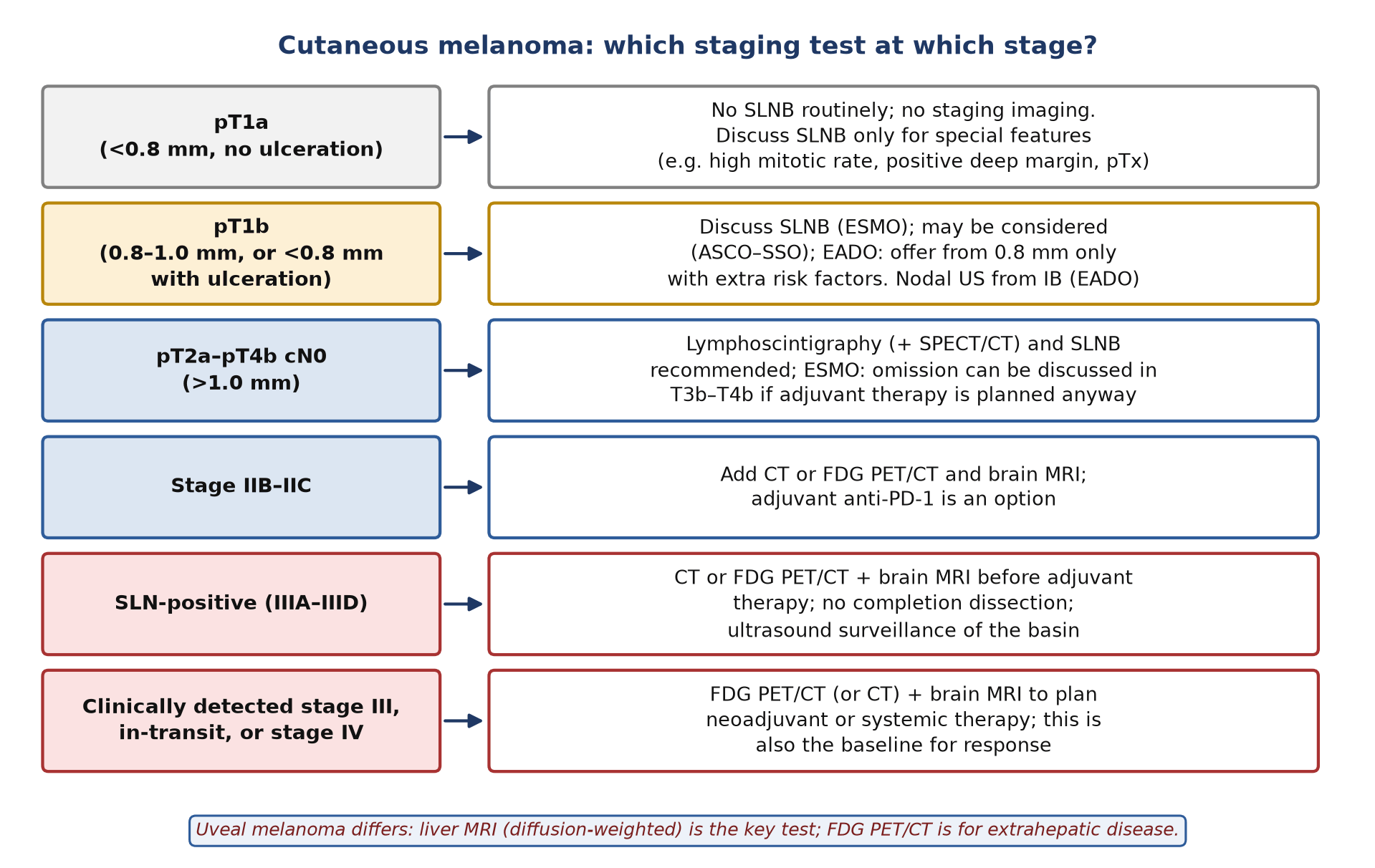
- Sentinel node biopsy is the staging test for the node-negative patient. It is recommended from pT2a (>1.0 mm) and discussed for pT1b; lymphoscintigraphy with single-photon emission computed tomography/CT (SPECT/CT) is the map the surgeon follows.
- Completion dissection is no longer routine. MSLT-II and DeCOG-SLT showed no survival gain from immediate dissection after a positive sentinel node.
- FDG PET/CT is for stage IIB and above. In early-stage disease it rarely finds anything that sentinel node biopsy misses (nodal sensitivity 21% in one prospective study); in clinically detected stage III it changed management in 37%.
- PET does not replace brain magnetic resonance imaging (MRI). From stage IIB, whole-body staging is paired with contrast-enhanced brain MRI.
- Read immune checkpoint inhibitor (ICI) scans by pattern. New symmetric hilar nodes, diffuse colitis or thyroiditis are usually immune effects, not metastases; confirm doubtful progression 4–8 weeks later if the patient is well.
- A complete metabolic response (CMR) at 1 year predicts durable control. In 104 patients on anti-PD-1 therapy, 5-year progression-free survival after the 1-year scan was 90% with CMR versus 54% without.
- Uveal melanoma is different. Liver MRI is the key test; FDG PET/CT misses many small liver metastases.
1. The clinical problem
Cutaneous melanoma spreads first to regional lymph nodes in most patients, but it can also skip to any organ, including the brain. The oncologist needs imaging to answer four questions: are the regional nodes involved, is there distant disease, is systemic treatment working, and has the disease come back? Nuclear medicine answers the first with lymphoscintigraphy and sentinel lymph node biopsy (SLNB), and the other three largely with ¹⁸F-fluorodeoxyglucose positron emission tomography/computed tomography (FDG PET/CT).
Staging system in force: AJCC eighth edition
Current European Society for Medical Oncology (ESMO 2024), European Association of Dermato-Oncology (EADO 2024) and uveal melanoma (ESMO with the European Reference Network for rare adult solid cancers, ESMO–EURACAN 2026) guidelines stage melanoma with the eighth edition of the American Joint Committee on Cancer (AJCC8) system. Its key features for the imager are:
| Category | Definition (AJCC8) | Why it matters for imaging |
|---|---|---|
| T1a | Breslow thickness <0.8 mm, no ulceration | Nodal risk low: no SLNB or staging imaging routinely |
| T1b | 0.8–1.0 mm, or <0.8 mm with ulceration | SLNB discussed with the patient |
| T2 / T3 / T4 | >1.0–2.0 mm / >2.0–4.0 mm / >4.0 mm; suffix a = no ulceration, b = ulceration | SLNB recommended; T3b and above (stage IIB–IIC) also need cross-sectional staging |
| N | Clinically occult (found at SLNB) versus clinically apparent nodes; microsatellite, satellite or in-transit metastases counted as N1c–N3c | Clinically apparent nodes and in-transit disease need whole-body staging |
| M | M1a skin, soft tissue or distant nodes; M1b lung; M1c other visceral sites; M1d central nervous system; each with a lactate dehydrogenase (LDH) suffix | Brain disease is its own category, so brain MRI is part of staging |
| Stage III | Four subgroups (IIIA–IIID) built from N and T categories | 5-year melanoma-specific survival 93% (IIIA) to 32% (IIID) |
Mitotic rate is no longer a T criterion, and pathological stage IA now includes T1bN0. Stage IIC (T4b N0) has a 5-year melanoma-specific survival of about 82%, lower than stage IIIA, which is why thick ulcerated primaries are now staged and treated almost like node-positive disease.
Where nuclear medicine changes management
- SLNB: the most accurate nodal staging test; the status of the sentinel lymph node (SLN) sets stage III and the eligibility for adjuvant therapy.
- FDG PET/CT staging: decides between surgery, neoadjuvant therapy and systemic therapy when nodes are clinically involved or distant disease is suspected.
- Response and duration of therapy: FDG PET/CT is widely used to judge response to ICIs (anti-PD-1, programmed cell death protein 1; anti-CTLA-4, cytotoxic T-lymphocyte-associated protein 4) and BRAF/MEK inhibitors, and a complete metabolic response is increasingly used to support stopping therapy.
2. Tracers and why they work
Radiocolloids and receptor-targeted tracers for lymphatic mapping
Particles injected into the dermis enter the initial lymphatics and are trapped in the first draining node by macrophage phagocytosis or by particle size. Small particles move fast but pass on to second-echelon nodes; large particles move slowly and stay at the injection site. The choice is largely regional (European Association of Nuclear Medicine, EANM, guideline, endorsed by the Society of Nuclear Medicine and Molecular Imaging, SNMMI):
| Tracer | Typical size | Where used | Notes |
|---|---|---|---|
| ⁹⁹ᵐTc-human serum albumin nanocolloid | 5–80 nm | Europe | Labels within 10 min at room temperature |
| ⁹⁹ᵐTc-antimony trisulphide | 5–30 nm | Australia, Canada | Fast drainage; imaging usually complete in 1–3 h |
| ⁹⁹ᵐTc-rhenium sulphide | 50–200 nm | Europe | Nodes may need 4–6 h or next-day images |
| ⁹⁹ᵐTc-sulphur colloid (filtered) | 100–200 nm after 100–200 nm filtration | United States | Unfiltered particles up to 5,000 nm stay at the injection site |
| ⁹⁹ᵐTc-tilmanocept | about 7 nm | United States (Food and Drug Administration approval 2013) | Binds the CD206 mannose receptor on macrophages; rapid injection-site clearance and little second-echelon uptake |
FDG
Cutaneous melanoma metastases are usually intensely FDG-avid, which makes FDG PET/CT the most sensitive single test for distant spread. It fails in predictable places: micrometastases in nodes (below the resolution of PET, which is why SLNB remains the nodal test), the brain (high cortical glucose use), small lung nodules (lost to partial-volume effects, although CT shows them) and the liver in uveal melanoma, where many metastases have low uptake. In the ESMO–EURACAN uveal guideline FDG PET/CT sensitivity for liver metastases ranges from 41% to 88% and falls to 11% for small lesions found on MRI.
What replaces FDG when it fails
- Occult nodal disease: SLNB with lymphoscintigraphy; nodal ultrasound for surveillance.
- Brain: contrast-enhanced MRI.
- Small lung nodules: the CT component, reported in lung windows.
- Uveal melanoma liver metastases: liver MRI with diffusion-weighted sequences.
3. Indications by clinical scenario
| Scenario | Nuclear medicine test | Guideline position (strength where given) |
|---|---|---|
| pT1a primary | None | SLNB not routinely recommended (ESMO II, E; ASCO–SSO); may be discussed for special cases such as a high mitotic rate, a positive deep margin or unmeasurable thickness (ESMO III, D). No staging imaging (EADO) |
| pT1b primary | Lymphoscintigraphy and SLNB if chosen | ESMO: should be discussed (III, B). ASCO–SSO: may be considered after discussion. EADO: offer at ≥0.8 mm only with additional histological risk factors |
| pT2a–pT4b, clinically node-negative | Lymphoscintigraphy with SPECT/CT, then SLNB | Recommended (ESMO I, A; ASCO–SSO for T2–T3; T4 'may be recommended'). ESMO: in T3b–T4b patients who will receive adjuvant therapy anyway, omitting SLNB can be discussed (V, C) |
| Stage IIB–IIC | CT or FDG PET/CT, plus brain MRI | ESMO: ultrasound, CT and/or PET and brain MRI from stage IIB (III, B). EADO: CT or PET/CT with brain MRI from IIB/C |
| Positive SLN (stage IIIA–IIID) | FDG PET/CT or CT, plus brain MRI | Detailed staging before adjuvant therapy (ESMO); completion dissection not recommended (ESMO I, E) |
| Clinically detected nodes, in-transit or satellite disease | FDG PET/CT plus brain MRI | Staging before surgery or neoadjuvant therapy; EADO 2024: neoadjuvant immunotherapy can be offered for resectable macroscopic disease |
| Stage IV, before systemic therapy | Baseline FDG PET/CT (at least skull base to mid-thigh; vertex to feet is appropriate in melanoma), brain MRI | Baseline for response; joint EANM/SNMMI/Australian and New Zealand Society of Nuclear Medicine (ANZSNM) immunotherapy guideline 2022 |
| Response during ICI or BRAF/MEK inhibitors | FDG PET/CT | Used widely; criteria not yet validated in randomised trials (EANM/SNMMI/ANZSNM 2022) |
| Surveillance after resection | Imaging per national guidance | ESMO: no consensus on schedule or imaging (IV, B); tailor to stage and risk |
| Uveal melanoma | FDG PET/CT or CT for extrahepatic disease | Liver ultrasound or MRI with diffusion weighting for staging; high-risk surveillance with liver MRI every 4–6 months for ≥10 years (ESMO–EURACAN III, A) |
- pT1b: ESMO says SLNB should be discussed, ASCO–SSO that it may be considered, and EADO offers it only from 0.8 mm with additional risk factors, so a 0.9 mm non-ulcerated melanoma without risk features may or may not be referred depending on the guideline followed.
- Thick primaries: ESMO allows omitting SLNB in T3b–T4b patients who will receive adjuvant therapy anyway; the older EANM and ASCO–SSO documents do not address this.
- Stage IIB imaging: ESMO lists ultrasound, CT and/or PET; EADO names CT or PET/CT. Neither makes FDG PET/CT mandatory over CT.
4. Evidence at a glance
| Study | Design and n | Key finding | What it changed |
|---|---|---|---|
| MSLT-I (Morton 2014) | RCT, 2,001 patients: SLNB versus nodal observation | No overall melanoma-specific survival difference; 10-year disease-free survival 71.3% vs 64.7% for 1.20–3.50 mm (HR 0.76); in node-positive intermediate-thickness melanoma, melanoma-specific survival HR 0.56 | Established SLNB as standard staging |
| MSLT-II (Faries 2017) | RCT, 1,934 SLN-positive patients: completion dissection versus ultrasound observation | 3-year melanoma-specific survival 86% in both arms; regional control 92% vs 77%; non-sentinel metastases in 11.5%; lymphoedema 24.1% vs 6.3% | Ended routine completion dissection; nodal ultrasound surveillance instead |
| DeCOG-SLT (Leiter 2019) | RCT, 483 SLN-positive patients, median follow-up 72 months | 5-year distant metastasis-free survival 64.9% dissection vs 67.6% observation (HR 1.08) | Confirmed MSLT-II |
| Stoffels 2012 | Cohort, 403 patients, SLNB with vs without SPECT/CT | More SLNs (2.40 vs 1.87) and more positive SLNs per patient (0.34 vs 0.21); 4-year disease-free survival 93.9% vs 79.2% | Supported routine SPECT/CT (non-randomised) |
| Wagner 2005 | Prospective, 144 patients >1 mm, clinically node-negative | PET sensitivity 21% for nodal disease; no distant PET finding confirmed at presentation | No routine PET in early-stage disease |
| Aukema 2010 | Prospective, 70 patients with palpable nodes | PET/CT sensitivity 87%, specificity 98%; changed planned dissection in 37%; brain MRI found metastases in 7% | PET/CT plus brain MRI before surgery for clinical stage III |
| Xing 2011 | Meta-analysis, 74 studies, 10,528 patients | Nodes: ultrasound best (sensitivity 60%, specificity 97%). Distant staging: PET/CT best (80%, 87%); surveillance PET/CT 86%, 91% | Ultrasound for nodes, PET/CT for distant disease |
| Tan 2018; Dimitriou 2022 | Retrospective, 104 patients with PET at 1 year of anti-PD-1 | CMR in 75% (68% of CT partial responders); 5-year PFS after the 1-year scan 90% CMR vs 54% non-CMR | PET used to support stopping therapy |
| Ito 2019 (imPERCIST5) | Retrospective, 60 patients on ipilimumab | 2-year OS 66% responders vs 29% non-responders; new lesions alone did not predict outcome | New lesions should not define progression on their own |
| Anwar 2018 (PERCIMT) | Retrospective, 41 patients on ipilimumab | Four or more new lesions: sensitivity 84%, specificity 100% for lack of clinical benefit; standardised uptake value (SUV) change did not predict response | Lesion counting as a progression rule |
Abbreviations: RCT, randomised controlled trial; HR, hazard ratio; PFS, progression-free survival; OS, overall survival; CMR, complete metabolic response.
5. Patient preparation and acquisition
Lymphoscintigraphy (EANM guideline, Bluemel 2015)
- History and examination: prior excision (with histology), surgery or trauma in the region, pregnancy or breastfeeding. A clinically suspicious node needs fine-needle aspiration first; a node replaced by tumour may not take up tracer and can give a false-negative map.
- Timing: same-day or day-before protocols have similar detection and false-negative rates. Do not inject in theatre, because drainage in melanoma can be delayed, aberrant or to several basins.
- Injection: intradermal wheals, usually four or more aliquots of 0.1–0.2 ml around the primary or on each side of the excision scar, within 1 cm of it; 25–27 gauge needle. Avoid subcutaneous injection. In head and neck melanoma use four deposits around the lesion; on a limb, at least medial and lateral deposits.
- Activity: no consensus; published activities range from about 5 to 120 MBq depending on the interval to surgery. Aim for a residual activity at operation that still gives good probe counts (above about 10 MBq); prepare albumin nanocolloid at ≥100 MBq/ml so that 20 MBq fits in 0.2 ml.
- Dynamic images: start immediately, 10–20 min at one frame per minute (128 × 128), with the patient positioned for the region; for hand, forearm, foot or leg primaries follow the channels to the elbow or knee to catch epitrochlear or popliteal nodes.
- Early and delayed statics: 5-min anterior and lateral views (256 × 256) over all possible basins; for the trunk, both axillae and both groins, or a neck-to-groin sweep. Delayed 3–5 min views at 1–3 h identify the nodes to mark.
- SPECT/CT: should be done for head and neck primaries, is highly recommended for the groin and recommended for the axilla; it finds interval nodes, separates nodes close to the injection site and gives depth.
- Skin marking: mark every sentinel node in each basin in the operating position, with depth; do not mark nodes clearly identified as second echelon.
- Trunk and midline primaries often drain to several basins, including the opposite axilla or groin; image both sides.
- Interval (in-transit) nodes lie between the primary and the basin, in the chest wall, flank or limb (epitrochlear, popliteal). A node with its own channel from the primary is a sentinel node wherever it lies.
- Head and neck drainage is complex and nodes can be masked by the injection site; SPECT/CT is part of the standard study.
- Pregnancy SLNB is possible; use a 1-day protocol and SPECT without CT for the axilla or groin. Blue dye is avoided.
FDG PET/CT in melanoma
- Standard preparation: fasting for at least 4 h (EANM; SNMMI 4–6 h) and imaging at 60 min (55–75 min) after injection, as in the EANM and SNMMI tumour guidelines.
- Field of view: include the skull base on every response study so that hypophysitis is not missed; the guideline allows extended vertex-to-feet imaging in melanoma, and it is essential when the primary or known disease is on the scalp or limbs (EANM/SNMMI/ANZSNM 2022).
- Serial studies: same scanner, same uptake time and same reconstruction, ideally on a system accredited by EANM Research Ltd (EARL), so that changes in SULpeak (peak standardised uptake value normalised to lean body mass) are real.
- Record for the reader: dates of surgery, SLNB, radiotherapy and the start of ICI, BRAF/MEK inhibitors or steroids, and any symptoms of colitis, thyroiditis or arthritis.
- Brown fat: common in young, slim patients and in cold rooms; keep the patient warm before injection, because supraclavicular and paravertebral brown fat can mimic nodal disease.
6. Interpretation
Reading a lymphoscintigram
- Sentinel node: a node receiving a lymphatic channel directly from the primary (the strongest criterion), or the first node to appear. It is often, but not always, the hottest.
- Late nodes in another basin are also sentinel nodes, unless the dynamic images show that they fill from an earlier node.
- False positives: skin or urine contamination (very hot and focal; check with a second view or SPECT/CT), second-echelon nodes read on late images only, lymphatic lakes.
- False negatives: two adjacent nodes read as one, a node masked by the injection site, poor drainage (massage, warming or limb exercise can help; drainage is slower in patients over 50).
- Report: tracer, activity, injection sites, image times; number, basin and depth of each sentinel node; second-echelon nodes; interval nodes; any node enlarged on CT without uptake.
Reading FDG PET/CT
Report distant disease by AJCC M category, because M1d (brain) and M1c (visceral) change treatment. Look specifically at skin and subcutaneous tissue (in-transit and satellite disease), bowel wall, bone, adrenals, spleen and muscle. Check the CT for sub-centimetre lung nodules that are too small to show uptake, and state that brain assessment needs MRI.
| Pitfall or mimic | What it looks like | How to tell |
|---|---|---|
| Recent excision, SLNB or wide local excision | Linear or rim uptake along the scar, seroma, drain track or dissected basin | Match to surgical dates; reassess later rather than calling nodal disease |
| Brown fat | Symmetric supraclavicular, axillary and paravertebral uptake in fat on CT | Fat density on CT; keep the patient warm and repeat if needed |
| Sarcoid-like reaction on ICI | Symmetric mediastinal and bilateral hilar nodes, sometimes skin or lung nodules, while metastases respond | Symmetry and timing; biopsy (for example endobronchial ultrasound) if management depends on it |
| Immune colitis, thyroiditis, arthritis | Diffuse colonic, diffuse thyroid or periarticular uptake | Pattern and symptoms; correlate with the clinical team |
| Splenic and marrow activation | Diffuse increase in spleen and marrow uptake, equalising or reversing the spleen-to-liver ratio | Diffuse, not focal; a raised spleen-to-liver ratio was an unfavourable sign in several studies, not a sign of response |
| Small lung nodules | FDG-negative nodules below about 1 cm | Negative PET does not exclude metastasis; follow on CT |
| Brain metastases | Masked by grey-matter uptake | Brain MRI |
| Injection-site or skin contamination | Hot focus on the skin | Check the injection arm; CT shows no lesion |
Immune-related adverse events on FDG PET/CT
Immune-related adverse events (irAEs) are common with ICIs, particularly with ipilimumab plus nivolumab. In 31 patients on this combination, 36 PET findings suggested an irAE and 29 (80%) were confirmed clinically; the PET finding came first in 7%, and endocrinopathies (36%) and enterocolitis (35%) were the commonest.
| irAE | FDG pattern | Report it as |
|---|---|---|
| Colitis / enterocolitis | Diffuse or segmental uptake along the colon, often with wall thickening | Probable immune colitis; correlate with diarrhoea |
| Thyroiditis | New diffuse, symmetric thyroid uptake | Probable thyroiditis; check thyroid function |
| Hypophysitis | Increased pituitary uptake (skull base must be in the field) | Possible hypophysitis; check pituitary hormones |
| Sarcoid-like reaction | Symmetric mediastinal and hilar nodes ± skin, subcutaneous or lung nodules | Likely sarcoid-like reaction rather than progression; biopsy if it changes management |
| Pneumonitis | Uptake in ground-glass or consolidative change | Possible pneumonitis; urgent clinical review |
| Arthritis | Periarticular uptake in several joints | Probable immune arthritis |
| Other | Diffuse pancreatic, hepatic or adrenal uptake | Possible pancreatitis, hepatitis or adrenalitis |
Wording the conclusion
- Answer the clinical question first (stage, response category and the criteria used).
- Separate tumour findings from immune findings: 'new symmetric hilar and mediastinal nodes in a patient whose metastases are responding: favour sarcoid-like reaction'.
- When progression is doubtful early on ICIs, write 'unconfirmed progressive metabolic disease' and recommend a repeat scan in 4–8 weeks if the patient is clinically stable.
7. Response assessment and follow-up
Criteria in use
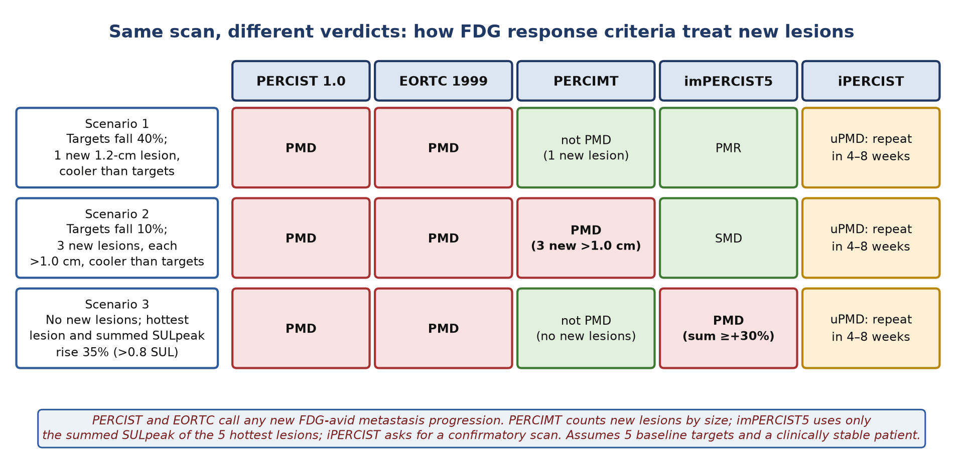
Five sets of FDG criteria are in use: the 1999 European Organisation for Research and Treatment of Cancer (EORTC) criteria, PET Response Criteria in Solid Tumours (PERCIST 1.0), and three immunotherapy adaptations: PET Response Evaluation Criteria for Immunotherapy (PERCIMT), immunotherapy-modified PERCIST (imPERCIST5) and immune PERCIST (iPERCIST). SUL is the SUV normalised to lean body mass.
| Criteria | Measurement | Response | Progression | New lesions |
|---|---|---|---|---|
| EORTC 1999 | SUV in tumour regions | Partial response: fall of 15–25% after one cycle, >25% after more | >25% SUV rise, >20% increase in extent, or new FDG-avid metastases | Progression |
| PERCIST 1.0 (2009) | SULpeak of the single hottest lesion (≥1.5 × mean liver SUL + 2 standard deviations); PERCIST5 sums up to 5 lesions, 2 per organ | CMR: disappearance of all metabolically active tumour. Partial metabolic response (PMR): ≥30% and ≥0.8 SUL fall | Progressive metabolic disease (PMD): ≥30% and ≥0.8 SUL rise, >75% rise in total lesion glycolysis, or new FDG-avid lesions | Progression |
| PERCIMT (2018) | Number and functional size of new lesions | Clinical benefit if progression thresholds not met | ≥4 new lesions <1 cm, or ≥3 >1.0 cm, or ≥2 >1.5 cm | Counted; one or two small new lesions are not progression |
| imPERCIST5 (2019) | Sum of SULpeak of up to 5 hottest lesions (2 per organ) | PMR: ≥30% fall in the sum | ≥30% rise in the sum | Not progression on their own; added to the sum only if hotter than the targets or fewer than 5 targets at baseline |
| iPERCIST (2019) | PERCIST with confirmation | As PERCIST | Unconfirmed PMD (uPMD) until a repeat scan confirms it | Trigger uPMD; confirm in 4–8 weeks |
PERCIST and EORTC were built for cytotoxic therapy and treat any new FDG-avid metastasis as progression. The immunotherapy-adapted criteria were each derived in small retrospective cohorts (41 to 60 patients; iPERCIST in non-small cell lung cancer), and the EANM/SNMMI/ANZSNM guideline states that there are not yet enough data to choose between them. In practice, state which criteria you used and apply them consistently.
Atypical response patterns on ICIs
- Pseudoprogression: a transient increase in lesion size, uptake or number followed by response, seen in up to about 10% of patients, most often with anti-CTLA-4 therapy in melanoma and usually in the first 4–6 weeks.
- Dissociated response: some lesions respond while others grow; local treatment of the growing lesions with continued ICI can be appropriate.
- Hyperprogression: accelerated growth after starting ICIs; no agreed definition.
- Guideline advice: if progression versus pseudoprogression is uncertain, especially on the first scan, repeat FDG PET/CT 4–8 weeks later in a clinically stable patient and continue treatment meanwhile (EANM/SNMMI/ANZSNM 2022).
The 1-year scan and stopping anti-PD-1 therapy
In 104 patients with baseline and 1-year PET and CT on anti-PD-1-based therapy, CT showed complete response in only 28%, but PET showed CMR (EORTC criteria) in 75%, including 68% of CT partial responders. Five years after the 1-year scan, progression-free survival was 90% with CMR versus 54% without, and 88% versus 59% among CT partial responders. A residual mass on CT without FDG uptake therefore usually means inactive tissue.
This evidence is retrospective. The ECOG-ACRIN Cancer Research Group PET-Stop trial (NCT04462406) is testing prospectively whether stopping anti-PD-1 therapy at 12 months after a negative FDG PET/CT, or a negative biopsy of a residual avid lesion, is safe; primary completion is expected in 2027. The UK DANTE trial of stopping at 1 year versus continuing has so far been reported only as a conference abstract; it closed early, was underpowered, and its investigators advised that 2 years remain the standard. Report 1-year scans with a clear CMR or non-CMR statement.
BRAF/MEK inhibitors and combinations
PERCIST applies to targeted therapy as to any systemic treatment. The immunotherapy-adapted rules for new lesions were derived in patients on ICIs, so they should not be borrowed for patients on BRAF/MEK inhibitors alone; state the criteria used in every report.
Surveillance
There is no consensus on imaging surveillance after resection (ESMO IV, B). In the Xing meta-analysis, PET/CT had the best accuracy for distant recurrence (sensitivity 86%, specificity 91%) and ultrasound for nodal recurrence (96%, 99%). ESMO leaves the schedule to national guidance, EADO proposes a stage-based schedule, and neither mandates PET over CT; nodal ultrasound is the test for an observed sentinel-node-positive basin. Uveal melanoma follows its own schedule: liver ultrasound every 6–12 months for at least 5 years in low-risk patients and liver MRI every 4–6 months for at least 10 years in high-risk patients.
8. Theranostics and emerging tracers
| Agent | Target | Evidence level (2026) |
|---|---|---|
| ⁹⁹ᵐTc-colloids, ⁹⁹ᵐTc-tilmanocept | Lymphatic mapping | Approved; guideline-endorsed |
| ¹⁸F-FDG | Glucose metabolism | Guideline-endorsed for stage IIB–IV staging and response |
| ¹⁸F-PFPN, ¹⁸F-P3BZA and other benzamide derivatives | Melanin | Investigational. In 65 patients with mucosal melanoma, [¹⁸F]PFPN had higher lesion sensitivity than FDG (91% vs 83.2%) with no false positives; amelanotic lesions are a predictable blind spot |
| ²⁰³Pb/²¹²Pb-VMT01 | Melanocortin-1 receptor | Phase I/IIa image-guided alpha therapy, alone or with nivolumab (NCT05655312); investigational |
| ⁸⁹Zr-atezolizumab | Programmed death-ligand 1 (PD-L1) | First-in-human (22 patients with bladder, non-small cell lung or triple-negative breast cancer): uptake predicted response better than immunohistochemistry; not melanoma-specific; investigational |
| ⁸⁹Zr-Df-IAB22M2C | CD8 (T cells) | Phase I, 15 patients including melanoma: uptake in tumours and CD8-rich tissues peaking at 24–48 h; investigational |
No radioligand therapy is approved for melanoma. Melanin and melanocortin-1 receptor targets are attractive because they are specific to melanocytic tissue, but both depend on differentiation, and uveal and mucosal melanomas are the main populations under study.
9. Structured report example
- Clinical question: Stage IV (M1b, normal LDH) cutaneous melanoma, BRAF wild type, on pembrolizumab for 12 months. Is there residual active disease?
- Technique: ¹⁸F-FDG after a 6-h fast (glucose 5.4 mmol/l), imaging at 62 min (baseline 60 min), vertex to toes, same scanner and reconstruction as baseline. Low-dose CT.
- Comparison: Baseline and 12-week studies.
- Findings: The two left lower lobe nodules (baseline SULpeak 7.8 and 5.1) now measure 6 and 4 mm with no uptake above blood pool. The left axillary node measures 11 mm (was 24 mm) with uptake at blood-pool level. No new FDG-avid lesion. Diffuse colonic uptake seen at 12 weeks has resolved. Physiological uptake elsewhere. Brain not assessed: needs MRI.
- Conclusion: Complete metabolic response (PERCIST 1.0). Residual CT abnormalities are without metabolic activity. Resolved immune colitis.
- Recommendation: Suitable for multidisciplinary discussion of treatment duration; continue brain MRI surveillance as scheduled.
10. Take-home points
- Stage with AJCC8; Breslow thickness, ulceration and nodal status drive every imaging decision.
- Lymphoscintigraphy is a dynamic study: watch the channels, image every possible basin, use SPECT/CT, and mark every sentinel node including interval nodes, but not second-echelon nodes.
- After a positive sentinel node, stage with CT or FDG PET/CT and brain MRI; the basin is followed with ultrasound, not dissected.
- Do not use FDG PET/CT for stage I–IIA disease; use it from stage IIB, for clinically detected nodes and for stage IV.
- On ICIs, symmetric hilar nodes, diffuse colitis, thyroiditis and splenic activation are usually immune effects. Confirm doubtful progression 4–8 weeks later and name the criteria used.
- A CMR at 1 year of anti-PD-1 therapy predicts long-term control and is used to support stopping treatment, pending prospective trial results.
- In uveal melanoma, liver MRI comes first.
Test yourself
5 quick questions. Pick an answer to see the explanation.
References
- Gershenwald JE, Scolyer RA, Hess KR, et al. Melanoma staging: evidence-based changes in the American Joint Committee on Cancer eighth edition cancer staging manual. CA Cancer J Clin. 2017;67(6):472-92.
- Amaral T, Ottaviano M, Arance A, et al. Cutaneous melanoma: ESMO Clinical Practice Guideline for diagnosis, treatment and follow-up. Ann Oncol. 2025;36(1):10-30.
- Garbe C, Amaral T, Peris K, et al. European consensus-based interdisciplinary guideline for melanoma. Part 1: Diagnostics - Update 2024. Eur J Cancer. 2025;215:115152.
- Garbe C, Amaral T, Peris K, et al. European consensus-based interdisciplinary guideline for melanoma. Part 2: Treatment - Update 2024. Eur J Cancer. 2025;215:115153.
- Wong SL, Faries MB, Kennedy EB, et al. Sentinel lymph node biopsy and management of regional lymph nodes in melanoma: American Society of Clinical Oncology and Society of Surgical Oncology clinical practice guideline update. J Clin Oncol. 2018;36(4):399-413.
- Piperno-Neumann S, Angi M, Ascierto PA, et al. Uveal melanoma: ESMO-EURACAN Clinical Practice Guideline for diagnosis, treatment and follow-up. ESMO Open. 2026;11(4):106888.
- Bluemel C, Herrmann K, Giammarile F, et al. EANM practice guidelines for lymphoscintigraphy and sentinel lymph node biopsy in melanoma. Eur J Nucl Med Mol Imaging. 2015;42(11):1750-66.
- Stoffels I, Boy C, Pöppel T, et al. Association between sentinel lymph node excision with or without preoperative SPECT/CT and metastatic node detection and disease-free survival in melanoma. JAMA. 2012;308(10):1007-14.
- Morton DL, Thompson JF, Cochran AJ, et al. Final trial report of sentinel-node biopsy versus nodal observation in melanoma. N Engl J Med. 2014;370(7):599-609.
- Faries MB, Thompson JF, Cochran AJ, et al. Completion dissection or observation for sentinel-node metastasis in melanoma. N Engl J Med. 2017;376(23):2211-22.
- Leiter U, Stadler R, Mauch C, et al. Final analysis of DeCOG-SLT trial: no survival benefit for complete lymph node dissection in patients with melanoma with positive sentinel node. J Clin Oncol. 2019;37(32):3000-8.
- Xing Y, Bronstein Y, Ross MI, et al. Contemporary diagnostic imaging modalities for the staging and surveillance of melanoma patients: a meta-analysis. J Natl Cancer Inst. 2011;103(2):129-42.
- Wagner JD, Schauwecker D, Davidson D, et al. Inefficacy of F-18 fluorodeoxy-D-glucose-positron emission tomography scans for initial evaluation in early-stage cutaneous melanoma. Cancer. 2005;104(3):570-9.
- Aukema TS, Valdés Olmos RA, Wouters MW, et al. Utility of preoperative 18F-FDG PET/CT and brain MRI in melanoma patients with palpable lymph node metastases. Ann Surg Oncol. 2010;17(10):2773-8.
- Lopci E, Hicks RJ, Dimitrakopoulou-Strauss A, et al. Joint EANM/SNMMI/ANZSNM practice guidelines/procedure standards on recommended use of [18F]FDG PET/CT imaging during immunomodulatory treatments in patients with solid tumors version 1.0. Eur J Nucl Med Mol Imaging. 2022;49(7):2323-41.
- Wahl RL, Jacene H, Kasamon Y, Lodge MA. From RECIST to PERCIST: evolving considerations for PET response criteria in solid tumors. J Nucl Med. 2009;50 Suppl 1:122S-50S.
- Anwar H, Sachpekidis C, Winkler J, et al. Absolute number of new lesions on 18F-FDG PET/CT is more predictive of clinical response than SUV changes in metastatic melanoma patients receiving ipilimumab. Eur J Nucl Med Mol Imaging. 2018;45(3):376-83.
- Ito K, Teng R, Schöder H, et al. 18F-FDG PET/CT for monitoring of ipilimumab therapy in patients with metastatic melanoma. J Nucl Med. 2019;60(3):335-41.
- Goldfarb L, Duchemann B, Chouahnia K, et al. Monitoring anti-PD-1-based immunotherapy in non-small cell lung cancer with FDG PET: introduction of iPERCIST. EJNMMI Res. 2019;9(1):8.
- Tan AC, Emmett L, Lo S, et al. FDG-PET response and outcome from anti-PD-1 therapy in metastatic melanoma. Ann Oncol. 2018;29(10):2115-20.
- Dimitriou F, Lo SN, Tan AC, et al. FDG-PET to predict long-term outcome from anti-PD-1 therapy in metastatic melanoma. Ann Oncol. 2022;33(1):99-106.
- Iravani A, Osman MM, Weppler AM, et al. FDG PET/CT for tumoral and systemic immune response monitoring of advanced melanoma during first-line combination ipilimumab and nivolumab treatment. Eur J Nucl Med Mol Imaging. 2020;47(12):2776-86.
- Lin Z, Tang C, Liu T, et al. Comparison of [18F]PFPN and [18F]FDG PET in mucosal melanoma: diagnostic performance, staging impact, and correlation with molecular markers. Clin Cancer Res. 2026;32(10):2036-46.
- Bensch F, van der Veen EL, Lub-de Hooge MN, et al. 89Zr-atezolizumab imaging as a non-invasive approach to assess clinical response to PD-L1 blockade in cancer. Nat Med. 2018;24(12):1852-8.
- Farwell MD, Gamache RF, Babazada H, et al. CD8-targeted PET imaging of tumor-infiltrating T cells in patients with cancer: a phase I first-in-humans study of 89Zr-Df-IAB22M2C, a radiolabeled anti-CD8 minibody. J Nucl Med. 2022;63(5):720-6.
- Melanoma on combination immunotherapy · Intermediate