Nuclear Medicine in Medullary Thyroid Cancer
- A C-cell tumour, not a follicular one. Medullary thyroid cancer (MTC) does not concentrate iodine, so radioiodine has no role in imaging or treating it (except rare mixed tumours with a follicular component).
- Two blood tests steer every scan. Calcitonin tracks tumour burden; the calcitonin and CEA doubling times track tempo. A calcitonin doubling time under 6 months carried 8% 10-year survival; over 2 years, no deaths in the study period.
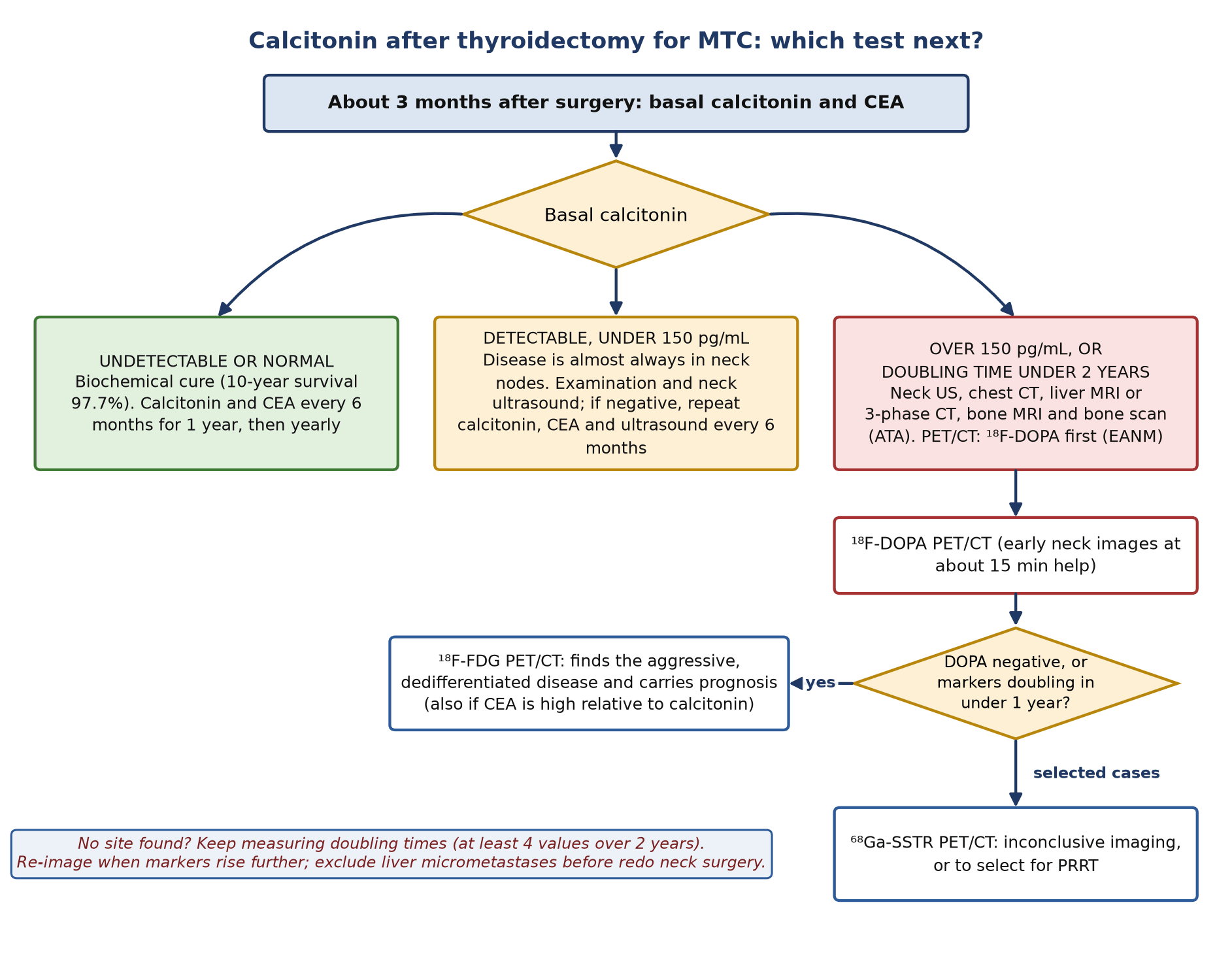
- Neck ultrasound first. After thyroidectomy, a detectable calcitonin below 150 pg/mL almost always means neck disease: examine and scan the neck. Above 150 pg/mL, image the whole body.
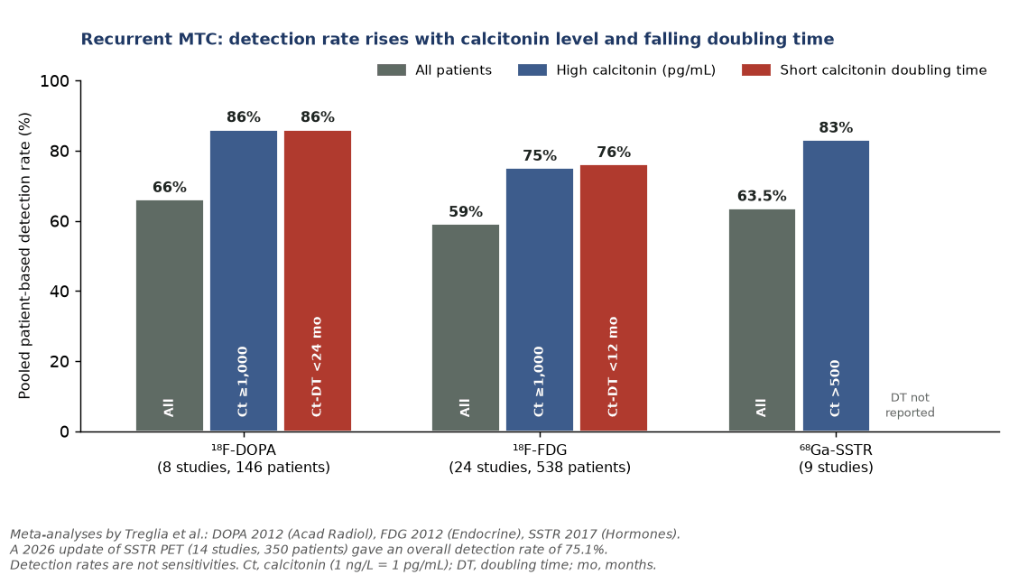
- ¹⁸F-DOPA is the most sensitive PET tracer. Pooled detection rate 66%, rising to 86% when calcitonin is at least 1,000 pg/mL or its doubling time is under 24 months; it beat FDG in a network meta-analysis of 14 head-to-head studies.
- FDG measures aggression. FDG-avid disease goes with short doubling times and poorer survival. Add FDG when DOPA is negative, markers double within a year, or CEA is high relative to calcitonin.
- SSTR PET is a second-line and theranostic tool. Detection is lower than in other neuroendocrine tumours (pooled 63.5% in 2017, 75.1% in a 2026 update); its main job is selecting patients for PRRT.
- Guidelines disagree. ATA 2015 recommends against FDG or DOPA PET/CT to look for distant metastases (grade E); the EANM 2020 practice guideline makes DOPA the first-line PET when calcitonin exceeds 150 pg/mL or doubles in under 2 years.
- Therapy has moved on. Selective RET inhibition (selpercatinib) beat cabozantinib or vandetanib in LIBRETTO-531 (hazard ratio for progression 0.28). PRRT and gastrin-receptor (CCK2R) theranostics remain small-series or investigational.
1. The clinical problem
MTC arises from the parafollicular C cells, which derive from the neural crest and secrete calcitonin. It accounts for 1–2% of thyroid cancers in the United States. It behaves as a neuroendocrine tumour: it makes calcitonin and carcinoembryonic antigen (CEA), expresses the machinery for amine-precursor uptake and decarboxylation, and variably expresses somatostatin receptors (SSTR). It does not express the sodium–iodide symporter, so ¹³¹I is neither a diagnostic nor a therapeutic option. The American Thyroid Association (ATA) advises against postoperative radioiodine, except in the rare patient whose metastases contain MTC mixed with papillary or follicular carcinoma (grade E recommendation against routine use).
Sporadic and hereditary disease: RET
- Hereditary MTC: virtually all patients with multiple endocrine neoplasia type 2 (MEN2A, which makes up about 95% of MEN2, and MEN2B) carry a germline RET mutation. About 95% of MEN2B is codon M918T. Because 1–7% of apparently sporadic MTC is hereditary, every patient with MTC should have germline RET testing.
- Sporadic MTC: about half have a somatic RET mutation (M918T is the commonest and marks aggressive disease), and 18–80% of the RET-negative tumours carry a somatic RAS mutation.
- ATA risk levels (2015): highest risk (ATA-HST) for M918T, with thyroidectomy in the first year of life; high risk (ATA-H) for codon C634 and A883F, with thyroidectomy at or before 5 years; moderate risk (ATA-MOD) for other codons, with ultrasound and calcitonin from about age 5 and surgery timed by calcitonin. Nuclear medicine has no role in timing prophylactic surgery.
- Before surgery in MEN2: phaeochromocytoma must be excluded before thyroidectomy in hereditary MTC; that is a biochemical question first, with adrenal imaging to follow.
Markers, grade and stage
- Calcitonin: serum level reflects C-cell mass. In 300 untreated patients, nodal metastases appeared in the ipsilateral central and lateral neck above a basal calcitonin of 20 pg/mL, the contralateral central neck above 50 pg/mL, the contralateral lateral neck above 200 pg/mL and the upper mediastinum above 500 pg/mL. The ATA cites the same threshold: no distant metastases were found below 500 pg/mL.
- CEA: not specific, but useful for progression. A CEA that is high relative to calcitonin, or rising faster, suggests dedifferentiation.
- Doubling times: in 65 patients with persistent disease, 5- and 10-year survival were 25% and 8% when the calcitonin doubling time was under 6 months, 92% and 37% at 6–24 months, and all patients with doubling times over 24 months were alive at the end of the study. Only the calcitonin doubling time was independent in multivariate analysis. The ATA asks for markers every 6 months while they are detectable, and at least four values over 2 years for a reliable estimate.
- Grade: the International Medullary Thyroid Carcinoma Grading System (2022) calls a tumour high grade if it has at least one of: 5 or more mitoses per 2 mm², Ki-67 index of 5% or more, or tumour necrosis. High grade (25% of 327 tumours) predicted worse overall, disease-specific and metastasis-free survival.
- Stage: AJCC/UICC 8th edition TNM, with no age cut-off (unlike differentiated thyroid cancer). Stage I is T1N0; II is T2–T3N0; III is T1–T3 with N1a (level VI or VII nodes, so upper mediastinal nodes count as N1a); IVA is N1b (lateral neck or retropharyngeal) or T4a; IVB is T4b; IVC is any M1.
What the team needs from imaging, and where nuclear medicine changes management
Surgery is the only curative treatment: total thyroidectomy with compartment-oriented neck dissection. Before surgery the team needs the neck mapped (ultrasound) and, when calcitonin is high, distant disease excluded, because metastatic disease makes extensive neck surgery futile. After surgery, a persistent or rising calcitonin is common, and the questions become: where is it, is it resectable, and how fast is it growing? This is where PET changes management: finding a resectable neck or mediastinal recurrence, finding liver, bone or lung disease that turns a planned re-operation into surveillance or systemic therapy, and separating indolent from aggressive disease.
2. Tracers and why they work
| Tracer | Mechanism in MTC | Strength | Weakness |
|---|---|---|---|
| ¹⁸F-DOPA (6-[¹⁸F]fluoro-L-DOPA) | Taken up by L-type amino acid transporters (LAT) and decarboxylated by aromatic L-amino acid decarboxylase (AADC), both upregulated in MTC | Highest detection rate in recurrent disease; high specificity; best map of disease extent | Rapid washout from MTC lesions; misses small and dedifferentiated lesions; expensive and not available everywhere |
| ¹⁸F-FDG | Glucose transporter overexpression and hexokinase activity; uptake rises with proliferation and loss of differentiation | Marks aggressive, progressive disease; prognostic; widely available | Often negative in slow-growing MTC (pooled detection 59%) |
| ⁶⁸Ga-DOTA-somatostatin analogues (DOTATATE, DOTATOC, DOTANOC) | Binding to SSTR, mainly subtype 2, whose expression in MTC is variable | Selects patients for PRRT; can add information when DOPA and FDG are inconclusive | Lower detection than in other neuroendocrine tumours; high physiological liver uptake hides liver metastases |
| ¹³¹I / ¹²³I | Needs the sodium–iodide symporter, which C cells lack | None in pure MTC | Ordering it is a recognised error |
Think of the tracers as reporting on biology rather than competing for the same lesions. Well-differentiated MTC keeps its amine-precursor pathway and so is DOPA-avid and usually FDG-negative. As the tumour dedifferentiates, DOPA uptake falls and FDG uptake rises, much as iodine and FDG swap places in differentiated follicular cancer. The EANM guideline puts it simply: DOPA is the most accurate way to assess extent, and FDG is the stronger prognostic tool.
Older single-photon agents (¹¹¹In-pentetreotide, ⁹⁹ᵐTc-(V)-DMSA, ¹²³I-MIBG and radiolabelled anti-CEA antibodies) have been superseded by PET because of low sensitivity. A ⁹⁹ᵐTc-diphosphonate bone scan still has a place: the ATA pairs it with axial MRI, because the bone scan is better for peripheral bone lesions and MRI for the spine and pelvis.
3. Indications by clinical scenario
| Scenario | Recommended imaging | Guideline position (grade where given) |
|---|---|---|
| Suspicious nodule, cytology or calcitonin suggesting MTC | Neck ultrasound; calcitonin, CEA, germline RET testing | ATA R21 (B) and R22 (C): neck ultrasound in all patients |
| Pre-operative staging, calcitonin over 500 pg/mL, extensive neck disease or signs of metastases | Contrast CT of neck and chest; three-phase liver CT or contrast liver MRI; axial MRI and bone scan | ATA R22 (C). ATA R23 (E): FDG or DOPA PET/CT not recommended to look for distant metastases. EANM 2020: insufficient evidence to recommend PET for staging before treatment |
| After thyroidectomy, calcitonin undetectable or normal | No imaging; calcitonin and CEA at 3 months, 6-monthly for a year, then yearly | ATA R46 (C) |
| After thyroidectomy, calcitonin detectable but under 150 pg/mL | Examination and neck ultrasound; repeat markers and ultrasound every 6 months if negative | ATA R47 (C). EANM agrees: neck nodes are the usual site |
| After thyroidectomy, calcitonin over 150 pg/mL | Neck ultrasound, chest CT, liver MRI or three-phase CT, bone scan and MRI of the pelvis and axial skeleton, plus PET/CT | ATA R48 (C) lists conventional imaging only. EANM 2020: PET/CT when calcitonin exceeds 150 pg/mL or its doubling time is under 24 months, with ¹⁸F-DOPA first line |
| DOPA negative or not available; markers doubling in under a year; CEA disproportionately high | ¹⁸F-FDG PET/CT | EANM 2020 |
| Inconclusive anatomical, DOPA and FDG imaging; candidate for PRRT | ⁶⁸Ga-SSTR PET/CT | EANM 2020: selected cases |
| Before redo neck surgery with persistent calcitonin | Exclude distant disease; consider laparoscopic or open liver assessment | ATA (C): occult miliary liver metastases are missed by CT |
| Response to systemic therapy | RECIST on CT or MRI plus marker doubling times | EANM 2020: insufficient evidence to recommend PET for response |
Where the guidelines disagree
The ATA 2015 guideline leaned on a prospective series (Giraudet 2007) in which FDG PET/CT was less sensitive than neck ultrasound, chest CT, liver MRI and bone imaging, and it recommended against FDG or DOPA PET/CT to detect distant metastases (recommendation 23, grade E). Its own text acknowledges that later studies found both PET tracers better than conventional imaging, but cites cost and the need for selection criteria. In 2016 EANM declined to endorse the ATA guideline, arguing that the evidence for nuclear medicine had been marginalised, and the EANM 2020 practice guideline recommends PET/CT to restage patients with rising markers. The practical compromise used in most European centres: neck ultrasound for low calcitonin, then conventional imaging plus DOPA PET/CT once calcitonin exceeds 150 pg/mL or the doubling time shortens, with liver MRI kept because small liver deposits are hard for every PET tracer.
The ATA 150 pg/mL threshold also holds for PET. In a prospective comparison, DOPA detected disease in 90.9% of patients with calcitonin of 150 pg/mL or more but only 28.6% below it (FDG 72.7% and 14.3%). Below 150 pg/mL the pre-test probability of a PET-visible lesion outside the neck is low, and ultrasound does the work.
4. Evidence at a glance
| Study | Design and size | Key finding | What it changed |
|---|---|---|---|
| Barbet 2005 | Retrospective, 65 patients with persistent calcitonin after thyroidectomy and bilateral neck dissection | Calcitonin doubling time under 6 months: 10-year survival 8%; 6–24 months: 37%; over 24 months: all alive; the only independent predictor | Doubling time became the prognostic yardstick and the trigger for imaging and treatment |
| Giraudet 2007 | Prospective imaging comparison, 55 patients with elevated calcitonin | Neck: ultrasound 56% vs FDG 32%; liver: MRI 49% vs FDG 27%; bone: bone scan 40%, MRI 40%, FDG 35%; 10 patients had no site found | Basis of the ATA conventional work-up and its scepticism about PET |
| Treglia 2012 (DOPA meta-analysis) | 8 studies, 146 patients, suspected recurrence | Per-patient detection 66%, per-lesion 71%; 86% if calcitonin 1,000 pg/mL or more; 86% if calcitonin doubling time under 24 months | Set DOPA as the reference PET tracer; tied its yield to marker level and tempo |
| Treglia 2012 (FDG meta-analysis) | 24 studies, 538 patients | Per-patient detection 59%; 75% if calcitonin 1,000 pg/mL or more; 76% if calcitonin doubling time under 12 months; 91% if CEA doubling time under 24 months | FDG is for aggressive disease |
| Treglia 2012 (head to head) | Retrospective, 18 patients, all three tracers | Patient sensitivity DOPA 72.2%, SSTR 33.3%, FDG 16.7%; lesions detected 85%, 20% and 28%; management changed in 44% | DOPA first; FDG to complement in aggressive tumours |
| Verbeek 2012 | 47 patients with FDG and/or DOPA PET | 77% of FDG-positive patients had doubling times under 24 months vs 12% of FDG-negative; DOPA found 75% of lesions vs 47% for FDG; FDG positivity predicted poorer survival | The complementary model: DOPA for extent, FDG for prognosis |
| Lee 2020 (network meta-analysis) | 14 direct-comparison studies, 306 patients, 5 tracers | DOPA beat FDG (patient-based odds ratio 2.44; lesion-based 5.74) and ranked first whatever the calcitonin, CEA or doubling time | Strongest comparative evidence for DOPA |
| Treglia 2017; Tasevski 2026 (SSTR meta-analyses) | 9 studies; 14 studies with 350 patients | Detection 63.5% (83% if calcitonin over 500 pg/mL); updated 75.1% | SSTR PET is second line, and for PRRT selection |
| Rasul 2018 | 32 patients before surgery, DOPA PET with contrast CT | Primary detected in 88% (misses were all pT1a); nodal sensitivity central 53% and lateral 73% vs ultrasound 20% and 39% | Evidence for pre-operative DOPA, not yet in guidelines |
| Terroir 2026 | Prospective multicentre, 24 patients, calcitonin over 150 pg/mL after surgery (median 824 pg/mL) | DOPA found 1.5 times more metastatic nodes than conventional imaging; management changed in 58.3%; central-review disagreement in 20.8% | Current prospective support for the EANM approach; reading needs experience |
| LIBRETTO-531 (Hadoux 2023) | Phase 3, 291 patients, progressive RET-mutant MTC | Selpercatinib vs cabozantinib or vandetanib: progression-free survival not reached vs 16.8 months (hazard ratio 0.28); response 69.4% vs 38.8% | First-line selective RET inhibition |
5. Patient preparation and acquisition
What follows is taken from the EANM 2020 practice guideline for PET/CT in MTC and lists only what differs from routine oncological FDG practice.
| ¹⁸F-DOPA | ¹⁸F-FDG | ⁶⁸Ga-SSTR analogue | |
|---|---|---|---|
| Fasting | At least 4 h (water allowed), to avoid competition from dietary amino acids | At least 4 h; glucose under 11 mmol/L; no strenuous exercise for 6 h | Not needed |
| Adult activity | 2–4 MBq/kg | 2–5 MBq/kg | 100–200 MBq (under 50 μg peptide) |
| Premedication | Carbidopa 1 h before injection is optional: no consensus, and no proven gain in detection in MTC | Keep warm and quiet during uptake (brown fat, muscle) | Discontinuing cold somatostatin analogues is debated |
| Uptake time | 30–60 min; add early images of the neck at about 15 min, because MTC lesions often wash out quickly | About 60 min (45–90) | 45–90 min |
| Field | Skull base to mid-thigh, or whole body as needed | As for DOPA | As for DOPA |
- Early DOPA imaging: EANM notes that, compared with the standard 30–60 min acquisition, images at about 15 min improved detection in some studies. At minimum, image the neck and upper mediastinum early.
- Contrast CT: a diagnostic contrast CT in the same session is worthwhile when neck or vascular invasion is in question or when a lesion sits in an organ with physiological uptake.
- Liver: no PET tracer reliably shows miliary liver metastases. Keep contrast liver MRI (or three-phase CT) in the work-up, and remember that the ATA found laparoscopy detected sub-5-mm liver deposits in 8 of 41 patients with elevated calcitonin, only one of whom had a positive CT.
- Request information: the most recent calcitonin and CEA values and their doubling times, previous surgery and systemic therapy, and all earlier imaging. EANM asks for these to be recorded in the report.
- Breastfeeding: EANM suggests stopping for 12 h after PET/CT.
6. Interpretation
Normal distribution
- ¹⁸F-DOPA: striatum, pancreas, liver (mild), gallbladder, biliary tract and bowel, kidneys and urinary tract; adrenals faint. The thyroid bed should be empty after thyroidectomy.
- ¹⁸F-FDG: brain, Waldeyer ring, salivary glands, muscle, brown fat, myocardium, liver, kidneys and bladder, gut, and the uterus and ovaries before menopause.
- ⁶⁸Ga-SSTR: liver, spleen, pituitary, thyroid, kidneys, adrenals, salivary glands, stomach wall, bowel, pancreas (including the uncinate process), prostate and breast.
How to read
- Start with the markers: the calcitonin level tells you how much disease to expect, the doubling time how aggressive it is. A negative scan at a calcitonin of 80 pg/mL is expected; a negative scan at 5,000 pg/mL needs an explanation (small liver deposits, a missed early-washout lesion, or dedifferentiated disease that needs FDG).
- Read the early DOPA neck images as well as the standard ones; small nodes may be visible only early.
- Map every node to its neck level (I–VII, with retropharyngeal nodes named separately) and give its short axis, because the surgeon plans a compartment-oriented operation. Say whether nodes lie in a previously operated compartment.
- Search the upper mediastinum, lungs, liver and skeleton. Correlate liver findings with MRI; a few visible liver lesions usually mean more that are not visible.
- If both DOPA and FDG are available, record which lesions are concordant and which are FDG-only: FDG-only lesions are the aggressive clone and the likely targets for local therapy or the reason to start systemic therapy.
- For SSTR PET done for therapy selection, grade uptake against liver and spleen (Krenning-style scale adapted for PET) and state whether all known disease is SSTR-positive.
Pitfalls and mimics
| Finding | Tracer | Why it misleads | How to avoid it |
|---|---|---|---|
| Uptake in gallbladder, bile ducts, duodenum and bowel | DOPA | Biliary excretion can resemble a liver or nodal lesion near the porta hepatis | Check CT anatomy; compare early and late images; excreted activity moves |
| Pancreatic uptake, especially uncinate process | DOPA (and SSTR) | Can mimic a peripancreatic node or a second neuroendocrine tumour | Physiological uptake follows the gland; correlate with CT |
| Ureteric activity | DOPA, FDG | Focal ureteric stasis mimics a retroperitoneal or pelvic node | Follow the ureter on sequential slices; delayed or post-void images |
| Small, miliary liver metastases | All tracers | Below PET resolution or masked by liver background (worst for SSTR) | Contrast liver MRI; report the limitation |
| Rapid washout from MTC | DOPA | Lesions fade by 60 min | Early neck acquisition at about 15 min |
| Dedifferentiated lesions | DOPA, SSTR | False negatives when uptake is lost | FDG PET/CT when markers rise fast or CEA is disproportionately high |
| Reactive or inflammatory nodes after neck surgery | FDG (occasionally DOPA) | Inflammatory cells take up FDG; macrophages can take up amino acids | Look for size and shape on CT; ultrasound and fine-needle aspiration with calcitonin in the washout |
| Brown fat and muscle | FDG | Symmetric neck and supraclavicular uptake hides or mimics nodes | Warm, quiet uptake; CT shows fat density |
| Residual thyroid tissue, jugulodigastric nodes, vertebral haemangioma, fractures, splenules | SSTR | Non-tumoural SSTR uptake | Correlate with CT and history |
| Other neuroendocrine tumours (phaeochromocytoma in MEN2) | DOPA, SSTR | A true positive, but not MTC | Remember MEN2: an adrenal focus raises the question of phaeochromocytoma; check metanephrines |
Wording the conclusion
State the answer to the surgical question first: whether disease is confined to the neck (and which compartments), or whether there is distant disease. Then add a comment on tempo if FDG was done: for example, "FDG-avid mediastinal node and liver lesions, not DOPA-avid: in keeping with dedifferentiated, aggressive disease". Where the scan is negative, say whether that is expected for the calcitonin level and suggest the next step (liver MRI, repeat when calcitonin has risen, or FDG if markers are doubling fast). Avoid calling a scan normal without the calcitonin in view.
7. Response assessment and follow-up
- Markers: calcitonin and CEA every 6 months while detectable, with doubling times calculated from at least four values over at least 2 years (the ATA provides an online calculator); doubling times under 6 months can be estimated reliably within the first 12 months.
- Structural response: RECIST 1.1 on CT or MRI is the endpoint used in the tyrosine kinase inhibitor trials (ZETA, EXAM, LIBRETTO-531).
- PET for response: FDG has been used to assess response to targeted therapy, and DOPA has shown prognostic value, but the EANM guideline finds insufficient evidence to recommend PET for treatment response. Use it for problem-solving (for example, which lesion is progressing in mixed response) rather than routinely.
- When to start systemic therapy: not for rising markers alone, and not for stable low-volume disease with doubling times over 2 years (ATA, grade C). Imaging proof of progressive, measurable disease is the usual trigger.
- Local therapy: the ATA lists loco-regional control and palliation of symptomatic or threatening metastases (bone pain, fracture, airway or cord compression) among the goals; PET that shows which lesion is active helps target surgery, radiotherapy or ablation.
8. Treatment context, theranostics and emerging tracers
| Approach | Evidence | Status (September 2026) |
|---|---|---|
| Selpercatinib (selective RET inhibitor) | LIBRETTO-001: response 69% after vandetanib or cabozantinib (n = 55) and 73% without (n = 88). LIBRETTO-531 (n = 291): progression-free survival not reached vs 16.8 months with cabozantinib or vandetanib, hazard ratio 0.28; discontinuation for adverse events 4.7% vs 26.8% | Approved. US traditional approval (September 2024) for advanced RET-mutant MTC from age 2 years needing systemic therapy |
| Pralsetinib (selective RET inhibitor) | ARROW: response 71% (15 of 21) treatment-naive and 60% (33 of 55) after cabozantinib or vandetanib | US MTC indication voluntarily withdrawn in 2023 because the confirmatory trial was not feasible; still approved for other RET-altered cancers |
| Cabozantinib, vandetanib (multikinase inhibitors) | EXAM: progression-free survival 11.2 vs 4.0 months with placebo (hazard ratio 0.28); response 28%. Vandetanib prolonged progression-free survival against placebo in ZETA | Approved; now mostly second line or for RET-negative disease |
| SSTR PRRT (¹⁷⁷Lu-DOTATATE, ⁹⁰Y-DOTATOC) | 2026 meta-analysis: disease control 58% by imaging and 52% biochemically; objective response 17% by imaging. ¹⁷⁷Lu-DOTATATE had higher imaging disease control (64% vs 50%) and less toxicity (7% vs 24%) than ⁹⁰Y-DOTATOC. A ⁹⁰Y-DOTATOC phase II trial (n = 31) had 29% responders, who lived longer | Not approved for MTC; small uncontrolled series. Consider only for SSTR-positive progressive disease without better options, ideally in a trial |
| CCK2R (gastrin receptor) imaging: ¹¹¹In-CP04, ⁶⁸Ga-DOTA-MGS5 | GRAN-T-MTC phase I (n = 16): ¹¹¹In-CP04 safe, effective dose about 7 mSv for 200 MBq. ⁶⁸Ga-DOTA-MGS5 phase 1/2a: 4 of 6 MTC patients positive; 48 lesions vs 51 with DOPA, with higher lesion uptake; imaging best at 1–2 h | Investigational |
| CCK2R therapy: ¹⁷⁷Lu-labelled minigastrin analogues | The therapeutic partners of the imaging agents. In GRAN-T-MTC the highest absorbed doses (after the bladder) went to the kidneys and stomach wall, the organs most likely to limit therapy | Investigational (early-phase dosimetry only) |
| Anti-CEA pretargeted radioimmunotherapy | Phase II (n = 42): disease control 76.2%, grade 3–4 haematological toxicity 54.7%, 2 cases of myelodysplasia; lengthened doubling time predicted survival | Historic; not available |
Two practical consequences for the nuclear medicine physician. First, with RET inhibitors producing deep and durable responses, the imaging question has shifted from "is there disease?" towards "which lesion is progressing?" and "is there a RET-independent, FDG-avid clone?". Second, PRRT decisions need an SSTR PET showing uptake in all or nearly all known lesions; a DOPA- or FDG-positive lesion that is SSTR-negative will not be treated by PRRT.
9. Structured report example
- Clinical question: restaging. Sporadic MTC, total thyroidectomy and central neck dissection 2 years ago. Calcitonin 640 pg/mL (doubling time 14 months), CEA 12 ng/mL. Neck ultrasound equivocal; chest CT normal.
- Technique: ¹⁸F-DOPA 220 MBq (about 3 MBq/kg) after a 4-hour fast, no carbidopa. Early neck and upper-chest acquisition at 15 min; skull base to mid-thigh at 45 min; low-dose CT.
- Findings: Two DOPA-avid nodes in the left lateral neck, level III (8 mm short axis, SUVmax 6.1 early, 4.2 late) and level IV (6 mm). No uptake in the thyroid bed or central compartment. No mediastinal, lung or bone lesion. Liver: no focal uptake above background. Physiological uptake in the striatum, pancreas, biliary tract and urinary tract.
- Conclusion: Recurrent MTC in two left lateral neck nodes (levels III and IV), with no distant disease on DOPA PET. PET cannot exclude small liver metastases: suggest contrast liver MRI before any re-operation. Consider ultrasound-guided fine-needle aspiration with calcitonin in the washout if histology is needed.
10. Take-home points
- MTC is a C-cell neuroendocrine tumour: no radioiodine; calcitonin, CEA and their doubling times drive every imaging decision.
- Calcitonin detectable but under 150 pg/mL after surgery: neck examination and ultrasound. Over 150 pg/mL, or doubling time under 2 years: conventional whole-body imaging and ¹⁸F-DOPA PET/CT.
- DOPA shows the extent of disease best; image the neck early because lesions wash out.
- FDG shows aggressive, dedifferentiated disease and predicts survival; use it when DOPA is negative, markers double in under a year or CEA runs ahead of calcitonin.
- SSTR PET is second line for detection but essential before PRRT.
- Keep liver MRI in the work-up: miliary liver metastases escape every PET tracer.
- Know the guideline split: ATA 2015 recommends against PET for distant staging; EANM 2020 recommends DOPA PET for restaging.
Test yourself
5 quick questions. Pick an answer to see the explanation.
References
- Wells SA Jr, Asa SL, Dralle H, et al. Revised American Thyroid Association guidelines for the management of medullary thyroid carcinoma. Thyroid. 2015;25(6):567-610.
- Giovanella L, Treglia G, Iakovou I, et al. EANM practice guideline for PET/CT imaging in medullary thyroid carcinoma. Eur J Nucl Med Mol Imaging. 2020;47(1):61-77.
- Treglia G, Aktolun C, Chiti A, et al. The 2015 Revised American Thyroid Association guidelines for the management of medullary thyroid carcinoma: the "evidence-based" refusal to endorse them by EANM due to the "not evidence-based" marginalization of the role of nuclear medicine. Eur J Nucl Med Mol Imaging. 2016;43(8):1486-90.
- Machens A, Dralle H. Biomarker-based risk stratification for previously untreated medullary thyroid cancer. J Clin Endocrinol Metab. 2010;95(6):2655-63.
- Barbet J, Campion L, Kraeber-Bodéré F, et al. Prognostic impact of serum calcitonin and carcinoembryonic antigen doubling-times in patients with medullary thyroid carcinoma. J Clin Endocrinol Metab. 2005;90(11):6077-84.
- Xu B, Fuchs TL, Ahmadi S, et al. International Medullary Thyroid Carcinoma Grading System: a validated grading system for medullary thyroid carcinoma. J Clin Oncol. 2022;40(1):96-104.
- Giraudet AL, Vanel D, Leboulleux S, et al. Imaging medullary thyroid carcinoma with persistent elevated calcitonin levels. J Clin Endocrinol Metab. 2007;92(11):4185-90.
- Treglia G, Cocciolillo F, Di Nardo F, et al. Detection rate of recurrent medullary thyroid carcinoma using fluorine-18 dihydroxyphenylalanine positron emission tomography: a meta-analysis. Acad Radiol. 2012;19(10):1290-9.
- Treglia G, Villani MF, Giordano A, Rufini V. Detection rate of recurrent medullary thyroid carcinoma using fluorine-18 fluorodeoxyglucose positron emission tomography: a meta-analysis. Endocrine. 2012;42(3):535-45.
- Treglia G, Tamburello A, Giovanella L. Detection rate of somatostatin receptor PET in patients with recurrent medullary thyroid carcinoma: a systematic review and a meta-analysis. Hormones (Athens). 2017;16(4):362-72.
- Tasevski S, Imperiale A, Treglia G, Albano D. The evolving role of somatostatin receptor PET/CT in medullary thyroid carcinoma: an updated systematic review and meta-analysis. Cancers (Basel). 2026;18(13):2096.
- Lee SW, Shim SR, Jeong SY, Kim SJ. Comparison of 5 different PET radiopharmaceuticals for the detection of recurrent medullary thyroid carcinoma: a network meta-analysis. Clin Nucl Med. 2020;45(5):341-8.
- Treglia G, Castaldi P, Villani MF, et al. Comparison of 18F-DOPA, 18F-FDG and 68Ga-somatostatin analogue PET/CT in patients with recurrent medullary thyroid carcinoma. Eur J Nucl Med Mol Imaging. 2012;39(4):569-80.
- Verbeek HH, Plukker JT, Koopmans KP, et al. Clinical relevance of 18F-FDG PET and 18F-DOPA PET in recurrent medullary thyroid carcinoma. J Nucl Med. 2012;53(12):1863-71.
- Romero-Lluch AR, Cuenca-Cuenca JI, Guerrero-Vázquez R, et al. Diagnostic utility of PET/CT with 18F-DOPA and 18F-FDG in persistent or recurrent medullary thyroid carcinoma: the importance of calcitonin and carcinoembryonic antigen cutoff. Eur J Nucl Med Mol Imaging. 2017;44(12):2004-13.
- Rasul S, Hartenbach S, Rebhan K, et al. [18F]DOPA PET/ceCT in diagnosis and staging of primary medullary thyroid carcinoma prior to surgery. Eur J Nucl Med Mol Imaging. 2018;45(12):2159-69.
- Terroir M, Lusque A, Hindié E, et al. Prospective study on the [18F]FDOPA PET/CT role in the management of medullary thyroid cancer patients with high residual calcitonin level after surgery. Eur J Nucl Med Mol Imaging. 2026;53(11):6583-93.
- Hadoux J, Elisei R, Brose MS, et al. Phase 3 trial of selpercatinib in advanced RET-mutant medullary thyroid cancer. N Engl J Med. 2023;389(20):1851-61.
- Wirth LJ, Sherman E, Robinson B, et al. Efficacy of selpercatinib in RET-altered thyroid cancers. N Engl J Med. 2020;383(9):825-35.
- Subbiah V, Hu MI, Wirth LJ, et al. Pralsetinib for patients with advanced or metastatic RET-altered thyroid cancer (ARROW): a multi-cohort, open-label, registrational, phase 1/2 study. Lancet Diabetes Endocrinol. 2021;9(8):491-501.
- Elisei R, Schlumberger MJ, Müller SP, et al. Cabozantinib in progressive medullary thyroid cancer. J Clin Oncol. 2013;31(29):3639-46.
- Abdlkadir AS, Al-Adhami D, Shi H, et al. Safety and efficacy of peptide receptor radionuclide therapy for advanced medullary thyroid cancer: a systematic review and meta-analysis. Thyroid Res. 2026;19(1):7.
- Lezaic L, Erba PA, Decristoforo C, et al. [111In]In-CP04 as a novel cholecystokinin-2 receptor ligand with theranostic potential in patients with progressive or metastatic medullary thyroid cancer: final results of a GRAN-T-MTC phase I clinical trial. Eur J Nucl Med Mol Imaging. 2023;50(3):892-907.
- Uprimny C, von Guggenberg E, Sviridenko A, et al. Diagnostic performance of the 68Ga-labeled minigastrin analog DOTA-MGS5 in patients with advanced medullary thyroid cancer and other neuroendocrine tumors. J Nucl Med. 2025;66(10):1581-8.
- Salaun PY, Campion L, Bournaud C, et al. Phase II trial of anticarcinoembryonic antigen pretargeted radioimmunotherapy in progressive metastatic medullary thyroid carcinoma: biomarker response and survival improvement. J Nucl Med. 2012;53(8):1185-92.
- Rising calcitonin after thyroidectomy · Advanced