Role of PET-CT in Carcinoma of Unknown Primary
- CUP = biopsy-proven metastasis with no primary found after a standard work-up. Historically 2–5% of new cancers, and falling as more primaries are found.
- FDG PET-CT finds the primary in roughly 4 of 10 patients (37–41% pooled; 54% in the 2024 patient-level meta-analysis of extracervical CUP). Lung is the commonest primary found.
- Strongest indication: squamous cell carcinoma in a cervical node — PET-CT before panendoscopy or tonsillectomy finds the primary in ≈25% of conventionally negative patients and directs the biopsy.
- Changes management in ≈35%: finds the primary, finds extra metastases (14–27%), or confirms single-site disease that can be treated radically.
- FDG blind spots: breast, prostate, renal and low-grade neuroendocrine primaries — use SSTR PET, PSMA PET or FAPI PET as histology directs.
- A negative PET-CT should end the search, not prolong it.
1. Definition, Epidemiology and Natural History
1.1 Definition
Carcinoma of unknown primary is a histologically confirmed metastatic malignancy in which the primary site cannot be identified after a standardised diagnostic work-up consisting of:
- history and physical examination (including breasts, skin, thyroid, testes, pelvis and oropharynx),
- histopathology with immunohistochemistry,
- contrast-enhanced CT of chest, abdomen and pelvis, and
- context-directed tests — mammography, tumour markers, endoscopy — and, in some definitions, FDG PET-CT.
CUP is therefore a diagnosis of exclusion, made only after this work-up is complete.
1.2 Epidemiology
- Historically 2–5% of all new cancers, and falling because immunohistochemistry, routine CT and PET-CT find more primaries (under 1% of new diagnoses in the latest NCI summary).
- Median age in the seventh decade; slight male predominance; more common in smokers.
- Early, unpredictable dissemination is typical; most present with metastases in more than one site.
1.3 What the primary usually is
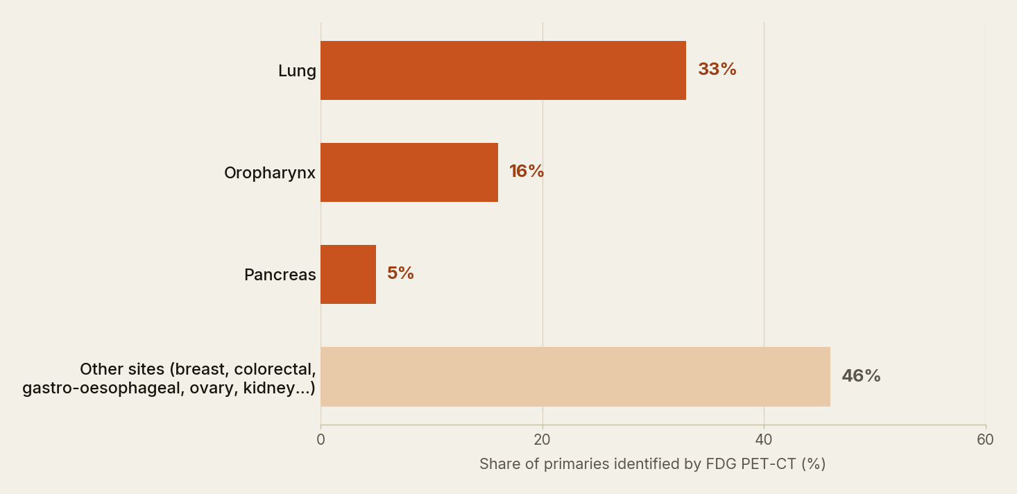
Autopsy series show that a primary exists in most patients. Lung and pancreas together account for about half; hepatobiliary, renal, colorectal and gastric primaries make up most of the rest. The primaries FDG PET-CT actually finds follow the same pattern (Figure 1).
1.4 Why is the primary occult?
- Too small to see: CUP tumours metastasise unusually early, so the primary can be below the resolution of CT and PET while the metastases are large.
- Regressed or eliminated: the primary may have involuted or been cleared by an immune response, leaving only its deposits.
- Already removed: a skin lesion or polyp excised years earlier and never recognised as the source.
1.5 Natural history and prognosis
- Favourable subsets (≈20%): treated like the cancer they resemble, with comparable outcomes; some are curable.
- Unfavourable CUP (≈80%): median survival about 6–12 months with empirical chemotherapy; about 30% alive at one year.
- How the subset is decided: three things are matched against the recognised favourable patterns (Section 2.2):
- histology and IHC — squamous, adenocarcinoma, poorly differentiated, neuroendocrine, germ-cell pattern;
- site and extent of metastases — a single nodal region or a single organ versus several organs;
- the patient — sex, age, and tumour markers such as PSA, AFP or β-hCG.
- If the combination fits one of the favourable patterns, the patient is treated as that cancer. If it fits none of them, the disease is unfavourable CUP.
- Adverse factors: poor performance status, raised LDH, liver metastases, multiple metastatic sites.
- Finding the primary is associated with longer survival — it unlocks site-specific therapy, and detectable primaries are a biologically different group.
2. Classification
2.1 By histology
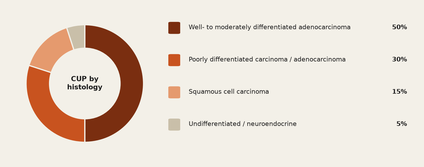
Histology decides the immunohistochemistry panel and the likely primaries (Figure 2).
| Histology | Share | Primaries to think of |
|---|---|---|
| Well- to moderately differentiated adenocarcinoma | ≈50% | Lung, pancreas, hepatobiliary, colorectal, gastric, breast, prostate, ovary |
| Poorly differentiated carcinoma / adenocarcinoma | ≈30% | As above; exclude germ-cell tumour, lymphoma and melanoma by IHC |
| Squamous cell carcinoma | ≈15% | Tonsil, tongue base, lung, oesophagus, cervix, anal canal, skin |
| Undifferentiated / neuroendocrine | ≈5% | Small bowel, pancreas, lung, rectum; high-grade NEC of any site |
2.2 By prognostic subset
Favourable subsets are defined by histology, site of metastasis and the patient. Each one is treated like a known cancer. Finding them is the most important job of the work-up. PET-CT helps by confirming that disease is limited to the site that defines the subset.
| Favourable subset | Treat as | What PET-CT adds |
|---|---|---|
| Squamous carcinoma in upper/mid cervical nodes | Head and neck cancer | Finds the primary before panendoscopy; excludes distant disease; shapes the radiotherapy field |
| Adenocarcinoma in axillary nodes (women) | Stage II–III breast cancer | Confirms isolated axillary disease (breast MRI is the better test for the primary) |
| Serous / papillary adenocarcinoma of the peritoneum (women) | Stage III ovarian cancer | Extent of peritoneal and extraperitoneal disease |
| Poorly differentiated midline carcinoma, young man, raised AFP / β-hCG (no longer an ESMO 2023 CUP subset — managed as a germ-cell tumour) | Extragonadal germ-cell tumour | Staging; residual mass after chemotherapy |
| Blastic bone metastases, raised PSA | Metastatic prostate cancer | PSMA PET rather than FDG |
| Well-differentiated neuroendocrine tumour (ESMO 2023 manages neuroendocrine neoplasms as such, whether or not a primary is found) | Metastatic NET | SSTR PET finds the primary and selects for PRRT |
| Single resectable metastasis, any histology | Oligometastatic disease | Proves it is truly solitary before surgery or SBRT |
| Squamous carcinoma in inguinal nodes | Anogenital / lower-limb skin primary | Locates the primary; excludes pelvic and distant disease |
| Adenocarcinoma with a colorectal IHC profile (CK20+, CDX2+, CK7−) | Metastatic colorectal cancer | Staging before site-specific therapy; colonoscopy looks for the primary |
| Carcinoma with renal-cell histology and IHC (renal-like CUP) | Metastatic renal cell carcinoma | Staging; clear-cell renal carcinoma is often FDG-poor, so contrast CT/MRI carry more weight |
| Melanoma in one nodal basin | Stage III melanoma | Whole-body staging including limbs |
Unfavourable CUP is everything else — the great majority:
- adenocarcinoma in liver, lung, bone or several sites,
- malignant ascites that is not papillary/serous,
- multiple brain metastases,
- squamous carcinoma outside the neck and groin.
3. Clinical Presentation and Pathological Work-up
3.1 Presentation
CUP presents through its metastases:
- neck lump; axillary or inguinal node
- hepatomegaly or abnormal liver function
- bone pain or pathological fracture
- pleural effusion or ascites
- brain lesion with neurological symptoms
- weight loss and malaise with an abnormal scan
Ask specifically about excised skin lesions, past biopsies or “benign” tumours, smoking and occupational exposure. Examine the breasts, skin, thyroid, testes and pelvis, and look at the oropharynx and larynx whenever the neck is involved.
3.2 Histopathology and immunohistochemistry
- Take a core biopsy from the most accessible metastasis. Fine-needle aspiration is not enough, because IHC and molecular profiling need adequate tissue.
- Immunohistochemistry (IHC) is the key step in suggesting the tissue of origin; save tissue for molecular profiling.
- Tier 1 — exclude mimics: lymphoma, melanoma, sarcoma, germ-cell tumour.
- Tier 2 — CK7 / CK20 pattern narrows the field.
- Tier 3 — organ-specific markers confirm the primary.
| CK7 / CK20 | Primaries suggested | Confirmatory markers |
|---|---|---|
| CK7+ / CK20− | Lung, breast, thyroid, endometrium, ovary (serous), pancreatico-biliary, upper GI | Lung: TTF-1, napsin A Breast: GATA3, GCDFP-15, mammaglobin, ER Ovary: PAX8, WT1 Thyroid: PAX8, thyroglobulin |
| CK7− / CK20+ | Colorectal; Merkel cell carcinoma | Colorectal: CDX2, SATB2 Merkel: dot-like CK20 with neuroendocrine markers |
| CK7+ / CK20+ | Urothelial, pancreatico-biliary, ovarian mucinous, gastric | Urothelial: GATA3, uroplakin, p63 Pancreatico-biliary / mucinous ovarian: CDX2 with CK7 |
| CK7− / CK20− | Prostate, renal, hepatocellular, adrenocortical, squamous carcinoma | Prostate: PSA, NKX3.1 Renal: PAX8, CAIX Liver: HepPar-1, arginase-1, glypican-3 Squamous: p40, p63, CK5/6 |
Two markers change the imaging plan directly:
- p16 (HPV surrogate) in a cervical nodal squamous carcinoma — points to the tonsil or tongue base; positive in roughly half of head and neck CUP overall (higher in North American series).
- EBER (Epstein–Barr virus RNA) — points to the nasopharynx.
- Neuroendocrine markers (synaptophysin, chromogranin, INSM1) with a low Ki-67 — switch from FDG to somatostatin-receptor PET.
3.3 Tumour markers and molecular profiling
| Marker | Order when | Points to |
|---|---|---|
| AFP and β-hCG | Young patient, poorly differentiated midline disease | Extragonadal germ-cell tumour |
| PSA | Man with bone metastases or adenocarcinoma of uncertain origin | Prostate |
| CA-125 | Woman with peritoneal disease or ascites | Ovarian / peritoneal serous carcinoma |
| Thyroglobulin, calcitonin | IHC suggests thyroid lineage | Differentiated / medullary thyroid carcinoma |
| CEA, CA 19-9, CA 15-3 | Not for localising the primary | Too non-specific — do not use to find the primary |
- Gene-expression (tissue-of-origin) classifiers suggest a tissue of origin in most cases, but their routine use is not yet supported by high-level evidence. The Fudan CUP-001 trial (2024) showed longer progression-free survival with therapy directed by a 90-gene classifier than with empirical chemotherapy.
- Comprehensive genomic profiling (NGS) is part of the ESMO 2023 work-up for unfavourable CUP to find targetable alterations and immunotherapy biomarkers (TMB, MSI); in CUPISCO, molecularly guided therapy improved PFS over continued chemotherapy in patients with disease control after induction (6.1 vs 4.4 months; HR 0.72).
4. Conventional Imaging Work-up
Imaging in CUP is stepwise. The aims: find a primary if one is findable, define extent, and identify a favourable subset — without exhausting a patient whose survival may be months.
| Modality | Role in CUP | Strengths | Limitations |
|---|---|---|---|
| Contrast CT chest, abdomen, pelvis | First-line in everyone; defines extent; often finds the primary | Fast, available; good for lung, liver, kidney, pancreas, nodes | Misses small primaries; no metabolic information; weak in head and neck and pelvis |
| Mammography ± ultrasound / breast MRI | Women with axillary adenocarcinoma or breast-lineage IHC | Breast MRI finds most mammographically occult primaries | Mammography poor in dense breasts |
| Ultrasound | Neck, thyroid, testes, pelvis; biopsy guidance | Cheap, no radiation, real-time biopsy | Operator-dependent; limited field |
| MRI | Brain, spine, pelvis, liver, breast | Best soft-tissue contrast; no radiation | Time and access; whole-body MRI not standard in CUP |
| Endoscopy (GI, bronchoscopy, panendoscopy) | Symptom- or histology-directed; panendoscopy is standard in cervical SCC CUP | Direct visualisation and biopsy | Invasive; blind biopsies have low yield |
| FDG PET-CT | When the above are negative and the result would change management | One whole-body study; finds primary in ≈40–55%; upstages 14–27% | False positives; blind to FDG-poor primaries; cost and access |
The order matters:
- Pathology and CT first — cheap, fast, decisive in most patients.
- PET-CT next, only if the primary is still unknown and one of the indications in Section 5 applies.
- In cervical squamous CUP, endoscopy and tonsillectomy should come after PET-CT, not before (NCCN; NICE CG104 still sequences PET after a negative panendoscopy) — biopsy-induced inflammation can hide the very site PET is meant to reveal.
5. When to Send for PET-CT — Indications in Short
Refer for FDG PET-CT when the standard work-up (Section 4) has not found the primary and one of the following applies:
- Squamous carcinoma in a cervical node with no mucosal primary on examination and CT/MRI — before panendoscopy, tonsillectomy or any biopsy of the upper aerodigestive tract.
- Single or few metastases where surgery or stereotactic radiotherapy with curative intent is being considered — to prove the disease is truly limited.
- Suspected favourable subset (axillary, peritoneal, inguinal, midline germ-cell pattern) — to confirm disease is confined to the site that defines the subset.
- No adequate tissue for IHC or molecular profiling — to choose the most active, most accessible site for re-biopsy.
- Staging is incomplete before systemic therapy, or CT is equivocal, and the result would change the plan.
- Histology points to another tracer: well-differentiated NET → SSTR PET; prostate lineage → PSMA PET; FDG-negative head and neck CUP → FAPI PET where available.
Do not refer when the patient is too unwell for any treatment (ECOG 3–4 with disseminated disease), or when the primary has already been found and PET-CT would only duplicate the CT.
6. Treatment Overview by Subset
| Group | Standard approach |
|---|---|
| Cervical squamous CUP | Chemoradiotherapy ± neck dissection, as for locally advanced head and neck cancer |
| Axillary adenocarcinoma (women) | Axillary clearance, breast radiotherapy and systemic therapy, as for breast cancer |
| Peritoneal papillary / serous adenocarcinoma (women) | Cytoreductive surgery and platinum–taxane chemotherapy, as for ovarian cancer |
| Extragonadal germ-cell pattern | Cisplatin-based chemotherapy |
| Prostate pattern (blastic bone, raised PSA) | Androgen deprivation therapy |
| Neuroendocrine tumour | Somatostatin analogues, PRRT, or surgery |
| Single metastasis | Surgery or stereotactic radiotherapy with radical intent |
| Unfavourable CUP, good performance status | Empirical platinum doublet (e.g. carboplatin–paclitaxel); genomic profiling with a view to molecularly guided therapy |
| Unfavourable CUP, ECOG ≥3 | Best supportive care; single-agent or no chemotherapy |
7. Radiotracers in CUP
| Tracer | Mechanism of action | Use in CUP · evidence · caveats |
|---|---|---|
| 18F-FDG | Glucose analogue. Enters the cell through GLUT transporters, is phosphorylated by hexokinase and trapped. Maps glycolytic activity, which is high in most carcinomas. | Use: default tracer for all histologies except NET and prostate lineage Evidence: primary found in 37–54% pooled; sensitivity 84%, specificity 84% (Kwee 2009) Caveats: misses breast (commonest false negative), prostate, renal, hepatocellular, mucinous and low-grade NET primaries; false positives ≈6% of patients (Willemse 2024) |
| 68Ga-DOTATATE / DOTATOC, 64Cu-DOTATATE (SSTR PET) | Somatostatin analogue. Binds somatostatin receptor subtype 2 (SSTR2), which is over-expressed on well-differentiated NET cells. | Use: well-differentiated NET of unknown primary; also selects for PRRT Evidence: primary found in ≈40–60% of NET-CUP (38% in the Menda DOTATOC series); ileum and pancreas commonest Caveats: high-grade or dedifferentiated NET needs FDG; pancreatic uncinate and splenule uptake are physiological |
| 68Ga- / 18F-PSMA ligands | Small-molecule inhibitor. Binds prostate-specific membrane antigen, a transmembrane enzyme over-expressed on prostate cancer cells; internalised after binding. | Use: adenocarcinoma with prostate-lineage IHC; blastic bone metastases with raised PSA Evidence: very high detection of prostate primary, nodes and bone disease Caveats: PSMA is also expressed in the neovasculature of other cancers — confirm lineage on IHC |
| 68Ga-FAPI-04 / -46, 18F-FAPI | Quinoline-based inhibitor. Binds fibroblast activation protein on cancer-associated fibroblasts in the tumour stroma; independent of tumour-cell metabolism. | Use: FDG-negative head and neck CUP; extracervical CUP under study Evidence: 91 HNCUP patients — primary found in 51% with FAPI vs 19% with FDG; PPV 98% vs 43%; management changed in 24% (Gu 2024) Caveats: uptake in fibrosis, healing tissue and some benign conditions; availability |
| 18F-FES | Oestradiol analogue. Binds oestrogen receptor-α in ER-positive tumour cells. | Use: suspected ER-positive / lobular breast primary when breast MRI is equivocal Evidence: small series Caveats: limited availability |
| 18F-fluorocholine, 18F-FDOPA | Choline: taken up by choline transporters and phosphorylated by choline kinase; reflects membrane phospholipid synthesis. FDOPA: amino-acid analogue taken up by LAT transporters and decarboxylated by AADC; reflects catecholamine and serotonin synthesis in neuroendocrine cells. | Use: HCC and prostate (choline); small-bowel NET and phaeochromocytoma-type tumours (FDOPA) Caveats: niche; largely replaced by PSMA and SSTR PET |
8. Preparation and Acquisition — Points Specific to CUP
Standard FDG preparation applies (fasting, glucose control, rest). The points below are the ones that matter when the diagnosis is CUP.
8.1 Before the scan
- Read the histology, IHC and the CT first. Choose the tracer from the IHC — SSTR PET for NET, PSMA PET for prostate lineage — before booking FDG.
- Schedule PET-CT before panendoscopy, tonsillectomy or any biopsy of the upper aerodigestive tract; if timing allows, leave an interval after a biopsy elsewhere, because biopsy sites take up FDG.
- Keep the patient warm and quiet, and ask them not to talk or chew after injection — brown fat, vocal-cord and masticator uptake obscure the neck.
8.2 Acquisition
- Contrast-enhanced diagnostic CT in the same session whenever renal function allows: one combined study completes the imaging work-up, although the Kwee meta-analysis found no significant effect of CT contrast on primary-tumour detection.
- Cervical nodal disease: add a dedicated head and neck bed position — arms down, small field of view, fine matrix, longer emission time — before the standard arms-up torso acquisition; the EANM tumour guideline notes that such a two-step protocol may be helpful for head and neck imaging.
- Some centres add a delayed neck acquisition to help separate a small primary from physiological lymphoid uptake; evidence for this in CUP is limited.
- Extend to the feet for melanoma, sarcoma-like histology or lower-limb disease; MRI remains the test for brain metastases.
9. Interpretation and Reporting
A CUP study is read differently from a staging study. The known metastases are the least interesting part. The search is for a small focus that could be the origin, and it is conducted with the IHC in mind.
- Start with the organs the histology makes plausible — lung and pancreas for adenocarcinoma, tonsils and tongue base for cervical squamous carcinoma, bowel for CK20-positive tumours — then survey the rest.
- Compare the two sides of the oropharynx. Asymmetric tonsillar uptake is the key sign in head and neck CUP, but it is common physiologically; use the CT and the side of the nodal disease to raise confidence.
- No SUV threshold separates a primary from inflammation. Recommend biopsy on pattern, anatomical correlate and fit with the histology.
- Look for the second reason for the scan: extra metastases, a solitary-metastasis picture, and the best site for a repeat biopsy.
- Know the mimics: lingual and palatine tonsils, salivary glands, vocal cords, thymus, brown fat, bowel (metformin), endometrium and ovaries mid-cycle, healing fractures and biopsy sites, granulomatous nodes.
9.1 FDG blind spots
- Breast carcinoma, especially lobular and low-grade disease — the commonest false negative (27% of missed primaries).
- Prostate adenocarcinoma, clear-cell renal carcinoma, hepatocellular carcinoma.
- Mucinous adenocarcinomas and well-differentiated neuroendocrine tumours.
- Very small primaries of any type.
9.2 The report
- Probable primary — yes / no, and how confident.
- Where a directed biopsy should be taken.
- Full extent of disease and any change in stage.
- Does the picture support a favourable subset or oligometastatic disease?
- If negative: state that no FDG-avid primary was found and whether a lineage-specific tracer is indicated.
- Cervical squamous CUP: compare the tonsils side by side on the dedicated neck acquisition; report the side and sub-site.
- A PET-CT that shows the known metastasis is the only site of disease is as useful as one that finds the primary: it allows surgery or stereotactic radiotherapy with curative intent.
- Neuroendocrine, prostate or lobular breast on IHC → the right tracer first, not FDG.
10. Role of PET-CT in CUP
10.1 Detecting the primary
PET-CT finds the primary in roughly two out of five patients overall (Figure 3). About a third remain true CUP even after PET-CT and further work-up (Willemse 2024).
Where the metastases are decides the yield (2024 individual-patient-data meta-analysis, 1,865 patients with extracervical CUP):
- Highest when the metastases are in the brain (primary found in ≈74%), liver (≈54%) or bone (≈49%) — lung was by far the commonest primary (29% of all patients).
- Lowest when the disease is in lymph nodes (≈37%), peritoneum (≈38%) or soft tissue (≈35%) — the primaries behind these are more often small, mucosal or FDG-poor.
10.2 Head and neck CUP — the strongest indication
- Squamous carcinoma in an upper or mid-cervical node with no mucosal primary: every major guideline recommends FDG PET-CT.
- The primary is usually oropharyngeal — the palatine tonsil or tongue base in most cases where it is found.
- p16 positivity (roughly half of cases overall) makes an oropharyngeal origin likely before any imaging.
- The 2004 meta-analysis of PET alone found the primary in 24.5% of patients whose conventional work-up (CT/MRI and, in most studies, panendoscopy) was negative; sensitivity 88%, specificity 75%, with most false positives in the tonsil.
- Timing is critical: PET-CT before panendoscopy, tonsillectomy or biopsy.
- Sequence: examination and p16/EBV → CT or MRI → FDG PET-CT with dedicated neck acquisition → panendoscopy with directed biopsies and ipsilateral tonsillectomy → TORS lingual tonsillectomy in selected PET-negative patients.
- If PET-CT does not show the primary, the surgical search follows. Removing the whole ipsilateral tonsil finds a hidden primary in about 30% of patients, whereas a deep biopsy of the tonsil finds it in only about 3%. So tonsillectomy, not biopsy, is the standard step.
- If the tonsil is clear, transoral robotic (TORS) tongue-base mucosectomy finds the primary in roughly 70% of patients in pooled series. It is reserved for selected patients because it carries morbidity.
- Finding the primary spares wide-field mucosal radiotherapy and its lifelong toxicity, and excludes distant disease.
- FAPI PET when FDG is negative: in 18 FDG-negative patients, 68Ga-FAPI found the primary in 7 (39%) (Gu 2022). In a prospective head-to-head trial of 91 head and neck CUP patients with a negative or equivocal conventional work-up, 68Ga-FAPI PET-CT found the primary in 51% versus 19% with FDG, with far fewer false positives (PPV 98% vs 43%). FAPI images the tumour stroma, so it is not defeated by the physiological lymphoid FDG uptake that hides small tonsillar and tongue-base primaries.
- Why it matters: in the same trial, patients whose primary was never found had a much higher risk of death (hazard ratio ≈5.8). A second tracer is therefore worth considering before a patient is committed to wide-field mucosal radiotherapy on a negative FDG scan.
10.3 Staging and additional metastases
- Extra metastases are found in 14–27% of patients.
- A favourable subset depends on disease being confined to one site. If PET-CT finds a second site — for example bone metastases in a woman thought to have isolated axillary nodes — she no longer belongs to the favourable subset, and the intent of treatment changes from curative to palliative.
- They can show that disease is too widespread for further searching to matter.
- Conversely, a thorough negative survey is what licenses radical local treatment of a single lesion.
10.4 Directing the biopsy
- Identifies the most metabolically active, most accessible lesion for re-biopsy when the first sample is inadequate for IHC or molecular profiling.
- Steers the needle away from necrotic or treated tissue, which is important now that genomic profiling is standard in unfavourable CUP.
10.5 Treatment planning
- Head and neck CUP: PET-CT maps the involved nodes for the radiotherapy plan. If it finds the primary, the mucosal field can be limited to that sub-site. If it does not, the radiation oncologist must choose between irradiating the whole pharyngeal mucosa and a smaller, selective volume; the PET findings guide that choice.
- Oligometastatic disease: surgery or stereotactic radiotherapy is offered only after PET-CT has shown no disease elsewhere.
- Unfavourable CUP: the baseline study for later response assessment; it also documents every disease site, which trials require.
10.6 Impact on management
- Management changes in ≈35% of patients — by finding the primary, upstaging, downstaging or redirecting biopsy.
- In that meta-analysis (Woo 2021, 38 studies, 2,795 patients; cited in the NCI summary), management changed because a primary was found in 22% and because extra metastases were found in 14%.
10.7 Response assessment and restaging
- Little CUP-specific evidence; practice follows the cancer the patient is treated as.
- Head and neck CUP after chemoradiotherapy: 12-week PET-CT with a structured response score guides the decision on neck dissection.
- Unfavourable CUP on systemic therapy: contrast CT with RECIST is standard; PET-CT when CT is equivocal, disease is bone-predominant, or a new symptom needs explaining.
- At relapse, PET-CT is worthwhile when the primary was never found — it occasionally declares itself later.
10.8 Lineage-directed PET
- NET of unknown primary: SSTR PET finds the primary in ≈40–60% (38% in the DOTATOC series; ileum and pancreas commonest) and establishes PRRT eligibility.
- Prostate lineage: PSMA PET identifies the primary and the extent of nodal and bone disease; confirm lineage on IHC because other tumours can be PSMA-positive.
- FDG-negative head and neck CUP: FAPI PET is the emerging problem-solver.
- Suspected lobular / ER-positive breast primary: breast MRI first; FES PET where available.
11. Prognostic PET Biomarkers
- Patients in whom PET-CT finds the primary live longer — they receive site-specific treatment and are a biologically different group. In the 2024 FAPI trial an unidentified primary carried a hazard ratio of ≈5.8 for death.
- Metabolic tumour volume and total lesion glycolysis are prognostic in many cancers, but CUP-specific data are sparse.
12. Guideline Recommendations
| Guideline | Recommendations on PET-CT in CUP |
|---|---|
| ESMO Clinical Practice Guideline — CUP (Krämer et al., Ann Oncol 2023) |
|
| NCCN — Occult Primary; Head and Neck Cancers (current versions) |
|
| NICE CG104 — Metastatic malignant disease of unknown primary origin (2010; reviewed 2023) |
|
| EANM / SNMMI FDG PET-CT procedure guidelines |
|
| ENETS / NANETS (NET); EAU / EANM (prostate) |
|
- Kwee & Kwee 2009 — 11 studies, 433 patients: FDG PET-CT found the primary in 37%; sensitivity 84%, specificity 84%; lung 33%, oropharynx 16%, pancreas 5% of primaries found; breast the commonest miss (27%).
- Willemse et al. 2024 — patient-level data from 32 studies, 1,865 patients: primary found in 54% overall; yield depends on the metastatic site — 74% with brain metastases, 35% with soft-tissue metastases; lung was the primary in 29% of all patients; one-third remained CUP after PET-CT and follow-up; cervical presentations excluded.
- Rusthoven et al. 2004 — 16 studies, 302 patients with cervical nodes, PET alone: primary found in 24.5% after negative conventional work-up.
- Gu et al. 2024 — 91 head and neck CUP patients (negative or equivocal conventional work-up): 68Ga-FAPI PET-CT found the primary in 51% vs 19% for FDG; PPV 98% vs 43%; management changed in 24%.
- Menda et al. 2017 — 40 NET patients: 68Ga-DOTATOC PET-CT found the primary in 38%; ileum and pancreas commonest.
13. Emerging Tracers and Future Directions
| Development | What it offers | Where it stands |
|---|---|---|
| FAPI PET-CT | Images tumour stroma; independent of glucose metabolism; very high tumour-to-background ratio | Prospective HNCUP data (51% vs 19% detection); small extracervical series; caveat — uptake in fibrosis and healing |
| Total-body / long-axial-field PET | Higher sensitivity; whole-body dynamic and dual-time-point imaging; lower dose | Early clinical use; may lift detection of small primaries |
| PET/MRI | Better soft-tissue contrast in pelvis, liver and head and neck | Not yet shown to raise detection in CUP |
| Molecular-imaging integration | IHC and gene-expression prediction choose the tracer and reading strategy; radiomic / deep-learning tissue-of-origin prediction | Under study |
| Theranostics | SSTR-avid NET can receive 177Lu-DOTATATE whether or not the primary is found; PSMA-avid prostate-lineage disease follows prostate-cancer indications for 177Lu-PSMA; FAP-targeted radioligands in trials | SSTR established; PSMA only within prostate-cancer indications; FAP early-phase |
| Better trials | Prospective studies with PET-CT in the defined baseline work-up and outcomes, not only detection rates, as endpoints | Needed — treatment trials (CUPISCO, CUP-001) now set the standard |
14. Pitfalls
- Biopsy or tonsillectomy before PET — iatrogenic inflammation hides the oropharynx for weeks.
- Calling tonsillar asymmetry a primary without CT correlation or the matching nodal side — physiological lymphoid uptake is the commonest false positive in head and neck CUP.
- Reassuring a patient with lobular breast, prostate, renal or low-grade NET histology on a negative FDG scan.
- Missing a small pancreatic or lung primary because only the known metastases were examined.
- Brown fat, muscle and vocal-cord uptake in a cold, anxious or talking patient — preparation matters more in CUP than in routine staging.
- Reporting an SUV cut-off as if it separated primary from inflammation.
- Ordering PET-CT in an ECOG 3–4 patient with disseminated disease where no result would change management.
- Forgetting that PSMA uptake occurs in the neovasculature of non-prostate tumours, and that FAPI accumulates in fibrosis and healing tissue.
15. Clinical Scenarios
Cervical node, p16-positive squamous carcinoma. 58-year-old non-smoker, 3 cm level II node; examination and CT show no primary.
→ FDG PET-CT with dedicated neck acquisition before panendoscopy: asymmetric uptake in the ipsilateral tongue base, no distant disease → directed biopsy and ipsilateral tonsillectomy confirm a 9 mm primary → treated as HPV-positive oropharyngeal cancer with a focused radiotherapy volume.
Liver metastases, adenocarcinoma, CK7+/CK20−, TTF-1 negative. 66-year-old smoker; CT shows no primary.
→ FDG PET-CT shows an avid 1.4 cm right upper-lobe nodule and mediastinal nodes → bronchoscopic biopsy: lung adenocarcinoma, EGFR-mutant → leaves the CUP pathway for targeted therapy. Lung is the commonest primary PET finds.
Bone and liver metastases, well-differentiated NET, Ki-67 4%. FDG PET-CT requested; only faint uptake in known lesions.
→ The right test was SSTR PET: 68Ga-DOTATATE shows intense uptake in all lesions and a 1.2 cm ileal primary → somatostatin analogue, primary resected for impending obstruction, PRRT eligibility documented. Histology should choose the tracer before the first PET is booked.
16. Frequently Asked Questions
Should every CUP patient have an FDG PET-CT?
No. Recommended in cervical squamous CUP; suggested where it would change management — limited disease that could be treated radically, biopsy targeting, or completing staging. Not in a frail patient with disseminated disease.
How often does it find the primary?
37–41% in the classic meta-analyses; 54% in the 2024 patient-level analysis. Higher with brain or liver metastases, lower with nodal or soft-tissue disease.
Why PET-CT before panendoscopy and tonsillectomy?
Biopsy and surgery cause FDG-avid inflammation for weeks, and a PET-directed biopsy has a far higher yield than a blind one.
Which primaries does FDG miss?
Lobular and low-grade breast, prostate, clear-cell renal, hepatocellular, mucinous and well-differentiated neuroendocrine tumours. IHC should redirect these patients to breast MRI, PSMA PET or SSTR PET.
What is the false-positive rate?
About 6% of patients in the 2024 patient-level analysis (1–22% across metastatic-site subgroups); in Kwee 2009 the lung and oropharynx were the commonest false-positive sites. No focus is a primary without anatomical correlation, and most need histological confirmation.
Is contrast-enhanced CT needed with the PET?
Preferred if not already done — one combined study completes the work-up, although the Kwee meta-analysis found no significant effect of CT contrast on primary-tumour detection.
Does finding the primary improve survival?
Patients with an identified primary live longer (an association — they are partly a different group), and in favourable subsets treatment changes fundamentally. For unfavourable CUP, molecularly guided therapy (CUPISCO) and gene-expression-directed therapy (CUP-001) now have randomised support.
Is FAPI PET ready for routine use?
For FDG-negative head and neck CUP the prospective data are compelling and some centres use it as a problem-solver; elsewhere it remains investigational. Availability and cost limit adoption.
17. Memory Box and High-Yield Pearls
P — Pathology first: core biopsy, IHC, p16/EBER, neuroendocrine markers.
R — Rule out the favourable subsets.
I — Image conventionally: contrast CT chest/abdomen/pelvis ± mammography/breast MRI.
M — Markers by context: AFP, β-hCG, PSA, CA-125.
A — Add FDG PET-CT when it changes the plan: cervical SCC (before endoscopy), oligometastatic disease, biopsy targeting.
R — Recognise the FDG blind spots (breast, prostate, renal, NET) and switch tracer.
Y — Yield ≈40–55%; when PET is negative, stop and treat as CUP.
- Pathology first, imaging second, PET third — and only when it will change the plan.
- Cervical squamous node with no primary: FDG PET-CT with a dedicated neck acquisition, before anyone touches the tonsils.
- The primary PET most often finds is lung; the one it most often misses is breast.
- Proving that a metastasis is solitary changes management as much as finding the primary: the patient becomes a candidate for radical local treatment.
- Let the IHC choose the tracer: NET → SSTR PET; prostate → PSMA PET; FDG-negative HNCUP → FAPI PET.
- Report extra metastases and the best re-biopsy site — molecular profiling needs good tissue.
- Once a good-quality PET-CT is negative, stop searching and treat as CUP.
18. Test Yourself
Six quick questions. Pick an answer to see the explanation.
19. Glossary
- CUP (carcinoma of unknown primary) — a histologically confirmed metastatic malignancy whose primary site cannot be identified after a standardised work-up.
- HNCUP — head and neck CUP — squamous carcinoma in a cervical node with no mucosal primary found.
- IHC (immunohistochemistry) — staining of tissue sections for lineage-specific proteins (CK7, CK20, TTF-1, GATA3, PAX8, PSA and others) to predict the tissue of origin.
- p16 — a surrogate marker of HPV-driven oropharyngeal carcinoma; positive in roughly half of head and neck CUP.
- EBER — in-situ hybridisation for Epstein–Barr virus RNA; positivity points to a nasopharyngeal primary.
- SSTR PET — PET with a somatostatin-receptor ligand (68Ga-DOTATATE, DOTATOC or 64Cu-DOTATATE) for well-differentiated neuroendocrine tumours.
- PSMA PET — PET with a ligand for prostate-specific membrane antigen; the imaging of choice for prostate-lineage disease.
- FAPI PET — PET with a fibroblast-activation-protein inhibitor that images tumour stroma; the emerging problem-solver in FDG-negative HNCUP.
- TORS — transoral robotic surgery — used for tongue-base mucosectomy when the tonsil is clear in HNCUP.
- IPD meta-analysis — individual-patient-data meta-analysis — pooled analysis of the raw patient records from many studies rather than their summary figures.
- PRRT — peptide receptor radionuclide therapy (for example 177Lu-DOTATATE) for SSTR-positive neuroendocrine tumours.
- SBRT — stereotactic body radiotherapy — high-dose, precisely targeted radiotherapy for a solitary or oligometastatic lesion.
- ECOG performance status — a 0–5 scale of a patient’s functional state; 3–4 means largely bed-bound.
20. References
- Krämer A, Bochtler T, Pauli C, et al. Cancer of unknown primary: ESMO Clinical Practice Guideline for diagnosis, treatment and follow-up. Ann Oncol. 2023;34(3):228-246.
- Kwee TC, Kwee RM. Combined FDG-PET/CT for the detection of unknown primary tumors: systematic review and meta-analysis. Eur Radiol. 2009;19(3):731-744.
- Burglin SA, Hess S, Høilund-Carlsen PF, Gerke O. 18F-FDG PET/CT for detection of the primary tumor in adults with extracervical metastases from cancer of unknown primary: a systematic review and meta-analysis. Medicine (Baltimore). 2017;96(16):e6713.
- Willemse JRJ, Lambregts DMJ, Balduzzi S, et al. Identifying the primary tumour in patients with cancer of unknown primary (CUP) using [18F]FDG PET/CT: a systematic review and individual patient data meta-analysis. Eur J Nucl Med Mol Imaging. 2024;52(1):225-236.
- Rusthoven KE, Koshy M, Paulino AC. The role of fluorodeoxyglucose positron emission tomography in cervical lymph node metastases from an unknown primary tumor. Cancer. 2004;101(11):2641-2649.
- Masuoka S, Hiyama T, Kuno H, et al. Imaging approach for cervical lymph node metastases from unknown primary tumor. RadioGraphics. 2023;43(3):e220071.
- Gu B, Yang Z, Du X, et al. Imaging of tumor stroma using 68Ga-FAPI PET/CT to improve diagnostic accuracy of primary tumors in head and neck cancer of unknown primary: a comparative imaging trial. J Nucl Med. 2024;65(3):365-371.
- Gu B, Xu X, Zhang J, et al. The added value of 68Ga-FAPI PET/CT in patients with head and neck cancer of unknown primary with 18F-FDG-negative findings. J Nucl Med. 2022;63(6):875-881.
- Menda Y, O’Dorisio TM, Howe JR, et al. Localization of unknown primary site with 68Ga-DOTATOC PET/CT in patients with metastatic neuroendocrine tumor. J Nucl Med. 2017;58(7):1054-1057.
- Krämer A, Bochtler T, Pauli C, et al. Molecularly guided therapy versus chemotherapy after disease control in unfavourable cancer of unknown primary (CUPISCO): an open-label, randomised, phase 2 study. Lancet. 2024;404(10452):527-539.
- Liu X, Zhang X, Jiang S, et al. Site-specific therapy guided by a 90-gene expression assay versus empirical chemotherapy in patients with cancer of unknown primary (Fudan CUP-001): a randomised controlled trial. Lancet Oncol. 2024;25(8):1092-1102.
- National Comprehensive Cancer Network. NCCN Clinical Practice Guidelines in Oncology: Occult Primary (Cancer of Unknown Primary); Head and Neck Cancers. Current versions, accessed September 2026.
- National Institute for Health and Care Excellence. Metastatic malignant disease of unknown primary origin in adults: diagnosis and management. Clinical guideline CG104. London: NICE; 2010 (surveillance review 2023).
- National Cancer Institute. Carcinoma of Unknown Primary Treatment (PDQ®) — Health Professional Version. Bethesda, MD: NCI; updated 2025.
- Boellaard R, Delgado-Bolton R, Oyen WJG, et al. FDG PET/CT: EANM procedure guidelines for tumour imaging: version 2.0. Eur J Nucl Med Mol Imaging. 2015;42(2):328-354.
- Pavlidis N, Pentheroudakis G. Cancer of unknown primary site. Lancet. 2012;379(9824):1428-1435.
- Waltonen JD, Ozer E, Schuller DE, Agrawal A. Tonsillectomy vs. deep tonsil biopsies in detecting occult tonsil tumors. Laryngoscope. 2009;119(1):102-106.
- van Weert S, Rijken JA, Plantone F, et al. A systematic review on transoral robotic surgery (TORS) for carcinoma of unknown primary origin: has tongue base mucosectomy become indispensable? Clin Otolaryngol. 2020;45(5):732-738.
- Woo S, Becker AS, Do RKG, et al. Impact of 18F-fluorodeoxyglucose positron emission tomography on management of cancer of unknown primary: systematic review and meta-analysis. Eur J Cancer. 2021;159:60-77.
- Losa F, Etxaniz O, Giménez A, et al. SEOM-GECOD clinical guideline for cancer of unknown primary (update 2025). Clin Transl Oncol. 2026;28(5):1610-1623.
- Boscolo-Rizzo P, Schroeder L, Romeo S, Pawlita M. The prevalence of human papillomavirus in squamous cell carcinoma of unknown primary site metastatic to neck lymph nodes: a systematic review. Clin Exp Metastasis. 2015;32(8):835-845.