Parathyroid Imaging
At a glance
- Localise, don't diagnose. Imaging finds the gland(s) in biochemically proven hyperparathyroidism so that surgery can be focused.
- MIBI. ⁹⁹ᵐTc-sestamibi is retained by mitochondria-rich parathyroid tissue; dual-phase or subtraction imaging with SPECT/CT is standard.
- Fluorocholine. ¹⁸F-fluorocholine PET/CT is more sensitive than MIBI and is increasingly used first line or after negative/discordant scans.
- Pitfalls. Multigland disease, small glands, rapid MIBI washout, thyroid nodules and ectopic glands (about 16%) cause errors.
- Beyond nuclear medicine. Ultrasound is always paired with scintigraphy; 4D-CT and venous sampling help before reoperation.
1. Anatomy and embryology
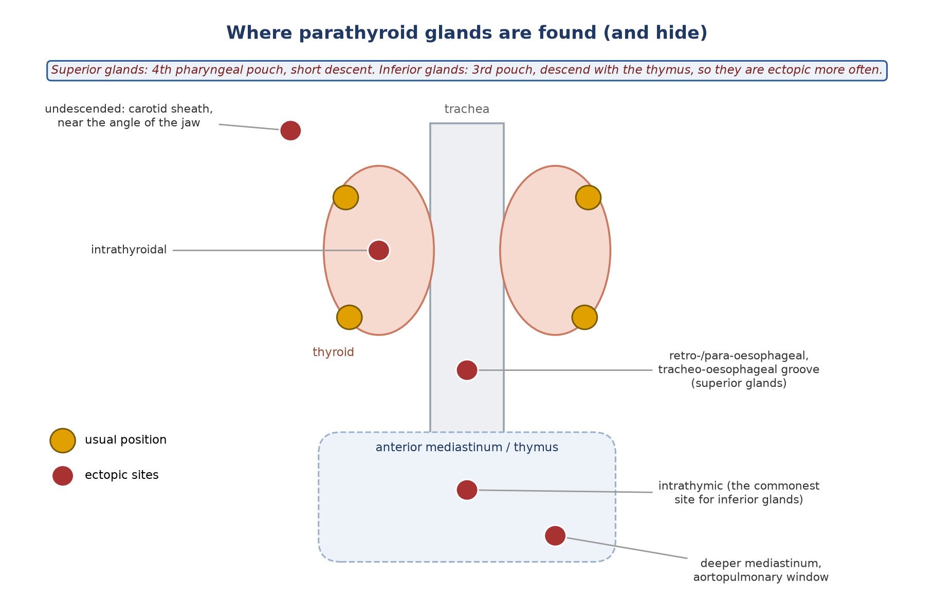
- Usually four glands: two superior (from the 4th pharyngeal pouch) and two inferior (from the 3rd pouch, descending with the thymus).
- The superior glands lie posterior to the thyroid near the cricothyroid junction, posterior to the recurrent laryngeal nerve; the inferior glands are anterior to it.
- Size: normal glands are about 3–5 mm and weigh roughly 30–40 mg; above ~60 mg is generally abnormal (some use 40 mg).
- Cells: chief cells and larger, mitochondria-rich oxyphil cells; the mitochondria explain MIBI retention.
- Ectopic glands: about 16% of operated patients. Inferior glands are ectopic more often than superior (about 60% vs 40% of ectopic glands); the thymus is the commonest inferior site.
- Supernumerary glands: about 3–13%. Ectopic and extra glands are a common cause of persistent or recurrent hyperparathyroidism.
2. What imaging is for
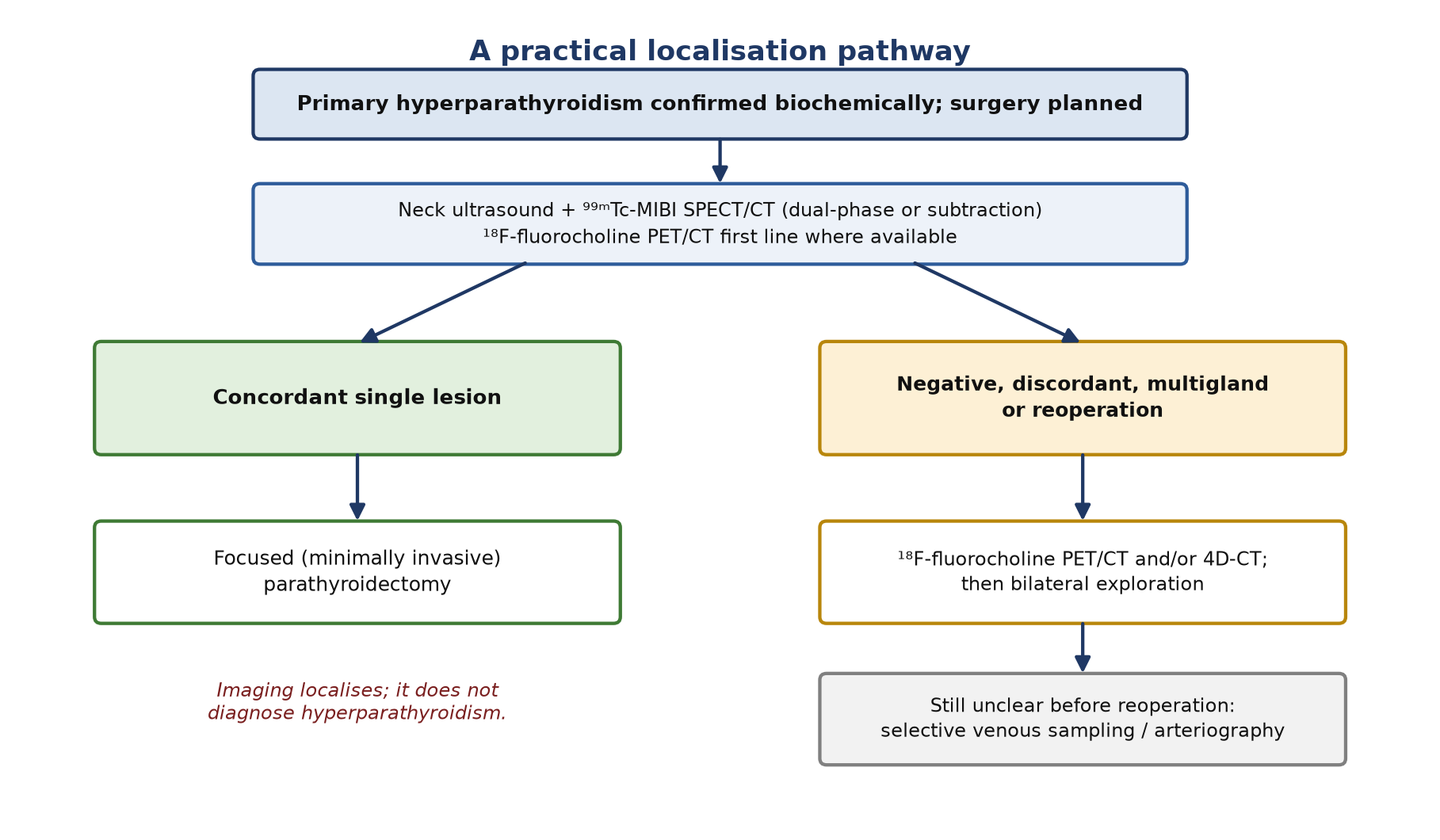
- Primary hyperparathyroidism is diagnosed biochemically (raised or inappropriately normal PTH with hypercalcaemia). Imaging does not make or exclude the diagnosis.
- Its job is localisation: a single, well-localised adenoma allows focused (minimally invasive) surgery instead of bilateral neck exploration.
- Main indications: before first surgery for primary hyperparathyroidism; persistent or recurrent disease after surgery; selected secondary or tertiary hyperparathyroidism.
Before the scan
- Bloods: PTH, calcium, 25-hydroxyvitamin D, phosphate, creatinine; 24-h urinary calcium where relevant.
- Previous imaging: review ultrasound, CT or MRI.
- Iodine (subtraction studies): recent iodinated contrast, iodine-containing drugs or thyroid hormone reduce thyroid uptake of pertechnetate or ¹²³I. In patients on thyroid hormone, prefer dual-phase MIBI.
- Drugs that lower MIBI uptake: active vitamin D analogues and calcimimetics (e.g. cinacalcet) — pause for 2 weeks if clinically possible. Calcium channel blockers also reduce uptake, but no withdrawal period has been established.
3. Tracers and how they work
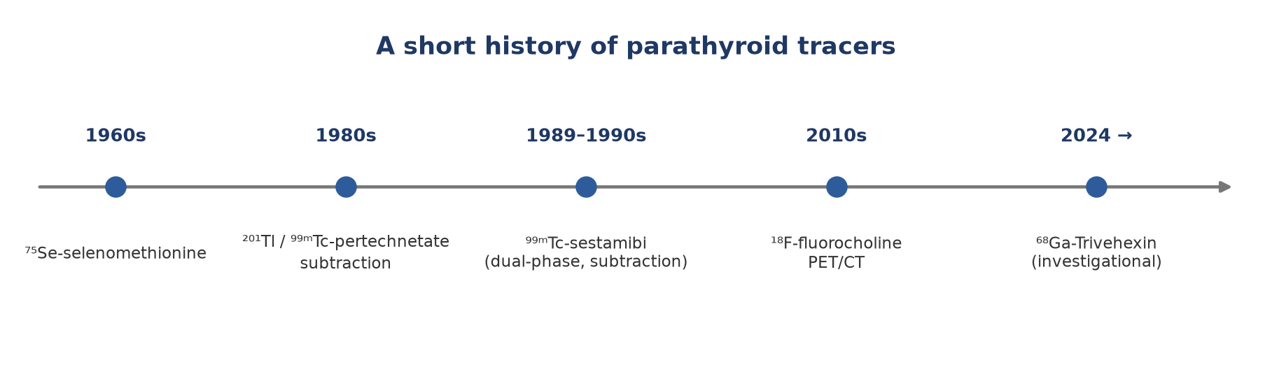
- ⁹⁹ᵐTc-sestamibi (MIBI): lipophilic cation concentrated in mitochondria. It usually washes out faster from thyroid than from hyperfunctioning parathyroid, which allows single-tracer dual-phase imaging. First used for parathyroid imaging in 1989; FDA-approved for cardiac imaging in 1990.
- ⁹⁹ᵐTc-tetrofosmin: no useful differential washout, so it is suitable only for subtraction protocols.
- ⁹⁹ᵐTc-pertechnetate: trapped (not organified) by thyroid; used for subtraction.
- ¹²³I: half-life ~13 h, 159 keV; trapped and organified by thyroid; used for subtraction with a separate energy window.
- Historical: ⁷⁵Se-selenomethionine (1960s) and ²⁰¹Tl/pertechnetate subtraction (1980s) were limited by poor image quality and, for thallium, a low-energy photon and high radiation dose.
4. Scintigraphy protocols
| Protocol | Activity | Imaging | Notes |
|---|---|---|---|
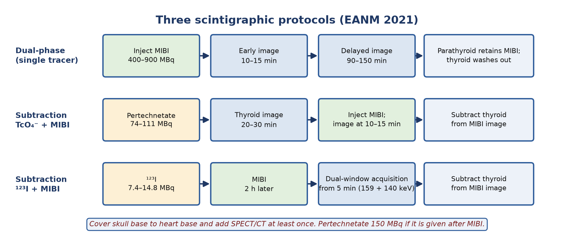
| Dual-phase MIBI | 400–900 MBq MIBI | 10–15 min and 90–150 min | Relies on differential washout; add SPECT/CT |
| MIBI + pertechnetate subtraction | Pertechnetate 74–111 MBq if first, 150 MBq if after MIBI | Pertechnetate at 20–30 min; MIBI at 10–15 min | Digital or visual subtraction; the tracers can also be given 2–3 days apart |
| MIBI + ¹²³I subtraction | ¹²³I 7.4–14.8 MBq; MIBI 2 h later | Simultaneous dual-window acquisition from 5 min after MIBI | Windows at 159 keV (10%) and 140 keV (15–20%) |
Table 1. EANM 2021 protocol summary.
- Camera: large field of view, LEHR collimator, 140 ± 10 keV window (15–20% for subtraction), 128² or 256² matrix; a pinhole view of the thyroid bed is optional.
- Field of view: the whole neck and chest down to the base of the heart, so that mediastinal ectopic glands are not missed.
- SPECT/CT: recommended at least once; it localises the gland in depth, separates thyroid from parathyroid and finds ectopic glands missed on planar images.
- Rapid parathyroid washout: add intermediate or dynamic images. Slow thyroid washout (e.g. nodules): subtraction helps.
5. Reading the scan
- Typical adenoma: focal uptake near the thyroid that persists or increases on delayed images while thyroid activity fades, or that remains after thyroid subtraction.
- Look for more than one focus (multigland disease), ectopic foci down to the heart base, and intrathyroidal lesions.
| Pitfall | Why |
|---|---|
| False positive | Thyroid adenoma or carcinoma, thyroiditis, lymph nodes, other tumours, brown fat |
| False negative | Small glands, multigland hyperplasia, rapid MIBI washout, few oxyphil cells, drugs (vitamin D analogues, calcimimetics, calcium channel blockers) |
| Missed ectopic gland | Field of view too small; a gland near the heart hidden by myocardial MIBI uptake |
| Subtraction artefact | Patient movement between acquisitions; poor thyroid uptake after iodine exposure |
6. PET tracers
| Tracer | Activity and timing | Evidence and role |
|---|---|---|
| ¹⁸F-fluorocholine | 100–300 MBq (1.5–3.2 MBq/kg); image at 60 min, ideally with an early 5-min acquisition; or one acquisition at 20 min with delayed images if negative | Most studied PET tracer. Head-to-head in 103 patients: sensitivity 92% vs 39–56% for single MIBI methods and 65% combined. Preferred after negative or discordant scans; first line where available |
| ¹¹C-choline | 200–650 MBq | Prospective pilot (40 patients): better image quality than MIBI, equal or better accuracy. Needs an on-site cyclotron |
| ¹¹C-methionine | 370–1100 MBq; best parathyroid/soft-tissue contrast at 10 min, parathyroid/thyroid at 40 min | Useful when MIBI is negative; on-site cyclotron needed |
| ¹⁸F-FDG | — | Not suitable for benign hyperfunctioning glands (sensitivity 0–94% across studies); may stage parathyroid carcinoma |
| ¹⁸F-FET, ¹⁸F-FDOPA | — | Faint or no uptake in adenomas: not recommended |
| ⁶⁸Ga-Trivehexin (integrin αvβ6) | — | First series 2024 (13 patients: lesion detection 94% vs 59% for MIBI); small, mostly single-centre studies. Investigational |
7. Other localisation tests
- Neck ultrasound: always paired with scintigraphy. Adenomas are hypoechoic, oval and separated from the thyroid by an echogenic line. Pooled sensitivity 76% and PPV 93% (meta-analysis, 19 studies), similar to MIBI SPECT (79% and 91%), but much lower in multigland disease (19% vs 67% in single-gland disease in one series).
- FNA with PTH washout: confirms parathyroid tissue in selected cases (sensitivity 70–100%, specificity 75–100%); no agreed cut-off.
- 4D-CT: non-contrast, arterial, venous and delayed phases; adenomas typically enhance early and wash out. In 45 reoperative patients sensitivity was 88% vs 54% for MIBI SPECT/CT and 21% for ultrasound. Higher radiation dose to the thyroid.
- Selective venous sampling: PTH gradient in neck and mediastinal veins; pooled sensitivity 74%, specificity 41% (12 studies). Reserved for reoperation when non-invasive tests fail.
- Selective arteriography with calcium stimulation: useful after previous surgery; PPV 92% in one series.
8. Challenges and what is coming
- Multigland disease and ectopic glands remain the main causes of failed localisation; PET performs better than SPECT here.
- Parathyroid carcinoma is rare; FDG and fluorocholine may help staging.
- Fluorocholine PET/MRI is promising (lower dose, better soft-tissue contrast), for example in children or secondary hyperparathyroidism.
- Research tracers include mitochondrial agents such as ¹⁸F-flurpiridaz.
Summary
- Confirm hyperparathyroidism biochemically before imaging.
- Pair ultrasound with MIBI SPECT/CT (dual-phase or subtraction), or use fluorocholine PET/CT first where available.
- Scan from skull base to heart base; about 16% of glands are ectopic.
- Use fluorocholine PET/CT (or 4D-CT) when scans are negative, discordant or before reoperation.
- Pause vitamin D analogues and calcimimetics for 2 weeks when possible.
Test yourself
5 quick questions. Pick an answer to see the explanation.
1. Why does sestamibi accumulate in parathyroid adenomas?
2. Which tracer is suitable only for a subtraction (dual-tracer) protocol, not single-tracer dual-phase imaging?
3. Before MIBI imaging, which drugs does EANM advise pausing for 2 weeks when possible?
4. MIBI SPECT/CT and ultrasound are negative in biochemically confirmed primary hyperparathyroidism. The best next imaging test is usually:
5. About how often are parathyroid glands ectopic in operated patients?
References
- Petranović Ovčariček P, Giovanella L, Carrió Gasset I, et al. The EANM practice guidelines for parathyroid imaging. Eur J Nucl Med Mol Imaging. 2021;48(9):2801-22.
- Cheung K, Wang TS, Farrokhyar F, Roman SA, Sosa JA. A meta-analysis of preoperative localization techniques for patients with primary hyperparathyroidism. Ann Surg Oncol. 2012;19(2):577-83.
- Phitayakorn R, McHenry CR. Incidence and location of ectopic abnormal parathyroid glands. Am J Surg. 2006;191(3):418-23.
- Ibraheem K, Toraih EA, Haddad AB, et al. Selective parathyroid venous sampling in primary hyperparathyroidism: a systematic review and meta-analysis. Laryngoscope. 2018;128(11):2662-7.
- Kuyumcu S, Denizmen D, Has-Simsek D, et al. ⁶⁸Ga-Trivehexin PET/CT: a promising novel tracer for primary hyperparathyroidism. Eur J Nucl Med Mol Imaging. 2024;51(13):3912-23.
Practise with a case
- Hypercalcaemia with a negative sestamibi scan · Intermediate
Spotted an error or something out of date? Report an error