Biological Effects of Ionising Radiation
At a glance
- Target. DNA is the critical target; the double-strand break is the lesion that kills cells or causes aberrations.
- Indirect action. About two-thirds of X- and γ-ray damage comes from free radicals made in water.
- Sensitivity. Dividing, undifferentiated cells are most sensitive; M and G2 phases most, late S phase least.
- LQ model. S = e^(−αD − βD²); α/β is about 10 Gy for tumours and early-reacting tissue, about 3 Gy for late-reacting tissue.
- LET. High-LET radiation (α) has a high RBE and an OER near 1.
- Two kinds of effect. Deterministic (tissue reactions) have thresholds; stochastic effects (cancer, heritable) are assumed to have none.
- Updated thresholds. Cataract 0.5 Gy (ICRP 118); LD50/60 about 3.3–4.5 Gy without treatment.
- In nuclear medicine. Diagnostic doses are far below every deterministic threshold; therapy is planned in absorbed dose (Gy).
1. From ionisation to the whole body
- Every exposure deposits energy by ionisation and excitation. Whether an effect follows, and how large, depends on dose, dose rate, radiation type and the tissue.
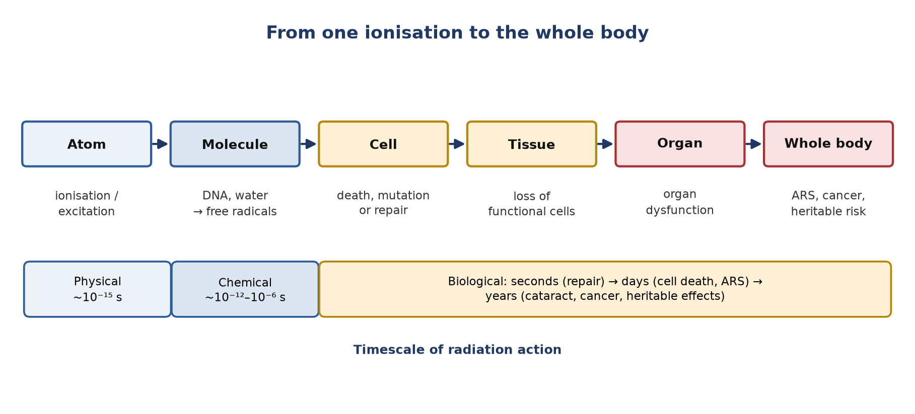
- Damage spreads upwards: atom → molecule → cell → tissue → organ → whole body.
- The events are fast at first (physical and chemical stages last fractions of a second) and slow later (repair within hours, cell death over days, cancer over years).
2. DNA damage and repair
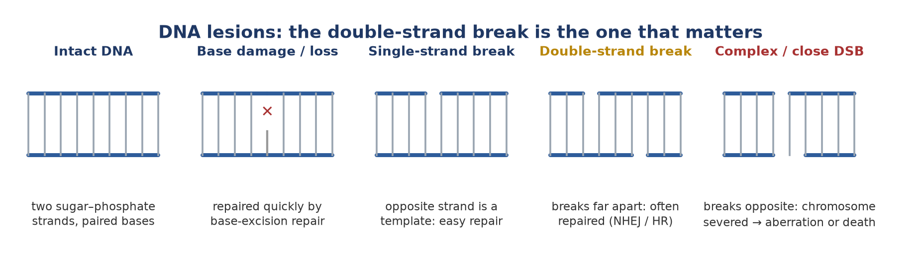
- DNA is the critical target: two sugar–phosphate strands joined by paired bases (A–T, G–C).
- Base damage and single-strand breaks: common and usually repaired accurately, because the intact opposite strand acts as a template.
- Double-strand breaks (DSBs): the key lethal lesion. Repaired by non-homologous end joining (fast, error-prone) or homologous recombination (accurate, needs a sister chromatid, so only in S/G2).
- Breaks close together, or clustered with other damage, are the hardest to repair.
Chromosome and chromatid aberrations
- Irradiation before DNA synthesis (G1) gives chromosome aberrations, because the damage is copied into both chromatids.
- Irradiation after DNA synthesis (G2) gives chromatid aberrations, affecting one arm only.
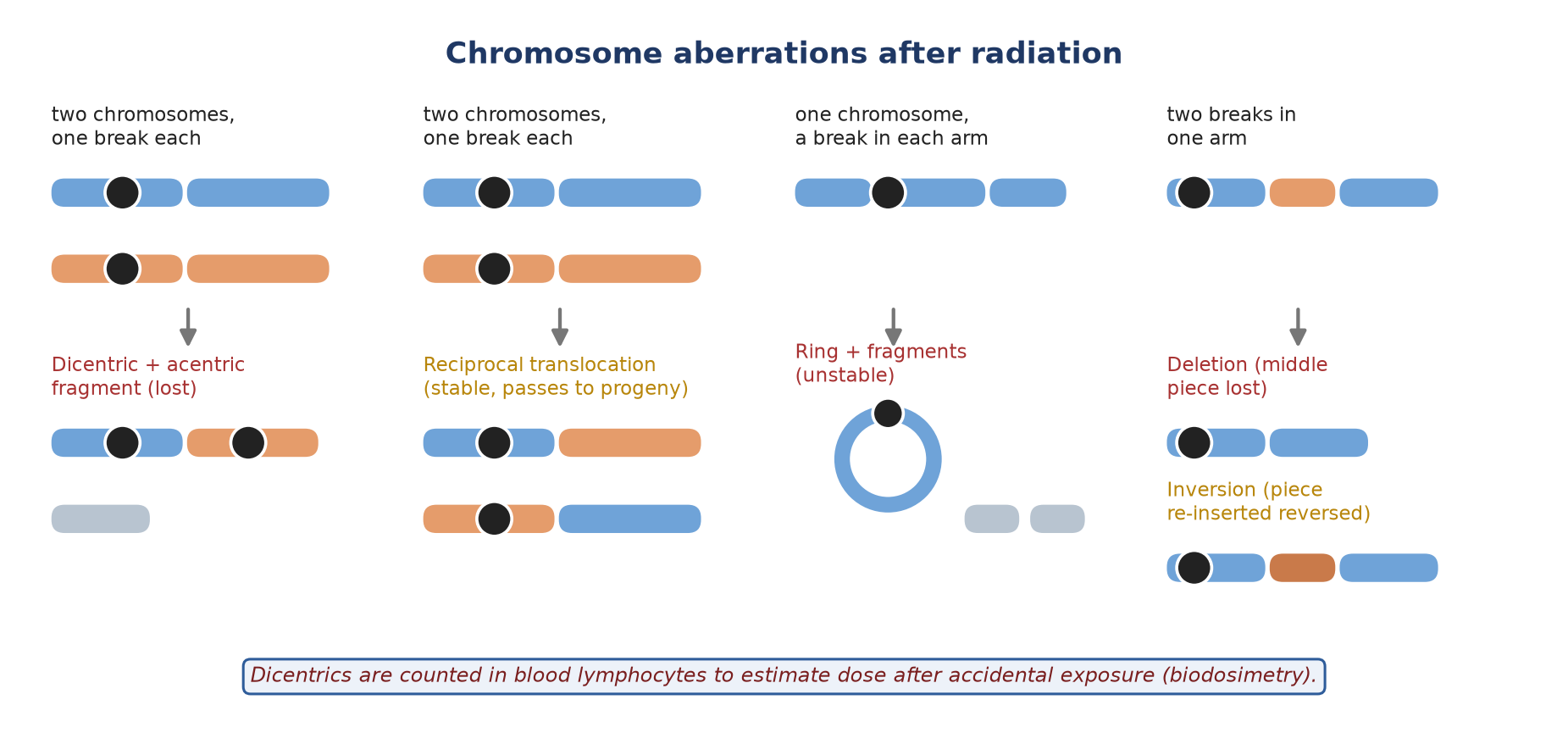
- A single break usually rejoins (restitution). Mis-rejoining produces aberrations:
| Aberration | How it forms | Consequence |
|---|---|---|
| Dicentric + acentric fragment | Breaks in two chromosomes; the two centromere-bearing pieces join | Unstable: cell usually dies at mitosis. Basis of dose estimation (biodosimetry) |
| Ring | A break in each arm of one chromosome; the ends join | Unstable; lethal to the cell |
| Reciprocal translocation | Two chromosomes swap broken ends | Stable; can activate oncogenes (e.g. in leukaemia) |
| Deletion | Two breaks in one arm; the middle piece is lost | Loss of genes; may be lethal or oncogenic |
| Inversion | Two breaks; the middle piece re-inserts upside down | Stable; gene order changed |
3. Direct and indirect action
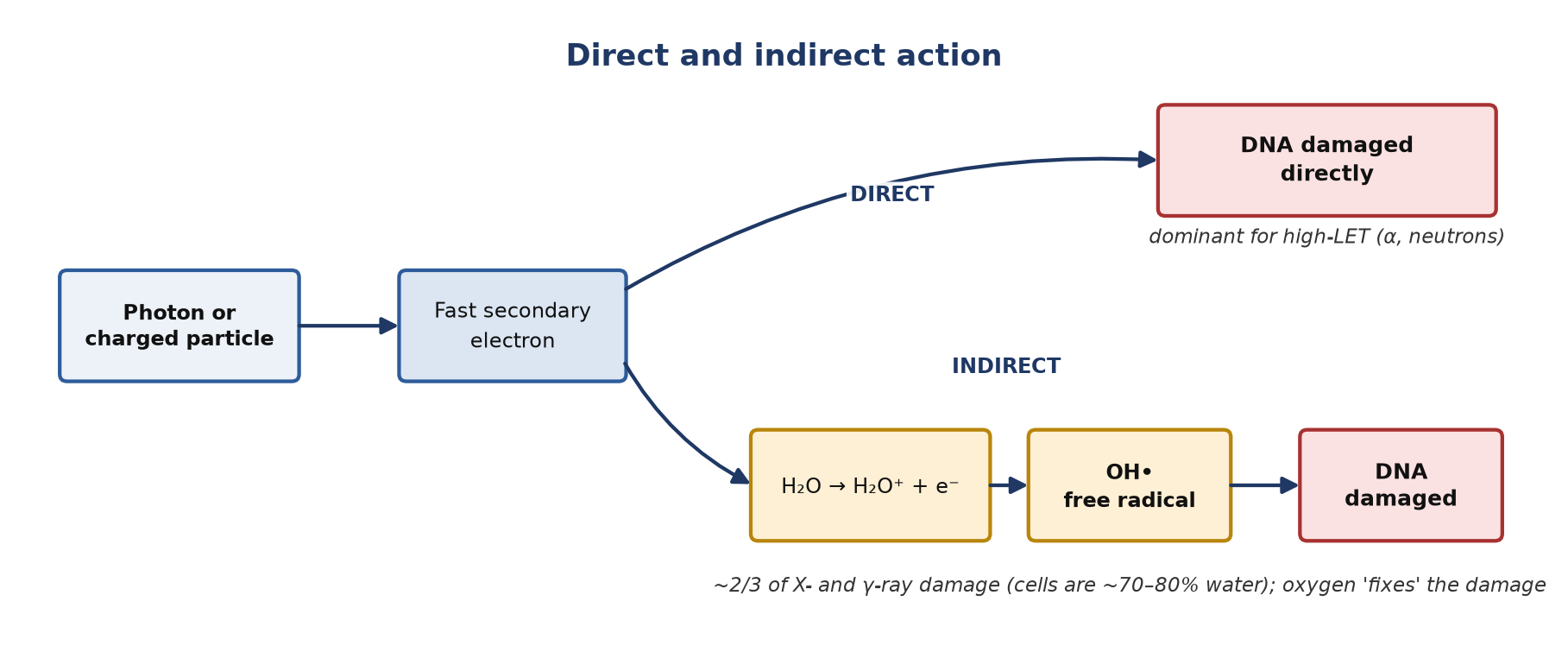
- Direct action: the radiation (or its secondary electron) ionises DNA itself. Dominant for high-LET radiation such as α particles and neutrons.
- Indirect action: radiation ionises water; the resulting free radicals, chiefly the hydroxyl radical (OH•), then damage DNA.
- Because cells are mostly water, about two-thirds of the damage from X- and γ-rays is indirect.
- Oxygen reacts with the DNA radical and 'fixes' the damage in a non-repairable form, which is why oxygenated cells are more sensitive (section 6).
4. Which cells are most sensitive?
Law of Bergonié and Tribondeau
- Radiosensitivity is greatest in cells that divide often, have a long dividing future and are undifferentiated.
- Most sensitive: lymphocytes (the exception: mature but very sensitive, dying by apoptosis), bone marrow stem cells, gut crypt cells, spermatogonia, oocytes, lens epithelium, the embryo.
- Least sensitive: non-dividing, differentiated cells such as nerve and muscle.
- Cell cycle: most sensitive in M and G2; most resistant in late S phase, when homologous-recombination repair is available.
- Cells often die at their next mitosis or the one after (mitotic death), so tissues with fast turnover show damage first.
Blood counts after whole-body exposure
- Lymphocytes fall first, within the first 1–2 days; the rate of fall is used to estimate dose.
- Granulocytes and then platelets fall over the following days to weeks as marrow production stops.
- Red cells fall last and least, because of their long (~120-day) lifespan.
5. Cell survival curves
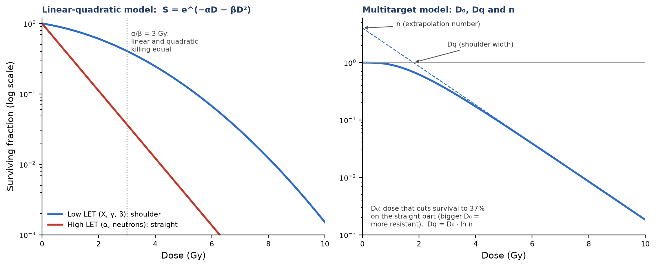
- Survival here means keeping the ability to divide (clonogenic survival). Curves plot dose on a linear axis against surviving fraction on a log axis.
- Low-LET (X, γ, β): a shoulder at low doses (repair of sublethal damage) followed by a steeper, straighter part.
- High-LET (α, low-energy neutrons): nearly straight from zero: little repairable damage.
Linear-quadratic (LQ) model
- S = e^(−αD − βD²)
- α (linear term): cell killing by a single track that causes a lethal, non-repairable lesion.
- β (quadratic term): killing by the interaction of two tracks whose separate sublethal lesions combine; this part can be repaired if the dose is spread out in time.
- α/β ratio: the dose at which both terms kill equally. About 10 Gy for tumours and early-reacting tissues, about 3 Gy for late-reacting tissues, which are therefore more spared by small fractions.
Multitarget model (older)
- D₀: the dose that reduces survival to 37% on the straight part of the curve; a larger D₀ means a more resistant cell.
- n (extrapolation number): where the straight part meets the y-axis; usually about 2–10 for mammalian cells.
- Dq (quasi-threshold dose): the width of the shoulder; Dq = D₀ · ln n.
6. Factors that modify the effect
Radiation type: LET, RBE and radiation weighting
| Feature | Low LET | High LET |
|---|---|---|
| Examples | X-rays, γ-rays, β particles (electrons) | α particles, neutrons, heavy ions |
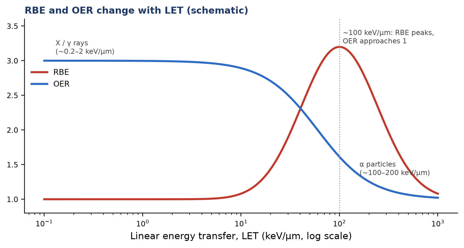
| Typical LET | ~0.2–2 keV/µm | ~10 to >100 keV/µm (α ~100–200) |
| Ionisation pattern | Sparse along the track | Dense along the track |
| Main mechanism | Mostly indirect (free radicals) | Mostly direct |
| Survival curve | Shoulder, then steeper | Straight from zero |
| RBE | Reference (≈1) | High (peaks near 100 keV/µm) |
| Oxygen effect (OER) | ≈2.5–3 | ≈1 |
| Dose-rate effect | Marked | Small or absent |
| Shielding | γ: lead; β: low-Z material (e.g. Perspex) to limit bremsstrahlung | α: stopped by paper or skin (an internal hazard); neutrons: hydrogen-rich material (water, polyethylene, concrete) |
- RBE: the dose of a reference radiation (classically 250 kVp X-rays) divided by the dose of the test radiation giving the same biological effect.
- Radiation weighting factor (wᵣ, ICRP 103): converts absorbed dose (Gy) to equivalent dose (Sv). Photons and electrons 1; protons 2; α particles and heavy ions 20; neutrons a continuous function of energy (about 2.5–20).
Dose rate and fractionation
- For low-LET radiation, the same dose given slowly kills fewer cells, because sublethal damage is repaired during exposure (the quadratic, β term shrinks).
- Radiotherapy uses this through fractionation. The four Rs: Repair (normal tissue repairs between fractions; at least ~6 h apart), Redistribution (cells move into sensitive phases), Repopulation (surviving cells divide) and Reoxygenation (hypoxic tumour cells become sensitive).
Oxygen
- Hypoxic cells are about 2.5–3 times more resistant to X- and γ-rays.
- OER = dose needed without oxygen ÷ dose needed with oxygen for the same effect: about 2.5–3 for low-LET radiation, about 1 for α particles.
Chemical modifiers
| Group | Examples | How they work |
|---|---|---|
| Radiosensitisers | Oxygen; hypoxic-cell sensitisers (metronidazole, misonidazole, etanidazole, nimorazole); halogenated pyrimidines (BUdR, IUdR); some cytotoxics (5-FU, cisplatin, actinomycin D) | Mimic oxygen in hypoxic cells, or are built into DNA in place of thymidine and make it easier to break |
| Radioprotectors | Sulfhydryl compounds (cysteine, cysteamine); amifostine (WR-2721) | Scavenge free radicals; must be present at the time of irradiation. Amifostine is used to reduce xerostomia in head and neck radiotherapy and cisplatin kidney toxicity |
7. Deterministic and stochastic effects
| Deterministic (tissue reactions) | Stochastic | |
|---|---|---|
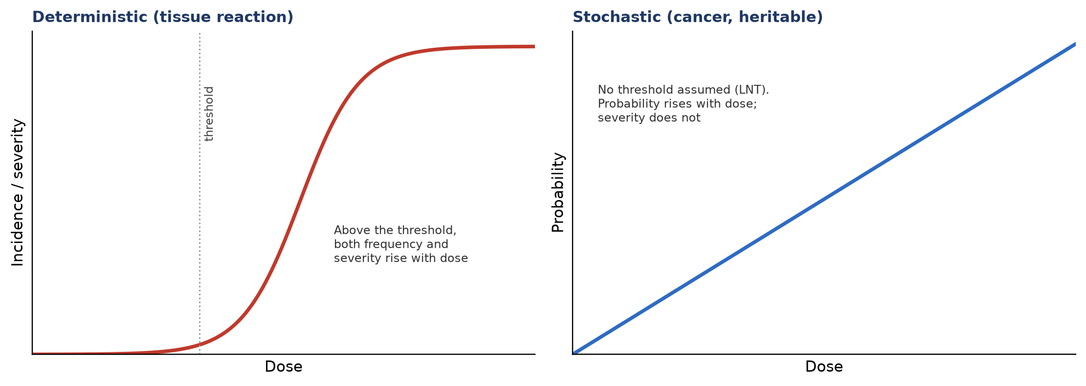
| Cause | Death or malfunction of many cells | A mutation in one or a few cells |
| Threshold | Yes (varies by tissue) | None assumed (linear no-threshold model) |
| Dose affects | Severity and frequency | Probability only; severity is the same |
| Onset | Hours to years | Years to decades |
| Examples | Skin erythema, hair loss, cataract, sterility, marrow failure, ARS | Cancer (e.g. leukaemia, thyroid cancer); heritable effects |
Threshold doses to know (ICRP 118, acute exposure, ~1% incidence)
| Effect | Tissue | Threshold (Gy) | Time to appear |
|---|---|---|---|
| Temporary sterility | Testes | 0.1 | 3–9 weeks |
| Depression of blood cell production | Bone marrow | 0.5 | 3–7 days |
| Cataract (visual impairment) | Lens | 0.5 | >20 years |
| Permanent sterility | Ovaries | 3 | <1 week |
| Temporary hair loss | Skin | 4 | 2–3 weeks |
| Main phase of skin reddening | Skin | <3–6 | 1–4 weeks |
| Permanent sterility | Testes | 6 | 3 weeks |
| Skin burns | Skin | 5–10 | 2–3 weeks |
Table 1. Selected ICRP 118 threshold doses. Values are approximate and apply to single acute exposures.
8. Acute radiation syndrome (ARS)
- Follows high-dose, whole-body (or large-volume) exposure over a short time. It is a deterministic effect.
- Four phases: prodromal (nausea, vomiting) → latent (apparent well-being) → manifest illness → recovery or death. Higher doses shorten every phase.
- LD50/60 (ICRP 118): about 3.3–4.5 Gy without medical treatment and 6–7 Gy with supportive care (antibiotics, blood products, fluids).
| Syndrome | Approx. dose | Target | Main features | Death (untreated) |
|---|---|---|---|---|
| Haematopoietic | ~1–10 Gy (signs from ~2 Gy) | Marrow stem cells | Infection, bleeding, anaemia after a latent period of days to 3 weeks | Weeks to months |
| Gastrointestinal | >6 Gy | Small-bowel crypt cells | Severe diarrhoea, dehydration, electrolyte loss, sepsis | About 1 week (6–9 days) |
| Neurovascular | Very high: from ~10–20 Gy | Brain and blood vessels | Onset within 1–72 h: hypotension, fever, confusion, neurological deficits, cardiovascular collapse | Within a few days |
Table 2. The acute radiation syndromes (dose ranges from ICRP 118). The ranges overlap, and survival depends heavily on medical care.
9. Late effects in the exposed person
Cancer
- Radiation acts mainly as an initiator (DNA damage); promotion and progression follow over years.
- Leukaemia appears first (minimum latency ~2 years, peak within about 5–10 years); solid cancers after 10 years or more.
- Risk is higher in children and younger adults.
- BEIR VII (LNT model): about 1 in 100 people would develop cancer from 0.1 Sv (100 mSv), against about 42 in 100 who develop cancer from other causes. A lifetime of natural background radiation (excluding radon) carries a similar ~1 in 100 risk.
- Below about 100 mSv the epidemiology cannot confirm or exclude a risk; LNT is used for protection because it is prudent, not because it is proven.
Other late effects
- Cataract: posterior subcapsular. ICRP 118 lowered the threshold to 0.5 Gy, and the occupational lens limit to 20 mSv per year (averaged over 5 years). Latency is years and shortens with higher dose.
- Skin: erythema, dry then moist desquamation, hair loss; later atrophy, telangiectasia and fibrosis (thresholds in Table 1).
- Gonads: spermatogonia and oocytes are highly sensitive; temporary male sterility from ~0.1 Gy; permanent sterility ~6 Gy (testes) and ~3 Gy (ovaries), lower in older women.
- Life-shortening and hormesis: non-specific life-shortening was seen in animals at high doses. Hormesis (benefit from low doses) is unproven and is not used in radiation protection.
Local radiotherapy: in brief
- Acute (weeks): effects in fast-renewing tissue in the field, e.g. skin reaction, mucositis, diarrhoea, cystitis.
- Late (months to years): effects in slowly renewing tissue, e.g. fibrosis, xerostomia, strictures, pneumonitis/fibrosis, necrosis. Late effects limit the dose and depend strongly on fraction size (low α/β).
10. Heritable effects
- Radiation can cause mutations in germ cells that could pass to children.
- No heritable effect has been demonstrated in humans, including the children of atomic-bomb survivors; the risk is inferred from animal studies.
- Radiation does not create new kinds of disease; it can only increase the frequency of mutations that occur spontaneously.
- Doubling dose: the dose that doubles the spontaneous mutation rate in a generation; about 1 Gy for humans (from mouse data).
- Genetically significant dose (GSD): the gonadal dose averaged over a population, weighted by each person's expected number of future children.
11. Embryo and fetus
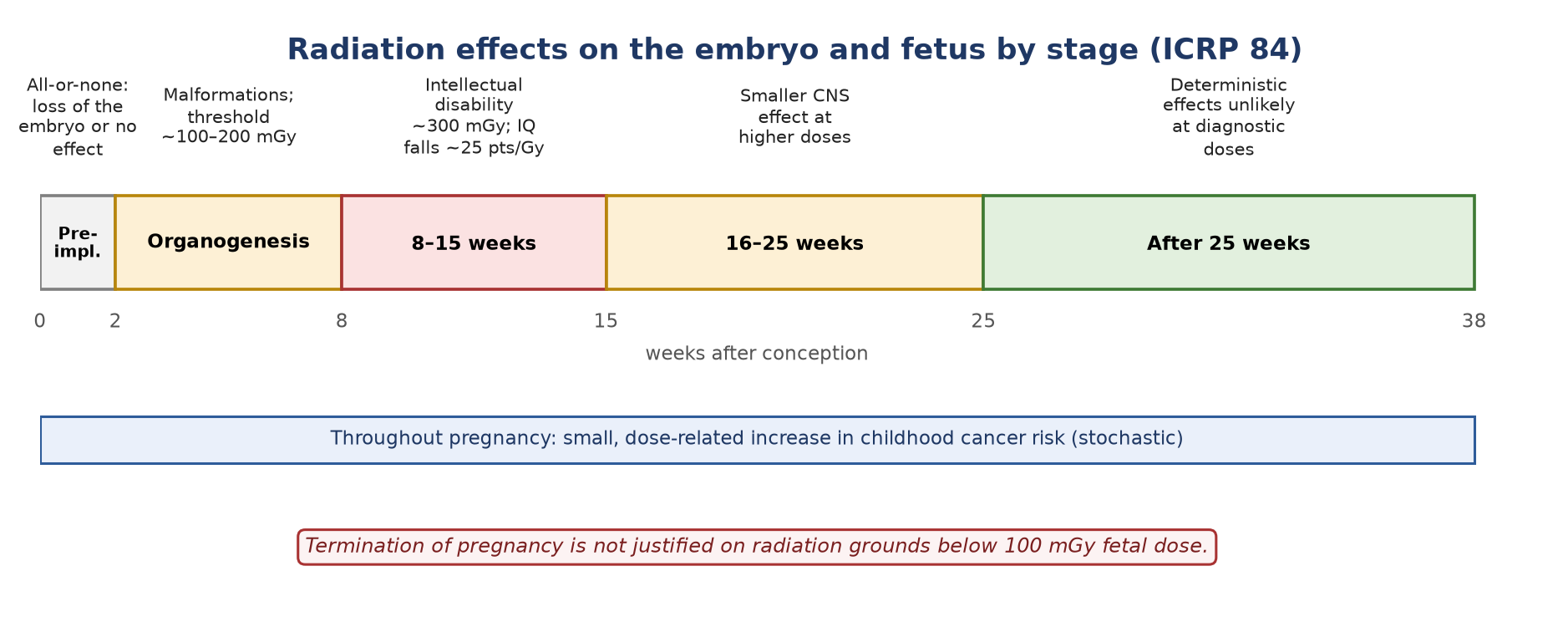
- The embryo is highly sensitive because its cells divide rapidly and are undifferentiated. The effect depends on gestational stage and dose.
- Pre-implantation (0–2 weeks): all-or-none: the embryo is lost or develops normally.
- Organogenesis (2–8 weeks): malformations, mainly CNS and skeleton, above about 100–200 mGy.
- 8–15 weeks: the most sensitive period for the brain: severe intellectual disability from about 300 mGy; IQ falls by about 25 points per Gy.
- Childhood cancer: a small, dose-related increase in risk after exposure at any stage.
Counselling rule (ICRP 84)
- Termination of pregnancy is not justified on radiation grounds at fetal doses below 100 mGy.
- Diagnostic nuclear medicine and CT fetal doses are almost always well below this.
12. What this means in nuclear medicine
- Diagnostic studies: effective doses are a few mSv (e.g. bone scan ~3–4 mSv, FDG PET/CT ~7–10 mSv). Organ doses are far below every deterministic threshold, so the only relevant risk is a small stochastic one.
- Therapy is planned in absorbed dose (Gy): the aim is deterministic cell killing in the target. Effective dose (Sv) is a radiation-protection quantity for stochastic risk and should not be used to prescribe therapy.
- β⁻ emitters (¹³¹I, ¹⁷⁷Lu, ⁹⁰Y): low LET, ranges of millimetres. The crossfire effect treats cells that do not take up the drug, and dose is delivered slowly over days, allowing repair.
- α emitters (²²³Ra, ²²⁵Ac): high LET, range of a few cell diameters (~50–100 µm). High RBE, little dependence on oxygen or dose rate, and effective against hypoxic or resistant cells.
- Low dose rate: radionuclide therapy is continuous, low-dose-rate irradiation. The LQ model is extended as the biologically effective dose (BED), which allows for repair during delivery when comparing it with external-beam treatment.
- Pregnancy and breastfeeding: check before every study; for therapy, exclude pregnancy and stop breastfeeding where required (see the Radiation protection page).
Summary
- DNA double-strand breaks, mostly caused indirectly by free radicals, are the key lesion.
- Dividing, undifferentiated cells are most sensitive; M and G2 phases most, late S least.
- The LQ model describes survival; α/β separates early- from late-reacting tissues.
- High-LET radiation has a high RBE, an OER near 1 and little dose-rate effect.
- Tissue reactions have thresholds (cataract 0.5 Gy); cancer and heritable risk are treated as having none.
- Fetal risk depends on gestational stage; below 100 mGy, termination is not justified on radiation grounds.
Test yourself
5 quick questions. Pick an answer to see the explanation.
1. In the linear-quadratic model, the α term represents cell killing by:
2. The oxygen enhancement ratio (OER) for α particles is approximately:
3. The ICRP 118 threshold for radiation cataract (acute or protracted exposure) is:
4. A pregnant patient's estimated fetal dose from an unplanned CT is 20 mGy at 10 weeks. What does ICRP 84 advise?
5. Which of these is a stochastic effect?
References
- ICRP. The 2007 Recommendations of the International Commission on Radiological Protection. ICRP Publication 103. Ann ICRP. 2007;37(2-4).
- Stewart FA, Akleyev AV, Hauer-Jensen M, et al. ICRP Publication 118: ICRP statement on tissue reactions and early and late effects of radiation in normal tissues and organs – threshold doses for tissue reactions in a radiation protection context. Ann ICRP. 2012;41(1-2):1-322.
- ICRP. Pregnancy and medical radiation. ICRP Publication 84. Ann ICRP. 2000;30(1).
- National Research Council. Health Risks from Exposure to Low Levels of Ionizing Radiation: BEIR VII Phase 2. Washington, DC: National Academies Press; 2006.
- Hall EJ, Giaccia AJ. Radiobiology for the Radiologist. 8th ed. Philadelphia: Wolters Kluwer; 2018.
- Saha GB. Physics and Radiobiology of Nuclear Medicine. 4th ed. New York: Springer; 2013.