Role of PET-CT in Vasculitis
- Best for: large-vessel vasculitis — giant cell arteritis (extracranial disease) and Takayasu arteritis — and the patient with unexplained fever, weight loss and raised CRP where vasculitis, infection and malignancy are all on the table.
- When to scan: before glucocorticoids, or within the first 3 days of starting them; sensitivity falls quickly after that.
- How to read it: compare arterial wall uptake with the liver (Meller grade 0–3; grade 3 is positive, grade 2 is equivocal), then put a number on it with TBR and PETVAS (nine territories, each 0–3, total 0–27).
- What it adds: wall inflammation before any luminal change, whole-body extent in one sitting, and an objective baseline for follow-up.
- Do not over-call: atherosclerosis (focal, eccentric, calcified), grafts and stents, steroid-treated disease, and the small cranial vessels, which PET images less reliably (it needs a dedicated head acquisition) — ultrasound comes first for those.
1. Introduction & Classification
Vasculitis, put simply, is inflammation of the blood vessel wall. The standard way to classify these disorders comes from the 2012 Revised International Chapel Hill Consensus Conference (CHCC), which groups them by the size of vessel predominantly involved.
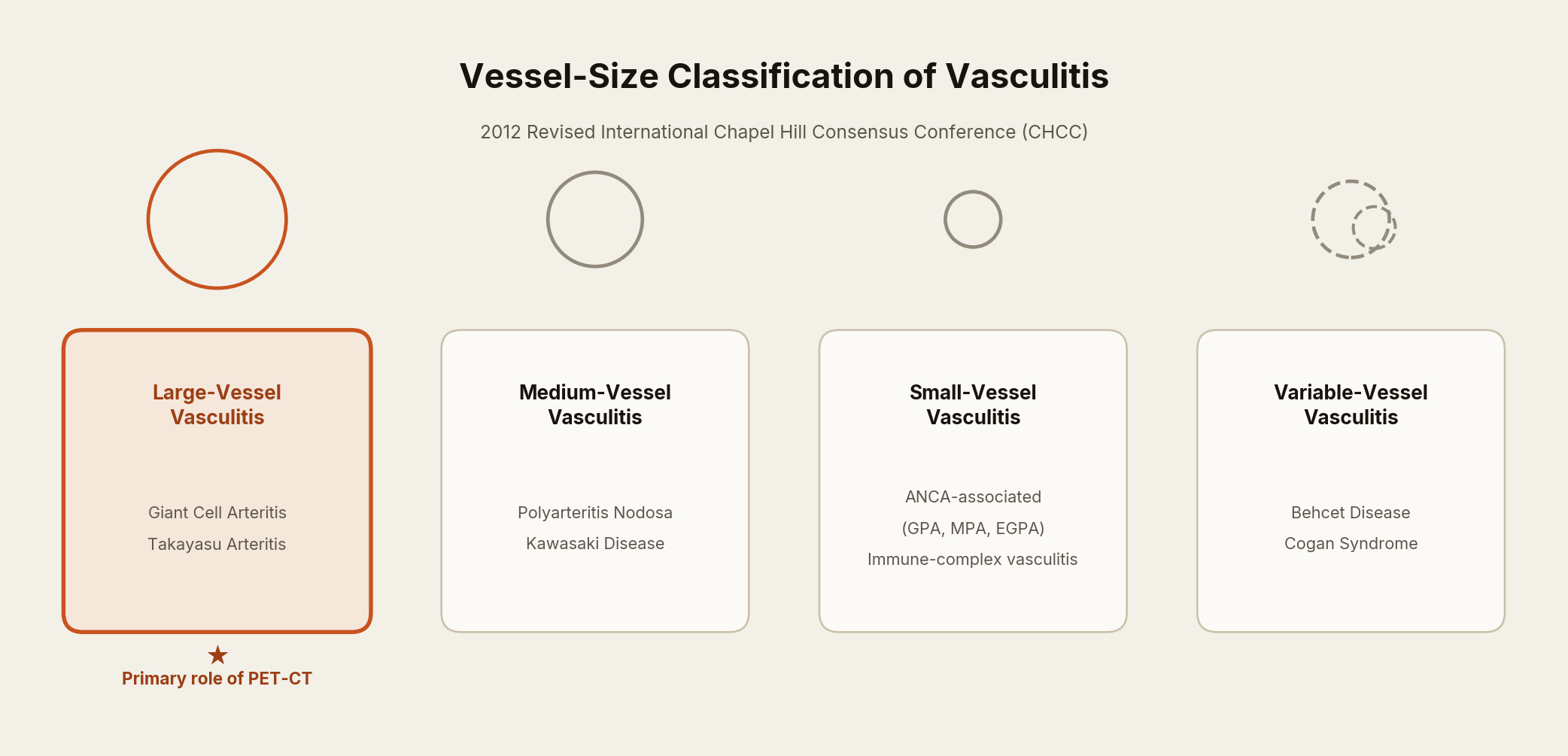
Of these categories, it is large-vessel vasculitis — giant cell arteritis and Takayasu arteritis — where PET-CT has really proven its worth. It picks up inflammation in the aorta and its major branches well before the vessel’s shape has changed enough to show up on a conventional angiogram, which is what the rest of this article is about.
2. Pathophysiological Basis of FDG Uptake
Vessel-wall inflammation in active large-vessel vasculitis is driven by activated macrophages, CD4+ T-lymphocytes and giant cells infiltrating the adventitia and media. These inflammatory cells markedly upregulate GLUT-1 and GLUT-3 glucose transporters and hexokinase activity, leading to avid uptake and trapping of 18F-fluorodeoxyglucose (18F-FDG), an analogue of glucose. The intensity of FDG uptake in the vessel wall therefore acts as a surrogate marker of the metabolic/inflammatory burden of active vasculitis, forming the biological basis for PET-CT imaging.
3. Patient Preparation & PET-CT Protocol
- Fasting: at least 6 hours; blood glucose ideally <126 mg/dL (7 mmol/L), with up to 180 mg/dL (10 mmol/L) acceptable, since hyperglycaemia competitively reduces FDG uptake.
- Rest: no strenuous exercise for 24 hours beforehand, and the patient rests quietly in a warm (20–22 °C) room around injection, to minimise skeletal-muscle and brown-fat uptake.
- Tracer & dose: 18F-FDG, ~2–3 MBq/kg (weight-based) IV for 3D acquisition, adjusted to the scanner.
- Uptake time: at least 60 minutes; EULAR 2023 prefers 90–120 minutes, and the interval should be kept the same on follow-up scans. Later (e.g. 3-hour) imaging improves vessel-to-blood-pool contrast but is not yet validated for LVV.
- Acquisition: vertex to at least the knees (ideally to the feet), arms by the side, PET with low-dose non-contrast CT for attenuation correction and anatomical localisation; a contrast-enhanced CT phase (PET/CTA) can be added in the same sitting to assess luminal/wall changes.
- Timing relative to steroids: scan should ideally be performed before starting glucocorticoids, or within the first 3 days of therapy — sensitivity falls progressively with duration of steroid treatment.
4. Image Interpretation Criteria
4.1 Qualitative (Visual) Grading — Meller Scale
The simplest way to read a scan is to compare arterial wall uptake against the liver, which serves as a convenient internal reference for background activity:
| Grade | Visual uptake | Interpretation |
|---|---|---|
| 0 | No uptake (≤ mediastinal blood pool) | Normal |
| 1 | Uptake less than liver | Normal / equivocal |
| 2 | Uptake equal to liver | Equivocal (borderline positive) |
| 3 | Uptake greater than liver | Positive for active vasculitis |
4.2 Semi-Quantitative Methods
- SUVmax: the highest standardised uptake value found in the arterial wall of interest — quick to report, but somewhat scanner- and patient-dependent.
- Target-to-Background Ratio (TBR): arterial wall SUV divided by venous blood-pool SUV (superior or inferior vena cava). It travels better than SUVmax alone across different scanners, which matters when comparing scans done months apart.
- PETVAS (PET Vascular Activity Score): grade FDG uptake 0–3 (the same liver-referenced scale as above) in nine standard arterial territories — the ascending aorta, aortic arch, descending thoracic aorta and abdominal aorta, plus the paired carotid and subclavian arteries and the brachiocephalic trunk — then add the nine grades together, giving a score from 0 to 27 (Grayson et al., 2018). Grayson and colleagues actually graded 15 territories, including the axillary, iliac and femoral arteries, but left those six out of PETVAS because their uptake did not differ between LVV patients and comparators (the iliofemoral arteries are also a common site of atherosclerosis).
A higher PETVAS mostly reflects how much of the vascular tree is inflamed — one intensely avid segment adds at most 3 points, so focal disease can be read as active with a low score. In active disease the score correlates moderately with ESR and CRP (r ≈ 0.36–0.52), but not during remission (Grayson et al., 2018); in TAK it also tracks ITAS-2010 clinical activity (Kang et al., 2020). A PETVAS of 10 or higher has been reported to separate active from inactive large-vessel vasculitis with roughly 61% sensitivity and 81% specificity overall, with specificity running a little higher in GCA and sensitivity a little higher in TAK (Galli et al., 2022) — though most centres weigh the PETVAS trend alongside TBR and the clinical picture rather than anchoring to a single cutoff.
5. Diagnostic Accuracy
Figures vary across series with patient selection, reference standard and positivity criteria. Pooled estimates from the main meta-analyses: for the diagnosis of GCA, sensitivity 80% and specificity 89% (Besson 2011) and 83% and 90% (Lee 2016); for large-vessel inflammation in GCA against controls, 90% and 98%; and for disease activity in Takayasu arteritis, 87% and 73% (both Soussan 2015). Uptake equal to or above liver was the criterion that best separated patients from controls.
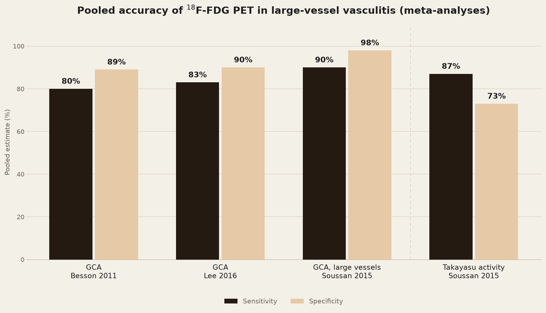
Sensitivity for GCA depends on the positivity criteria used and falls with glucocorticoid exposure and when disease is confined to the small cranial vessels. For TAK, PET-CT can show wall inflammation in early, pre-stenotic disease, before any luminal change is visible — although EULAR 2023 makes MRI the first imaging test in suspected TAK, with FDG-PET, CT or ultrasound as alternatives. Negative predictive value is high (~88% in GCA), making PET-CT particularly useful to exclude large-vessel vasculitis in patients with a low pre-test probability and non-specific systemic symptoms.
6. Clinical Role & Applications
6.1 Diagnosis
PET-CT earns its place in the diagnostic pathway exactly where things aren’t straightforward. In a patient with an atypical presentation, or a negative temporal artery biopsy where extracranial large-vessel disease is still suspected clinically, a positive scan — read alongside the clinical and laboratory picture — supports a clinical diagnosis of GCA/TAK, and when clinical suspicion is high a positive scan can spare the biopsy altogether (EULAR 2023). The 2022 ACR/EULAR criteria are classification (research) criteria, not diagnostic ones; in the GCA set, FDG-PET activity throughout the aorta scores +2 points.
The same logic carries over to pyrexia/inflammation of unknown origin (PUO/IUO): when a patient has unexplained fever, weight loss and raised inflammatory markers with no localising sign, occult large-vessel vasculitis sits on the differential right alongside infection and malignancy, and a single whole-body PET-CT can screen for all three at once.
A strongly negative scan, obtained before steroids are started, is just as useful clinically — its high negative predictive value helps confidently exclude active large-vessel disease and redirects the work-up toward other diagnoses.
6.2 Beyond Diagnosis — the Other Roles at a Glance
Once the diagnosis is made, the same scan keeps earning its keep. The roles below are the ones that come up in practice, with an honest note on how settled each one is.
| Role | What PET-CT adds | Where it stands |
|---|---|---|
| Mapping extent of disease | One whole-body scan maps the full distribution of vascular inflammation — aorta and branch vessels — and not infrequently turns up disease the referring clinician had not suspected. | Established. Maps the distribution used for angiographic TAK typing (Hata/Numano types I, IIa, IIb, III, IV and V), which is based on luminal changes seen on angiography. |
| Differentiating vasculitis from mimics | Pattern is the clue: vasculitis tends to be diffuse, smooth and circumferential, whereas atherosclerosis, IgG4-related aortitis, a mycotic aneurysm or peri-aortic lymphoma tend to look focal or eccentric. | Useful, but the overlap is real (see Limitations) — pattern is a clue, not a verdict. |
| Disease activity & treatment response | Serial PETVAS or TBR, acquired with the same protocol, can help assess response and a suspected relapse — particularly on tocilizumab, when CRP is no longer a reliable marker. Residual uptake often persists in genuine remission — remodelling rather than inflammation — and should not be read as failure on its own. | Not routine: EULAR 2023 recommends imaging only for suspected relapse, not for patients in clinical and biochemical remission; a single scan called “active” or “inactive” on uptake alone remains unreliable. |
| Detecting complications | A contrast CT angiography phase in the same sitting (PET/CTA) picks up aneurysm, dissection, stenosis and occlusion — structural information a purely metabolic scan will not give. | Established for structural assessment. Contrast CTA helps identify stenoses in TAK, but data are insufficient for its routine use in GCA; EULAR 2023 recommends MRA, CTA or ultrasound for long-term monitoring of structural damage. |
| Prognostication | Baseline aortic FDG uptake in GCA has been linked to later aortic dilatation (Blockmans et al., 2008), and a high PETVAS during clinical remission predicted relapse in one cohort (Grayson et al., 2018) but not in another (Galli et al., 2022). | Emerging — supportive evidence, not yet something to base decisions on alone. |
| Overlap with polymyalgia rheumatica (PMR) | Apparent PMR sometimes hides subclinical large-vessel vasculitis, which PET-CT unmasks. Periarticular and bursal uptake around the shoulders, hips and spinous processes also supports a PMR diagnosis in its own right (e.g. the Leuven score). | Established. A positive vascular scan in “isolated PMR” changes treatment intensity and duration. |
7. Comparison with Other Imaging Modalities
No single modality does everything, which is really why PET-CT is used alongside the others rather than instead of them:
| Modality | Key strength | Key limitation |
|---|---|---|
| Colour Doppler ultrasound | First-line for cranial GCA (halo sign); no radiation, bedside, cheap | Operator-dependent; limited for deep/thoracic vessels |
| MR angiography / vessel-wall MRI | No radiation; good wall detail; repeatable in young TAK patients | Limited access; less sensitive for early inflammation than PET |
| CT angiography | Excellent luminal/structural detail (stenosis, aneurysm, calcification) | Poor at detecting pure wall inflammation without luminal change; iodinated contrast, radiation |
| Conventional (catheter) angiography | Historic reference standard for luminal anatomy | Invasive; only shows luminal change, not wall inflammation; largely replaced |
| 18F-FDG PET-CT | Detects early wall inflammation before structural change; whole-body extent mapping; combines with CTA | Radiation, cost, limited availability; reduced sensitivity after steroids; atherosclerosis overlap |
- An early imaging test is recommended to support the clinical diagnosis of suspected GCA, assuming high expertise and prompt availability — and imaging must not delay treatment.
- Ultrasound of the temporal and axillary arteries is the first imaging test in suspected GCA; high-resolution MRI or FDG-PET are alternatives for the cranial arteries.
- Image before, or at the latest within 72 hours of, starting glucocorticoids — sensitivity falls with longer treatment.
- FDG-PET (alternatively MRI or CT) is used for the extracranial arteries in suspected GCA. In suspected TAK, MRI is the first imaging test, with FDG-PET, CT or ultrasound as alternatives.
- Imaging should be done by a trained specialist using appropriate equipment and standardised operating procedures; for FDG-PET, an uptake time of at least 60 minutes, preferably 90–120 minutes.
8. Advantages of PET-CT
- Detects inflammation at the metabolic level, often well before any structural change is visible — this is really PET-CT’s headline advantage over every angiographic technique.
- One whole-body, single-sitting look at the entire arterial tree — no other single modality offers that.
- Gives a number, not just an impression: PETVAS and TBR make activity scoring objective and repeatable across follow-up scans.
- Doubles as a screen for infection or malignancy in atypical presentations — no small thing in a sick, undiagnosed patient.
- Pairs naturally with contrast CT (PET/CTA) or MRI (PET/MRI) in one sitting, so metabolic and structural information arrive together.
9. Limitations & Pitfalls
- Sensitivity falls sharply with duration of glucocorticoid therapy — image before, or within 3 days of, starting steroids if at all possible.
- Atherosclerotic plaque, especially in older patients, can also take up FDG and mimic vasculitis — a focal/eccentric pattern favours atherosclerosis, a diffuse/circumferential one favours vasculitis.
- Physiological or reactive uptake in vascular grafts, recent stents, or post-surgical vessels can be mistaken for active disease.
- Hyperglycaemia and poor patient preparation quietly degrade both image quality and quantitative accuracy.
- Cranial vessels (temporal, occipital arteries) are small and lie next to intense brain uptake, so PET needs modern (digital or time-of-flight) scanners and a longer head acquisition to assess them; EULAR 2023 accepts FDG-PET as an alternative here, but ultrasound remains first-line for suspected cranial GCA.
- Radiation exposure, cost and limited availability mean it isn’t practical as a routine follow-up tool for every patient.
- Residual low-grade vascular uptake can persist despite clinical remission, which complicates calling a serial scan “active” or “inactive” on uptake alone.
10. Recent Advances & Emerging Tracers
FDG is still the workhorse, but a few newer tracers are worth knowing about, especially for the steroid-treated patient where FDG loses some of its edge:
| Tracer / technique | Target | Potential advantage |
|---|---|---|
| 68Ga/18F-FAPI (fibroblast activation protein inhibitor) | Activated fibroblasts in vessel wall | Shows fibroblast activation in the aortic wall even in long-standing clinical remission (pilot study, 8 patients) — may reflect ongoing remodelling rather than active inflammation; role still exploratory |
| 68Ga-DOTATATE / DOTATOC | Somatostatin receptor (SSTR2) on activated macrophages | Very low background in brain and heart, unlike FDG; signal fell with effective treatment in early studies, suggesting a monitoring role |
| Hybrid PET/MRI | Combines metabolic (PET) and vessel-wall/luminal (MRI) data | Lower radiation than PET/CT; excellent soft-tissue and wall detail; useful for young TAK patients needing serial imaging |
| Dual-time-point / extended (3-hour) FDG protocols | Improved lesion-to-background separation | Better vessel-to-blood-pool contrast; gains in diagnostic accuracy and in separating vasculitis from atherosclerosis are not yet validated in LVV |
- G — Glucose check & patient preparation before scanning.
- R — Rest at least 60 (preferably 90–120) minutes for tracer uptake.
- A — Assess visually against liver (Grade 0–3, Meller scale).
- D — Determine TBR / PETVAS for quantitative confirmation.
- E — Exclude mimics (atherosclerosis, graft, infection, malignancy) before labelling as active vasculitis.
- A PET-CT positive for large-vessel uptake in a patient with clinically isolated PMR should raise suspicion of underlying occult GCA/TAK — these patients often need more aggressive or prolonged immunosuppression than PMR alone would call for.
- Grade 2 (equal to liver) uptake is genuinely equivocal, not a coin toss to be called either way — correlate with TBR/PETVAS and the clinical context before committing to a verdict.
11. Conclusion
18F-FDG PET-CT has earned its place as a central, EULAR-endorsed imaging tool in the diagnostic and monitoring pathway for large-vessel vasculitis. By imaging the vessel wall’s metabolic activity directly, rather than waiting for its shape to change, it allows earlier diagnosis, maps the full extent of disease in a single sitting, and gives a reasonably objective way to track activity over time. None of this replaces ultrasound, MR/CT angiography or histopathology — it works alongside them, as one piece of a multimodality diagnostic algorithm rather than a stand-alone answer.
- 18F-FDG PET-CT is a validated, guideline-endorsed imaging tool for diagnosing and mapping the extent of large-vessel vasculitis (GCA and Takayasu arteritis).
- Its real strength is catching active vessel-wall inflammation before structural change occurs — a stage angiographic techniques simply can’t see.
- Diagnostic yield is highest when the scan is done before, or very early after, starting glucocorticoids.
- Interpretation combines qualitative visual grading (Meller scale) with semi-quantitative tools (TBR, PETVAS) for a more objective, reproducible report.
- Its role extends well beyond diagnosis: disease-extent mapping, activity/treatment-response monitoring, complication detection (with PET/CTA), and prognostication.
- The pitfalls worth remembering are atherosclerosis mimicry, steroid-related sensitivity loss, and limited spatial resolution for small cranial vessels.
12. Test Yourself
Five quick questions on the points that matter most. Pick an answer to see the explanation.
13. Glossary
- GCA (Giant Cell Arteritis) — a large-vessel vasculitis typically affecting patients over 50, classically involving the cranial branches of the carotid artery (temporal, occipital) but frequently extending to the aorta and its major branches.
- TAK (Takayasu Arteritis) — a large-vessel vasculitis predominantly affecting the aorta and its primary branches, usually presenting under age 40 and more common in women of Asian descent.
- SUV (Standardised Uptake Value) — a semi-quantitative measure of tracer concentration in a region of interest, normalised to injected dose and body weight (or lean body mass); SUVmax is the highest value within that region.
- TBR (Target-to-Background Ratio) — arterial wall SUV divided by venous blood-pool SUV, normalising arterial uptake against circulating background activity for more reproducible comparison across scans and scanners.
- PETVAS (PET Vascular Activity Score) — a composite score obtained by summing visual grades (0–3) of FDG uptake across nine standardised arterial territories, giving a single number that reflects total vascular inflammatory burden.
14. References
- Dejaco C, Ramiro S, Bond M, et al. EULAR recommendations for the use of imaging in large vessel vasculitis in clinical practice: 2023 update. Ann Rheum Dis. 2024;83(6):741-751.
- Grayson PC, Alehashemi S, Bagheri AA, et al. 18F-Fluorodeoxyglucose-Positron Emission Tomography As an Imaging Biomarker in a Prospective, Longitudinal Cohort of Patients With Large Vessel Vasculitis. Arthritis Rheumatol. 2018;70(3):439-449.
- Slart RHJA, Nienhuis PH, Glaudemans AWJM, Brouwer E, Gheysens O, van der Geest KSM. Role of 18F-FDG PET/CT in Large Vessel Vasculitis and Polymyalgia Rheumatica. J Nucl Med. 2023;64(4):515-521.
- Kang F, Han Q, Zhou X, et al. Performance of the PET vascular activity score (PETVAS) for qualitative and quantitative assessment of inflammatory activity in Takayasu’s arteritis patients. Eur J Nucl Med Mol Imaging. 2020;47(13):3107-3117.
- Marvisi C, Galli E, Ricordi C, et al. The Role of PET in the Diagnosis and Disease Activity Assessment in Large Vessel Vasculitis. Hemato. 2023;4(4):321-330.
- Röhrich M, Rosales JJ, Höppner J, et al. Fibroblast activation protein inhibitor-positron emission tomography in aortitis: fibroblast pathology in active inflammation and remission. Rheumatology (Oxford). 2024;63(9):2473-2483.
- Galli E, Muratore F, Mancuso P, et al. The role of PET/CT in disease activity assessment in patients with large vessel vasculitis. Rheumatology (Oxford). 2022;61(12):4809-4816.
- Slart RHJA, et al. FDG-PET/CT(A) imaging in large vessel vasculitis and polymyalgia rheumatica: joint procedural recommendation of the EANM, SNMMI, and the PET Interest Group (PIG), and endorsed by the ASNC. Eur J Nucl Med Mol Imaging. 2018;45(7):1250-1269.
- Blockmans D, Coudyzer W, Vanderschueren S, et al. Relationship between fluorodeoxyglucose uptake in the large vessels and late aortic diameter in giant cell arteritis. Rheumatology (Oxford). 2008;47(8):1179-1184.
- Ponte C, Grayson PC, Robson JC, et al. 2022 American College of Rheumatology/EULAR classification criteria for giant cell arteritis. Ann Rheum Dis. 2022;81(12):1647-1653.
- Ćorović A, Wall C, Nus M, et al. Somatostatin receptor PET/MR imaging of inflammation in patients with large vessel vasculitis and atherosclerosis. J Am Coll Cardiol. 2023;81(4):336-354.
- Besson FL, Parienti JJ, Bienvenu B, et al. Diagnostic performance of 18F-fluorodeoxyglucose positron emission tomography in giant cell arteritis: a systematic review and meta-analysis. Eur J Nucl Med Mol Imaging. 2011;38(9):1764-1772.
- Lee YH, Choi SJ, Ji JD, Song GG. Diagnostic accuracy of 18F-FDG PET or PET/CT for large vessel vasculitis: a meta-analysis. Z Rheumatol. 2016;75(9):924-931.
- Soussan M, Nicolas P, Schramm C, et al. Management of large-vessel vasculitis with FDG-PET: a systematic literature review and meta-analysis. Medicine (Baltimore). 2015;94(14):e622.
- Fever and weight loss in a 71-year-old woman · Intermediate