Foot ulcer over a Charcot midfoot
History
58-year-old man with type 2 diabetes and peripheral neuropathy. Charcot neuro-osteoarthropathy of the right midfoot was diagnosed 2 years ago and has left a rocker-bottom deformity. A plantar ulcer under the lateral midfoot has been present for 6 weeks; probe-to-bone is equivocal and CRP is 34 mg/L. Radiographs show chronic midfoot fragmentation. MRI shows marrow oedema throughout the midfoot that cannot be separated between active Charcot and osteomyelitis. Foot pulses are palpable.
Step 1 · FDG PET/CT
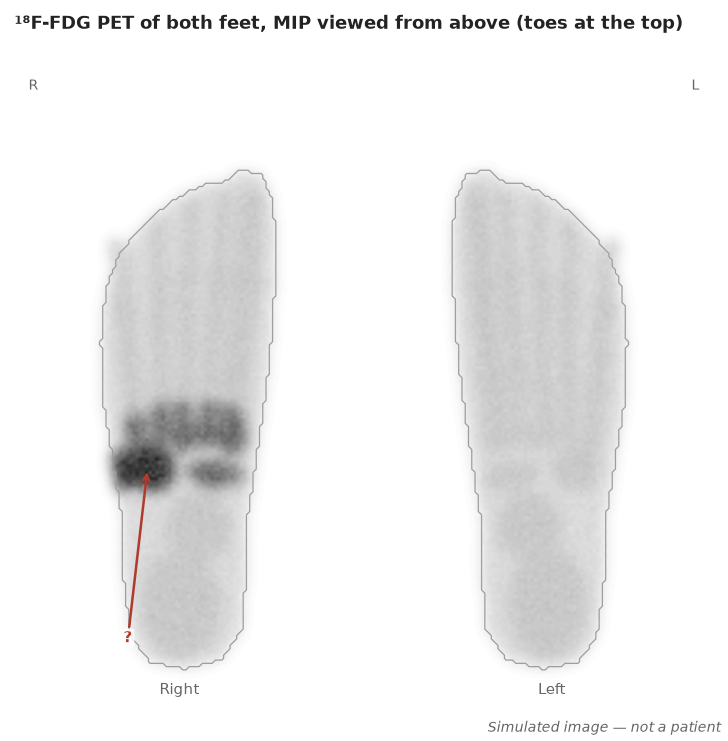
How should the FDG study be interpreted?