Interaction of Radiation with Matter
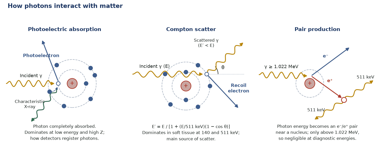
Image formation and its degradations both come from how photons interact with tissue and detectors. The photoelectric effect (full absorption) is how detectors register events; Compton scatter deflects photons and degrades contrast and localisation; and tissue attenuation removes photons along the path, requiring correction — especially in PET.
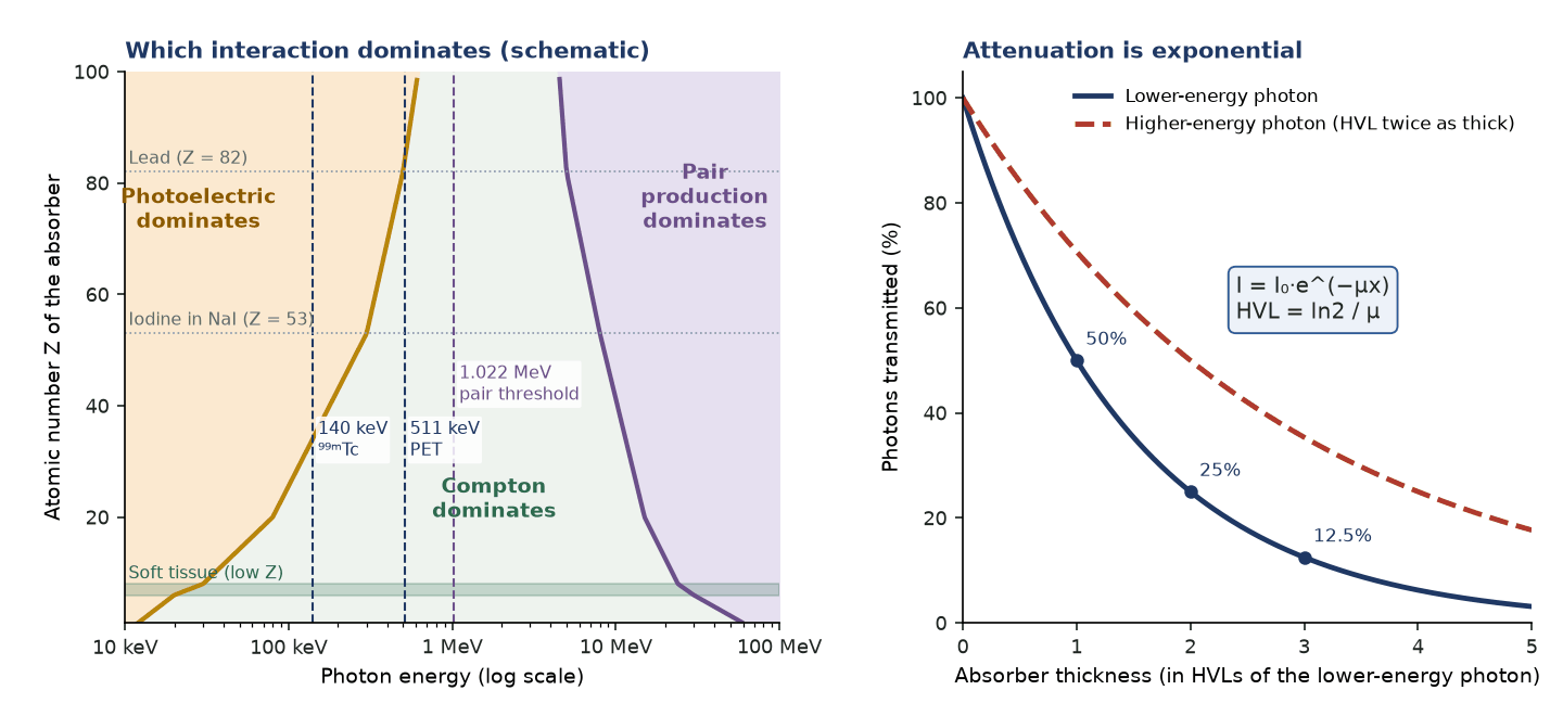
The probability of each interaction depends on photon energy and the atomic number of the medium. Photoelectric absorption dominates at low energy/high-Z; Compton scatter dominates at diagnostic energies in soft tissue; pair production only occurs above 1.022 MeV.
- Photoelectric absorption ∝ Z³/E³: steep rise with atomic number, steep fall with photon energy.
- Compton scatter depends on electron density, almost independent of Z per gram; in water it dominates from about 30 keV to about 20 MeV.
- Pair production: threshold 1.022 MeV, ∝ Z²; negligible at nuclear medicine energies.
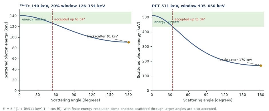
- Scattered energy E′ = E/[1 + (E/511)(1 − cos θ)]: 140 keV backscatters to 91 keV, 511 keV to 170 keV; the minimum approaches 256 keV only for very high energies.
- HVL = 0.693/µ; TVL = 3.32·HVL; I = I₀·e^(−µx).
- Narrow-beam µ in water: 0.15 cm⁻¹ at 140 keV (HVL 4.5 cm), 0.096 cm⁻¹ at 511 keV (HVL 7.2 cm).
- HVL of lead: about 0.3 mm at 140 keV; about 5 mm (broad beam) at 511 keV (AAPM TG-108).
- Mean energy to form one ion pair in air W ≈ 34 eV; bremsstrahlung yield rises with absorber Z, so shield β emitters with low-Z plastic first.
Lead needed to cut a ⁹⁹ᵐTc beam by 70%
Given. HVL(Pb) at 140 keV = 0.3 mm; want 30% transmitted.
- µ = 0.693/0.03 cm = 23.1 cm⁻¹.
- 0.30 = e^(−23.1x), so x = ln(1/0.3)/23.1 = 1.20/23.1 = 0.052 cm.
Answer. About 0.5 mm of lead removes 70% of 140 keV photons.
Attenuation in SPECT versus PET
Given. Source at 10 cm depth in water; body 30 cm thick along the PET line of response. µ = 0.15 cm⁻¹ (140 keV) and 0.096 cm⁻¹ (511 keV), narrow beam.
- SPECT: e^(−0.15 × 10) = 0.22, so 22% of 140 keV photons emerge towards the camera.
- PET: both photons must escape; e^(−0.096 × a) × e^(−0.096 × (30 − a)) = e^(−0.096 × 30) = 0.056 for any depth a.
- The PET correction factor (1/0.056 ≈ 18) is the same wherever the source lies on the line.
Answer. SPECT attenuation depends on depth; PET attenuation depends only on the total thickness along the line of response, which is why PET attenuation correction is exact once µ is known.
Key interactions
- Photoelectric effect: the photon is fully absorbed and an electron ejected — the basis of detection and of characteristic X-rays.
- Compton scatter: partial energy transfer with photon deflection — the main cause of scatter, reducing contrast and mis-positioning counts.
- Pair production: only above 1.022 MeV, negligible at diagnostic energies.
Attenuation
- Photon intensity falls exponentially through tissue: I = I₀·e^(−µx).
- The half-value layer (HVL) is the thickness that halves intensity; higher-energy photons are more penetrating.
- Attenuation is greatest for deep structures, causing artefactual reduced counts — corrected using a CT (or transmission) map.
Why it matters clinically
- Scatter degrades contrast and is reduced by a tight photopeak energy window and scatter-correction algorithms.
- Attenuation causes inferior-wall and deep-lesion drop-out on SPECT and quantitative error in PET.
- Collimator septal penetration by high-energy photons (e.g. ¹³¹I) produces star artefacts.
In the clinic — why the physics matters
- HVL(Pb) at 140 keV is only about 0.3 mm, so thin lead gives practical shielding and collimator septa.
- Photons scattered through less than about 54° keep more than 126 keV and are accepted by a 20% ⁹⁹ᵐTc window; backscattered photons (about 90 keV) fall well outside it.
- Photoelectric ∝ Z³ explains why iodine contrast, lead and detector crystals absorb low-energy photons far more than soft tissue.
- At 511 keV the lower window threshold (about 435 keV) accepts photons scattered by up to about 34°, so scatter correction remains essential in 3-D PET.
Photoelectric vs Compton vs pair production
| Interaction | Energy dependence | Z dependence | Nuclear medicine relevance |
|---|---|---|---|
| Photoelectric | ∝ 1/E³ (falls steeply); absorption edges | ∝ Z³ | Detection in NaI/LSO; contrast; dominant below ~30 keV in tissue |
| Compton | dominant in water ~30 keV to ~20 MeV | ≈ independent of Z per gram | Main source of scatter and attenuation in patients at 140–511 keV |
| Pair production | threshold 1.022 MeV | ∝ Z² | Negligible at 140–511 keV |
Common pitfalls & misconceptions
- Compton scatter is almost Z-independent per gram; do not assume it rises with Z like the photoelectric effect.
- The backscatter energy depends on the incident energy: 91 keV for 140 keV photons, 170 keV for 511 keV photons; 256 keV is only the high-energy limit.
- HVL is not a material constant: it grows with photon energy and is larger for broad beams (build-up) than narrow beams.
- Pair production cannot occur below 1.022 MeV and is negligible in tissue at nuclear medicine energies.
In depth
- Compton kinematics: E′ = E/[1 + (E/mec²)(1 − cos θ)]. For a 126–154 keV window, photons scattered through less than about 54° are accepted; for a 435 keV PET threshold, less than about 34°. Finite energy resolution accepts more.
- Broad-beam attenuation includes build-up of scattered photons, so the effective µ is lower than the narrow-beam value: Chang correction for ⁹⁹ᵐTc typically uses about 0.12 cm⁻¹ instead of 0.15 cm⁻¹.
- For PET the survival probability of a pair is e^(−∫µ dl) along the whole line of response, independent of the emission point; a 30 cm water path transmits about 6%.
- CT-based attenuation maps are measured at an effective energy of about 70 keV and scaled to 511 keV (or 140 keV) with bilinear or hybrid segmentation–scaling methods, one slope for soft tissue and another for bone. Iodinated contrast and metal are over-corrected because they are misread as bone.
- K-edges: absorption jumps just above the K-shell binding energy (iodine 33.2 keV, lead 88.0 keV). Lead K X-rays at about 75–85 keV appear in spectra from collimators with higher-energy sources, and NaI iodine escape peaks lie about 28–33 keV below the photopeak.
- ⁹⁰Y has a 32 ppm internal pair-production branch, a nuclear process unrelated to photon pair production, which allows post-radioembolisation ⁹⁰Y PET.
- Charged particles lose energy continuously: α particles (5–8 MeV) stop within about 0.1 mm of tissue with high LET, and electron ranges are set by E_max (⁹⁰Y up to about 11 mm).
Sources: Cherry, Sorenson & Phelps, 4th ed. (2012) · NIST XCOM cross-section data · Kinahan et al., Med Phys 1998 (PMID 9800714) · AAPM TG-108 (PMID 16485403) · Conti & Eriksson 2016 (PMID 27271304)
Sources
- Cherry SR, Sorenson JA, Phelps ME. Physics in Nuclear Medicine. 4th ed. Philadelphia: Elsevier Saunders; 2012.
- Bushberg JT, Seibert JA, Leidholdt EM Jr, Boone JM. The Essential Physics of Medical Imaging. 4th ed. Philadelphia: Wolters Kluwer; 2021.
- Saha GB. Physics and Radiobiology of Nuclear Medicine. 4th ed. New York: Springer; 2013.
- Berger MJ, Hubbell JH, Seltzer SM, et al. XCOM: photon cross sections database. NIST Standard Reference Database 8.
- Kinahan PE, Townsend DW, Beyer T, Sashin D. Attenuation correction for a combined 3D PET/CT scanner. Med Phys. 1998;25:2046-53.
- Madsen MT, Anderson JA, Halama JR, et al. AAPM Task Group 108: PET and PET/CT shielding requirements. Med Phys. 2006;33:4-15.
- Conti M, Eriksson L. Physics of pure and non-pure positron emitters for PET: a review and a discussion. EJNMMI Phys. 2016;3:8.