SPECT & SPECT/CT
SPECT rotates one or more detector heads around the patient to acquire projections that are reconstructed into cross-sectional images, adding depth and contrast over planar imaging. Iterative reconstruction (OSEM) with resolution recovery and CT-based attenuation correction is now standard, and SPECT/CT adds precise anatomical localisation and an attenuation map.
Projections acquired over 180° (cardiac) or 360° are reconstructed by filtered back-projection or, preferably, iterative algorithms that model the imaging physics and reduce noise and artefact.
- Typical acquisition: 60–128 projections (3–6° steps) over 360°, or 180° for cardiac; 64×64 or 128×128 matrix.
- Nyquist frequency = 0.5 cycle/pixel; pixel size should be no more than about half the system FWHM.
- OSEM: one pass through n subsets is roughly equivalent to n MLEM iterations (Hudson & Larkin 1994).
- Narrow-beam µ for 140 keV in water 0.15 cm⁻¹; broad-beam value used for Chang correction about 0.12 cm⁻¹.
- Routine QC (AAPM Report 177): daily flood, weekly bar phantom, monthly centre of rotation and multi-head registration, quarterly SPECT (or SPECT/CT) phantom.
- Centre-of-rotation offsets beyond about half a pixel blur a point into a ring on 360° studies.
- Triple-energy-window scatter estimate: C_s ≈ (C_low/W_low + C_up/W_up) × W_main/2.
Nyquist frequency of a SPECT matrix
Given. 64×64 matrix, pixel size 4.5 mm (0.45 cm).
- Nyquist = 0.5 cycle/pixel.
- = 0.5 / 0.45 cm.
Answer. 1.11 cycles/cm, the highest usable filter cut-off.
Attenuation correction by geometric mean (planar)
Given. Opposed anterior and posterior counts I_a and I_b through body thickness D, uniform µ.
- I_g = √(I_a·I_b) = I₀·e^(−µD/2), independent of source depth.
- Correction factor = e^(µD/2).
Answer. AC factor = e^(µD/2); for D = 20 cm and broad-beam µ = 0.12 cm⁻¹ it is e^1.2 = 3.3.
Triple-energy-window scatter correction
Given. Main window 126–154 keV (28 keV) holds 10 000 counts in a pixel; 3 keV sub-windows either side hold 300 (lower) and 30 (upper) counts.
- Scatter density: 300/3 = 100 and 30/3 = 10 counts/keV.
- Trapezoid estimate: (100 + 10) × 28/2 = 1 540 counts.
- Primary = 10 000 − 1 540 = 8 460 counts.
Answer. About 15% of the photopeak counts in this pixel are scatter.
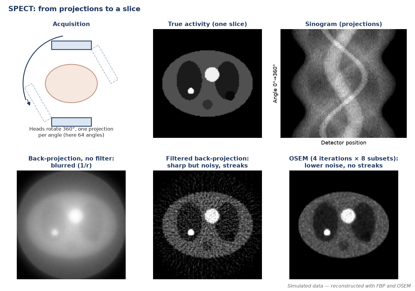
Acquisition
- Multiple projections (e.g. 60–120) over a 360° (or 180° cardiac) arc, step-and-shoot or continuous.
- Body-contouring orbits keep the detector close to improve resolution.
- ECG gating enables functional cardiac (EF, wall motion) SPECT.
Reconstruction
- Filtered back-projection is fast but noisy and streak-prone.
- Iterative OSEM models geometry and statistics, allowing resolution recovery and CT attenuation/scatter correction.
- Reconstruction filters trade noise against resolution.
SPECT/CT
- The CT provides an attenuation map and anatomical localisation, markedly improving specificity (bone, parathyroid, neuroendocrine, infection).
- Misregistration from motion/breathing can create artefacts — check fused images.
- Adds a small CT radiation dose.
Pitfalls
- Centre-of-rotation error causes blurring/ring artefacts — routine COR QC.
- Patient motion degrades reconstruction.
- Attenuation-correction artefacts from truncation, metal or contrast.
In the clinic — why the physics matters
- Iterative OSEM avoids the streak artefacts of filtered back-projection and gives better signal-to-noise in low-count regions.
- CT-based attenuation correction reduces apparent anterior-wall defects in women (breast) and inferior-wall defects in men (diaphragm) in cardiac SPECT, provided CT and SPECT are registered.
- A lower filter cut-off suppresses noise in low-count studies at the cost of resolution.
- Scatter is corrected with, or before, attenuation: attenuation correction alone amplifies scattered counts and over-corrects.
Filtered back-projection vs iterative OSEM
| Feature | FBP | Iterative (MLEM/OSEM) |
|---|---|---|
| Principle | Ramp filter × window, then back-project | Forward-project estimate, compare with measured projections, update |
| Typical artefacts | Streaks near hot structures; noise amplification | Edge overshoot with resolution recovery; noise grows with iterations |
| Detector non-uniformity and COR error | Rings and blurring | Rings and blurring (algorithm-independent) |
| Speed | Fast | Slower (subsets accelerate) |
| Physics modelling | Corrections applied outside reconstruction | Attenuation, scatter and collimator response modelled inside |
Common pitfalls & misconceptions
- A higher cut-off frequency isn't always better — past the optimum it reintroduces high-frequency noise.
- Simple backprojection is inadequate — 1/r blurring gives star artefacts, so a ramp filter is required.
- More OSEM subsets isn't free — it increases image variance (noise) relative to MLEM.
- Attenuation correction can create errors — contrast/metal/motion cause over-correction and misregistration.
In depth
- Filtered back-projection: the ramp filter |ν| undoes the 1/r blur of simple back-projection but amplifies high-frequency noise, so it is rolled off with a window (Butterworth order and cut-off, Hann, Hamming) expressed as a fraction of Nyquist.
- MLEM update: each voxel is multiplied by the back-projected ratio of measured to estimated projections. It preserves counts and non-negativity; OSEM applies this update subset by subset, accelerating convergence about n-fold for n subsets.
- Resolution recovery (modelling the distance-dependent collimator response) improves resolution and contrast but produces Gibbs-like overshoot at edges; it cannot recover detail lost to too few counts.
- Chang first-order correction assumes a convex outline and uniform µ; CT-based correction uses a measured µ map scaled to the emission energy and is required for the thorax and for quantitative SPECT.
- Triple-energy-window correction (Ogawa 1991) estimates scatter pixel by pixel from narrow windows either side of the photopeak and works for multi-peak nuclides such as ²⁰¹Tl and ¹⁷⁷Lu.
- SPECT magnifies non-uniformity because the same detector error is back-projected at every angle, producing rings; correction floods are therefore acquired with far more counts than daily QC floods.
- COR, multi-head registration and head tilt are checked monthly (or as the manufacturer advises); a SPECT/CT phantom with CT attenuation correction is imaged quarterly (AAPM Report 177).
- Quantitative SPECT/CT (Bq/mL, SUV) needs attenuation, scatter and resolution modelling plus a camera calibration factor; MIRD Pamphlet 26 sets out the method for ¹⁷⁷Lu dosimetry.
Sources: Cherry, Sorenson & Phelps, 4th ed. (2012) · Hudson & Larkin 1994 (PMID 18218538) · Ogawa et al. 1991 (PMID 18222843) · AAPM Report 177 (2019) · MIRD 26 (PMID 26471692)
Sources
- Cherry SR, Sorenson JA, Phelps ME. Physics in Nuclear Medicine. 4th ed. Philadelphia: Elsevier Saunders; 2012.
- Hudson HM, Larkin RS. Accelerated image reconstruction using ordered subsets of projection data. IEEE Trans Med Imaging. 1994;13:601-9.
- Ogawa K, Harata Y, Ichihara T, et al. A practical method for position-dependent Compton-scatter correction in single photon emission CT. IEEE Trans Med Imaging. 1991;10:408-12.
- Chang LT. A method for attenuation correction in radionuclide computed tomography. IEEE Trans Nucl Sci. 1978;25:638-43.
- Halama JR, Graham D, Harkness BA, et al. AAPM Report No. 177: acceptance testing and annual physics survey recommendations for gamma camera, SPECT and SPECT/CT systems. College Park, MD: AAPM; 2019.
- International Atomic Energy Agency. Quality Assurance for SPECT Systems. IAEA Human Health Series No. 6. Vienna: IAEA; 2009.
- Ljungberg M, Celler A, Konijnenberg MW, et al. MIRD Pamphlet No. 26: joint EANM/MIRD guidelines for quantitative ¹⁷⁷Lu SPECT applied for dosimetry of radiopharmaceutical therapy. J Nucl Med. 2016;57:151-62.