Other Lung Applications
Snapshot
⁹⁹ᵐTc-MAA does more than exclude PE. Because its particles lodge at first pass, any that reach the systemic circulation measure a right-to-left shunt, and particles injected into the hepatic artery measure the lung shunt fraction before radioembolisation. Quantitative perfusion also follows lung transplants and congenital pulmonary artery disease. ⁹⁹ᵐTc-DTPA aerosol clearance and functional lung avoidance in radiotherapy are research or niche tools.
Read the full article →In-depth, fully referenced version
2.7%MAA shunt in normal subjects
≥6%Brain shunt index in hepatopulmonary syndrome
30 / 50 GyLung dose limits: per session / cumulative
Reference values
- Whole-body method: shunt % = (whole-body − lungs) ÷ whole-body × 100, geometric-mean counts
- Brain method: shunt % = (brain ÷ 0.13) ÷ (brain ÷ 0.13 + lungs) × 100
- Lung shunt fraction (LSF) = lungs ÷ (lungs + liver) × 100
- Lung dose (Gy) = activity (GBq) × LSF × 50, assuming 1 kg of lung
- Resin microspheres: LSF >20% contraindicated (device label)
- Suspected shunt: inject only 100 000–200 000 particles
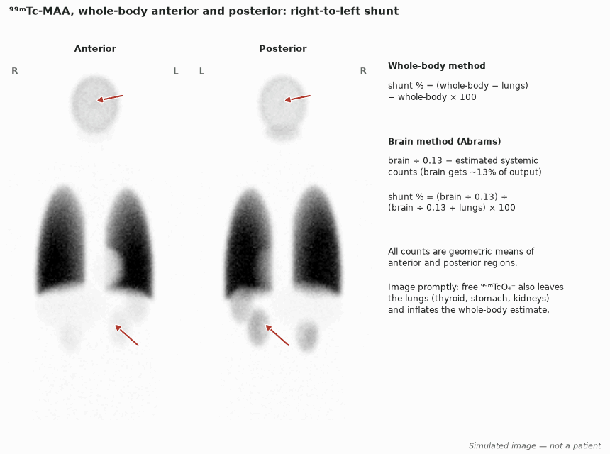
Right-to-left shunt
- MAA particles that bypass the pulmonary capillaries lodge in systemic capillary beds, most visibly the brain and kidneys. Reduce the dose to 100 000–200 000 particles (EANM, SNM).
- Whole-body method: acquire anterior and posterior whole-body images, draw lung and whole-body regions, and calculate the fraction of counts outside the lungs. In normal subjects the value was 2.7% (SD 1.2%); in pulmonary arteriovenous malformations it averaged 23%.
- Brain method: the brain receives about 13% of cardiac output, so brain counts ÷ 0.13 estimate total systemic counts. A head image is the most sensitive way to detect a small shunt (SNM).
- Image promptly. Free pertechnetate and MAA breakdown products leave the lungs and inflate the apparent shunt; check the thyroid and stomach.
- Agreement with the 100% oxygen method is poor (limits of agreement −32% to +45%), so the two measures are not interchangeable. MAA quantification also tracks the result of embolising arteriovenous malformations.
Hepatopulmonary syndrome
- Hepatopulmonary syndrome is liver disease with an increased alveolar–arterial oxygen gradient caused by intrapulmonary vascular dilatation.
- A brain shunt index of 6% or more supports the diagnosis, with an A–a gradient of at least 15 mmHg (20 mmHg over 64 years).
- Contrast echocardiography is the more sensitive screen; a positive MAA scan is specific for moderate to severe disease and helps judge how much a coexisting lung disease contributes to hypoxaemia.
- Before liver transplantation, PaO₂ of 50 mmHg or less, alone or with an MAA shunt fraction of 20% or more, was the strongest predictor of postoperative death.
Lung shunt fraction before radioembolisation
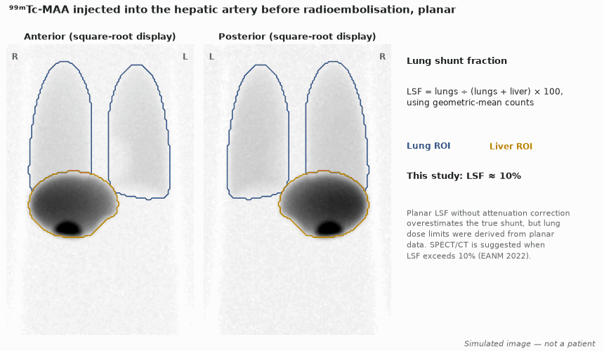
- About 150 MBq ⁹⁹ᵐTc-MAA is injected into the planned hepatic arterial branch at work-up angiography. Image as soon as possible, preferably within 1 hour, because MAA degrades.
- LSF = lung counts ÷ (lung + liver counts), using geometric means of anterior and posterior images. Lung dose (Gy) = activity (GBq) × LSF × 50 for a 1 kg lung.
- Limits: lung dose above 30 Gy per session or 50 Gy cumulatively (EANM 2022; the glass microsphere label uses the same values). The resin microsphere label contraindicates an LSF above 20%; its original US label reduced activity by 20% for LSF 10–15% and by 40% for 15–20%.
- Planar LSF without attenuation correction overestimates the true shunt, but the dose limits were derived from planar data. EANM suggests attenuation- and scatter-corrected SPECT/CT when LSF exceeds 10%.
- Therapy planning is covered in the Theranostics chapter.
Transplantation and congenital heart disease
- After single-lung transplantation the graft takes most of the perfusion: about 83% for emphysema recipients and 69% for pulmonary fibrosis. Use a reduced particle number (EANM).
- In single-lung recipients, graft perfusion at 1–3 months was 67% with a good outcome and 51% in those who developed chronic rejection; a 57% threshold gave sensitivity 83% and specificity 88%.
- After double-lung transplantation, a right–left perfusion difference of 10% or more at 3 months (49% of 340 patients) was associated with death or retransplantation and with chronic lung allograft dysfunction.
- Branch pulmonary artery stenosis: split perfusion measures the deficit and the gain from stenting (in one series the deficit fell from 22.7% to 10.3%).
- After Glenn or Fontan surgery, the injection site decides what is measured. Superior caval blood goes preferentially to the right lung (about 75%) while inferior caval blood divides equally; after a bidirectional Glenn each arm tends to feed its own side, so inject both arms.
Research and niche uses
- ⁹⁹ᵐTc-DTPA aerosol clearance measures alveolar–capillary permeability. Clearance half-time was 52.5 min in non-smokers and 28.3 min in smokers in one study; in ARDS about 15 min against 62 min in cardiogenic oedema.
- In fibrosing alveolitis, rapid DTPA clearance identified patients at risk of deterioration. Clearance is not a routine clinical test.
- Functional lung avoidance radiotherapy plans dose away from well-perfused or well-ventilated lung. The first prospective trial with ⁶⁸Ga V/Q PET/CT (18 patients) cut the functional mean lung dose by 12.4%; no randomised outcome data exist yet.
- During the COVID-19 pandemic, perfusion-only imaging was advised to avoid aerosols. In a registry of COVID-19 patients, perfusion alone confidently excluded PE in only 57%.
In depth
- The whole-body method assumes all non-lung counts are shunted particles. Free pertechnetate, urinary activity and particle breakdown all add counts outside the lungs, which is why prompt imaging was called crucial as early as 1977.
- The 6% HPS threshold is a brain index, not a whole-body shunt fraction; the two methods give different numbers for the same patient and must not be mixed.
- The lung dose equation uses 50 Gy·kg per GBq for ⁹⁰Y (complete decay in tissue). For 3 GBq and LSF 10%, lung dose = 3 × 0.10 × 50 ÷ 1 = 15 Gy, within the 30 Gy limit.
- EANM 2022 notes that a ¹⁶⁶Ho scout dose predicts lung shunt more accurately than ⁹⁹ᵐTc-MAA.
- In double-lung recipients a 55:45 right-to-left perfusion split is considered normal, matching the larger right lung.
- In smokers, clearance half-time rose from 15.8 min to 35.5 min within 7 days of stopping smoking, evidence that the test reflects epithelial permeability.
Sources: Gates 1977 (PMID 839272) · Abrams 1998 (PMID 9453490) · EANM 2022 radioembolisation (PMID 35146577) · Li 2023 (PMID 37291709) · Minty 1981 (PMID 6788126)
Sources
- Bajc M, et al. EANM guideline for ventilation/perfusion SPECT for diagnosis of pulmonary embolism and beyond. Eur J Nucl Med Mol Imaging. 2019;46:2429–51.
- Parker JA, et al. SNM practice guideline for lung scintigraphy 4.0. J Nucl Med Technol. 2012;40:57–65.
- Whyte MK, et al. Quantification of right to left shunt at rest and during exercise in patients with pulmonary arteriovenous malformations. Thorax. 1992;47:790–6.
- Abrams GA, et al. Use of macroaggregated albumin lung perfusion scan to diagnose hepatopulmonary syndrome: a new approach. Gastroenterology. 1998;114:305–10.
- Krowka MJ, et al. International Liver Transplant Society practice guidelines: diagnosis and management of hepatopulmonary syndrome and portopulmonary hypertension. Transplantation. 2016;100:1440–52.
- Arguedas MR, et al. Prospective evaluation of outcomes and predictors of mortality in patients with hepatopulmonary syndrome undergoing liver transplantation. Hepatology. 2003;37:192–7.
- Weber M, et al. EANM procedure guideline for the treatment of liver cancer and liver metastases with intra-arterial radioactive compounds. Eur J Nucl Med Mol Imaging. 2022;49:1682–99.
- Hardoff R, et al. The prognostic value of perfusion lung scintigraphy in patients who underwent single-lung transplantation for emphysema and pulmonary fibrosis. J Nucl Med. 2000;41:1771–6.
- Li DJ, et al. Relative lung perfusion on ventilation-perfusion scans after double lung transplant. Transplantation. 2023;107:2262–70.
- Pruckmayer M, et al. Scintigraphic assessment of pulmonary and whole-body blood flow patterns after surgical intervention in congenital heart disease. J Nucl Med. 1999;40:1477–83.
- Bucknell NW, et al. The HI-FIVE trial: a prospective trial using 4-dimensional ⁶⁸Ga ventilation-perfusion PET-CT for functional lung avoidance in locally advanced non-small cell lung cancer. Int J Radiat Oncol Biol Phys. 2023;117:887–92.
- Zuckier LS, et al. Diagnostic evaluation of pulmonary embolism during the COVID-19 pandemic. J Nucl Med. 2020;61:630–1.