Pre-resection Lung Quantification
Before lung resection, surgeons need the predicted postoperative (ppo) FEV₁ and DLCO. ACCP 2013 calculates them by anatomical segment counting for a lobectomy and by quantitative perfusion scintigraphy for a pneumonectomy, then sorts patients by ppo values into no further testing, a simple exercise test, or formal cardiopulmonary exercise testing. Planar anterior–posterior quantification is accurate for each whole lung but poor for individual lobes; SPECT/CT maps lobes anatomically, although better clinical outcomes have not been shown.
- Perfusion method: ppo = preoperative value × (1 − fraction of total perfusion in the lung to be removed)
- Segment method: ppo = preoperative value × (1 − y/z); y = unobstructed segments removed, z = total unobstructed segments
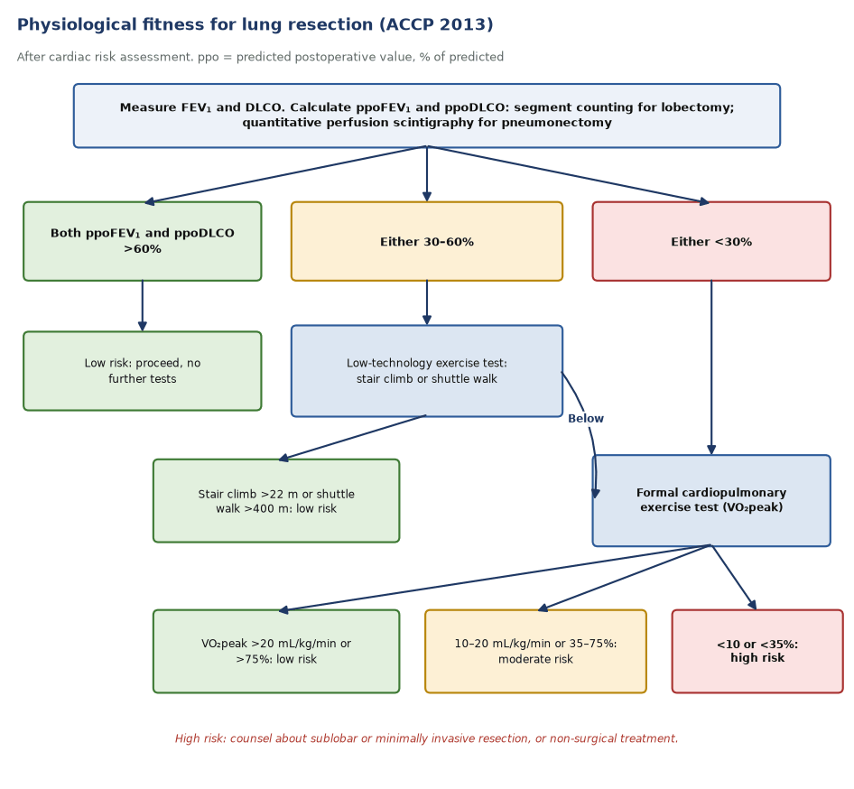
- Both ppoFEV₁ and ppoDLCO >60%: low risk; either 30–60%: stair climb (>22 m) or shuttle walk (>400 m); either <30%: CPET
- VO₂peak <10 mL/kg/min or <35% predicted: high risk; >20 mL/kg/min or >75%: low risk
- ERS/ESTS 2009: exercise testing if FEV₁ or DLCO <80%; ppoFEV₁ and ppoDLCO 30% are high-risk thresholds
- Planar method: anterior and posterior views, geometric mean, three equal zones per lung
ppoFEV₁ for a right pneumonectomy
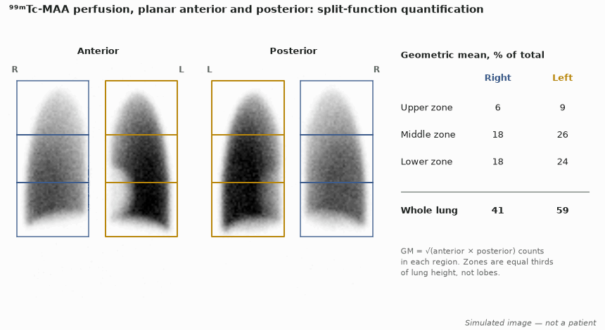
Given. Central right tumour; right pneumonectomy planned. FEV₁ 1.90 L (70% predicted), DLCO 64% predicted. Planar ⁹⁹ᵐTc-MAA quantification (figure below): right lung 41% of total perfusion.
- Fraction remaining after pneumonectomy = 1 − 0.41 = 0.59.
- ppoFEV₁ = 70% × 0.59 = 41% predicted (1.90 L × 0.59 = 1.12 L).
- ppoDLCO = 64% × 0.59 = 38% predicted.
- Both lie between 30% and 60%, so ACCP 2013 asks for a low-technology exercise test: stair climb >22 m or shuttle walk >400 m means low risk; below that, formal CPET.
- Compare a right upper lobectomy by segment counting (3 of 19 segments, none obstructed): ppoFEV₁ = 70% × (1 − 3/19) = 59% predicted.
Answer. ppoFEV₁ 41% and ppoDLCO 38% predicted: intermediate risk, needing an exercise test before pneumonectomy. Try other values in the ppoFEV₁ calculator.
The algorithms
- ACCP 2013 starts with cardiac risk assessment and spirometry with DLCO, then calculates ppoFEV₁ and ppoDLCO for every patient.
- If both are above 60% predicted, no further tests are needed (grade 1C). If either is 30–60%, a stair-climb or shuttle-walk test screens for risk. If either is below 30%, or the simple test is failed (shuttle walk under 400 m or stair climb under 22 m), formal cardiopulmonary exercise testing follows.
- VO₂peak below 10 mL/kg/min or 35% predicted means high risk: counsel about sublobar or minimally invasive resection, or non-operative treatment. Above 20 mL/kg/min or 75% predicted, risk is low.
- ERS/ESTS 2009 exercise-tests anyone with FEV₁ or DLCO below 80%. VO₂peak above 20 mL/kg/min (or 75%) allows pneumonectomy; below 10 (or 35%) means high risk for any resection. ppoFEV₁ or ppoDLCO below 30%, or ppo-VO₂peak below 10 mL/kg/min, marks high risk.
Anatomical or perfusion method
- Segment counting assumes every unobstructed segment contributes equally. ACCP counts 19 segments: right upper 3, middle 2, lower 5; left upper 5, lower 4. Segments that are totally obstructed are left out of both numerator and denominator.
- A central tumour, emphysema or previous surgery makes function uneven, and segment counting then misleads. Perfusion scintigraphy measures the actual share of each lung.
- ACCP recommends the perfusion method for pneumonectomy and segment counting for lobectomy. ERS/ESTS starts with segment counting and adds ventilation or perfusion scintigraphy before pneumonectomy when function is borderline.
- Scintigraphic predictions correlate with measured postoperative FEV₁ (r = 0.67–0.9), and performing both ventilation and perfusion adds nothing (ERS/ESTS).
- Use the ppoFEV₁ calculator to apply either method.
Planar zones or SPECT/CT lobes
- Planar technique: anterior and posterior images after ⁹⁹ᵐTc-MAA, a region over each lung split into upper, middle and lower thirds, and the geometric mean of the two views (which corrects for depth).
- Zones are not lobes. Lobes overlap in both views (the lower lobes rise high posteriorly; the middle lobe lies in front of the lower lobe), so zonal values misassign lobar function.
- For whole lungs, planar and SPECT/CT agree closely (r = 0.995). For lobes they do not: differences reach 20.7% for the right middle lobe and 22.9% for the right upper lobe.
- Posterior oblique views bring planar lobar estimates closer to SPECT/CT, but differences remain. SPECT/CT with CT-based lobar segmentation is anatomically the most accurate method.
- In 60 patients having lobectomy, segment counting, planar and SPECT/CT all predicted actual postoperative FEV₁ and DLCO similarly (r 0.6–0.76). Better anatomy has not yet shown better prediction.
Lung volume reduction and valves
- Before endobronchial valve treatment, the target lobe is chosen from CT emphysema destruction, the absence of collateral ventilation and perfusion.
- In more homogeneous emphysema, expert recommendations (2017, updated 2019) add perfusion scintigraphy to pick a target lobe with low perfusion relative to the untreated lobe on the same side.
- Report lobar or zonal perfusion percentages for each lung, and state the method, because zones and lobes are not interchangeable.
- In one valve series, high perfusion in the untreated ipsilateral lobe was associated with a larger gain in 6-minute walk distance.
In depth
- The geometric mean √(A × P) cancels most depth-dependent attenuation for a source between two opposed views, which is why single-view quantification is not used.
- ACCP: 'Lobectomy: the anatomic method'; 'Pneumonectomy: the perfusion method'. The same equations apply to DLCO.
- ERS/ESTS 2009 found scintigraphy little used before lobectomy 'because of the difficulty in interpreting the contribution of individual lobes', the problem SPECT/CT addresses.
- Lobar differences between planar and SPECT/CT are largest in the right lung; one study found no significant difference in the left lobes, but absolute differences still reached 12%.
- A newer ERS/ESTS guideline on fitness for curative treatment was published in 2025. The thresholds above are those of ACCP 2013 and ERS/ESTS 2009.
- Quantitative ventilation (Technegas or ⁸¹ᵐKr) can be used instead of perfusion, but ERS/ESTS found no benefit in doing both.
Sources: Brunelli, Chest 2013 (PMID 23649437) · Brunelli, ERJ 2009 (PMID 19567600) · Provost 2017 (PMID 28408698) · Brunelli, ERJ 2025 (PMID 41232938)
Sources
- Brunelli A, et al. Physiologic evaluation of the patient with lung cancer being considered for resectional surgery: ACCP evidence-based clinical practice guidelines, 3rd ed. Chest. 2013;143(5 Suppl):e166S–e190S.
- Brunelli A, et al. ERS/ESTS clinical guidelines on fitness for radical therapy in lung cancer patients. Eur Respir J. 2009;34:17–41.
- Toney LK, et al. Improved prediction of lobar perfusion contribution using ⁹⁹ᵐTc-MAA SPECT/CT with attenuation correction. J Thorac Cardiovasc Surg. 2014;148:2345–52.
- Provost K, et al. Reproducibility of lobar perfusion and ventilation quantification using SPECT/CT segmentation software in lung cancer patients. J Nucl Med Technol. 2017;45:185–92.
- Knollmann D, et al. Preoperative assessment of relative pulmonary lobar perfusion fraction in lung cancer patients. Nuklearmedizin. 2015;54:178–82.
- Suh M, et al. Comparison of two different segmentation methods on planar lung perfusion scan with reference to quantitative value on SPECT/CT. Nucl Med Mol Imaging. 2017;51:161–8.
- Arnon-Sheleg E, et al. Head-to-head prospective comparison of quantitative lung scintigraphy and segment counting in predicting pulmonary function in lung cancer patients undergoing video-assisted thoracoscopic lobectomy. J Nucl Med. 2020;61:981–9.
- Slebos DJ, et al. Endobronchial valves for endoscopic lung volume reduction: best practice recommendations from expert panel on endoscopic lung volume reduction. Respiration. 2017;93:138–50.
- Thomsen C, et al. Lung perfusion and emphysema distribution affect the outcome of endobronchial valve therapy. Int J Chron Obstruct Pulmon Dis. 2016;11:1245–59.