Diabetic Foot Infection
In the diabetic foot the central question is whether osteomyelitis is present — and whether bony change reflects infection or Charcot neuro-osteoarthropathy. MRI is the primary test (sensitivity ≈93%) but its specificity is limited (≈75%) because Charcot and infection overlap. ⁹⁹ᵐTc-HMPAO-labelled-WBC imaging and FDG PET/CT offer the highest specificity (≈92%), making them valuable problem-solvers — especially with SPECT/CT or PET/CT to separate bone from soft-tissue infection.
Diabetic foot osteomyelitis usually spreads contiguously from a soft-tissue ulcer, so localising infection (bone vs soft tissue) matters as much as detecting it. Charcot neuro-osteoarthropathy is the key mimic. Labelled-WBC imaging with a marrow scan, or FDG PET/CT, helps distinguish infection from uncomplicated Charcot when MRI is equivocal.
Clinical context & when to image
- Confirming or excluding osteomyelitis under a foot ulcer when MRI is equivocal, contraindicated or degraded by prior surgery/hardware.
- Distinguishing osteomyelitis from Charcot neuro-osteoarthropathy.
- Localising infection to bone versus soft tissue before debridement or surgery.
- Assessing treatment response in selected cases (evidence limited).
Imaging strategy
- MRI is the primary imaging test for its high sensitivity and anatomical detail.
- ⁹⁹ᵐTc-HMPAO-labelled-WBC imaging, ideally with a ⁹⁹ᵐTc-sulfur-colloid marrow scan, adds specificity.
- FDG PET/CT offers comparable sensitivity and high specificity; its ability to separate osteomyelitis from active Charcot is not yet established (insufficient evidence in EANM/SNMMI v2.0).
- Three-phase bone scan is sensitive but non-specific in the neuropathic foot.
- SPECT/CT or PET/CT localisation (bone vs soft tissue) is essential.
Protocol
- Labelled-WBC with paired ⁹⁹ᵐTc-colloid marrow imaging where Charcot or marrow-containing bone is in question (mandatory for Charcot per EANM).
- FDG PET/CT: standard whole-body/lower-limb acquisition with glycaemic control.
- SPECT/CT or PET/CT over the foot for precise localisation.
- Correlate with recent MRI and the site of any ulcer.
How to read it
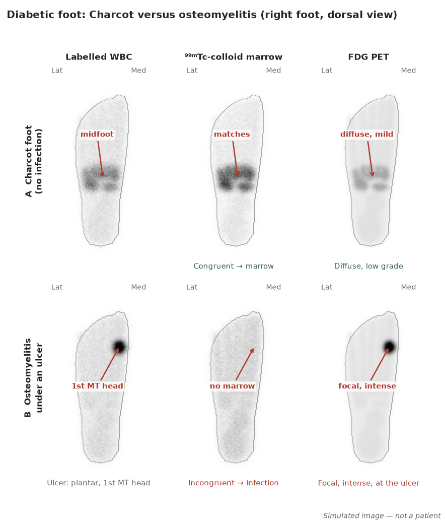
- Labelled WBC: focal marrow-incongruent uptake at bone favours osteomyelitis; uptake confined to soft tissue indicates soft-tissue infection.
- Charcot without infection tends to show congruent WBC/marrow uptake and, on FDG, more diffuse mild uptake.
- FDG: focal intense bone uptake favours osteomyelitis.
- Always localise with SPECT/CT before attributing uptake to bone.
Diagnostic performance
A meta-analysis of diabetic-foot osteomyelitis reported the following pooled performance; ⁹⁹ᵐTc-HMPAO-labelled WBC and FDG PET/CT gave the highest specificity while sensitivity was comparable across techniques.
| Modality | Sensitivity | Specificity |
|---|---|---|
| MRI | ≈93% | ≈75% |
| WBC — ¹¹¹In-oxine | ≈92% | ≈75% |
| WBC — ⁹⁹ᵐTc-HMPAO | ≈91% | ≈92% |
| FDG PET/CT | ≈89% | ≈92% |
Osteomyelitis versus Charcot
- Charcot neuro-osteoarthropathy is the classic mimic of pedal osteomyelitis and often coexists with ulceration.
- Infection favours focal, marrow-incongruent WBC uptake and focal intense FDG uptake, typically adjacent to an ulcer.
- Uncomplicated Charcot tends to show congruent WBC/marrow uptake and diffuse, lower-grade FDG uptake at joints.
- SPECT/CT and correlation with ulcer location are decisive.
Pitfalls
- MRI specificity is limited because Charcot and infection overlap.
- Soft-tissue infection (cellulitis, ulcer) can be mistaken for bone infection without SPECT/CT localisation.
- The neuropathic foot shows non-specific bone-scan uptake.
- Hyperglycaemia can degrade FDG image quality.
Evidence & guidelines
- Meta-analytic data: ⁹⁹ᵐTc-HMPAO WBC and FDG PET/CT offer the highest specificity for diabetic-foot osteomyelitis, with comparable sensitivity to MRI.
- IWGDF/IDSA 2023 recommends MRI when osteomyelitis remains in doubt after clinical assessment, radiographs and laboratory tests, and suggests PET, leucocyte scintigraphy or SPECT as alternatives to MRI (conditional recommendation, low-quality evidence); ACR Appropriateness Criteria take a similar approach.
- Technique choice depends on local availability, expertise and the Charcot question.
- Foot ulcer over a Charcot midfoot · Intermediate