Biological Effects of Ionising Radiation
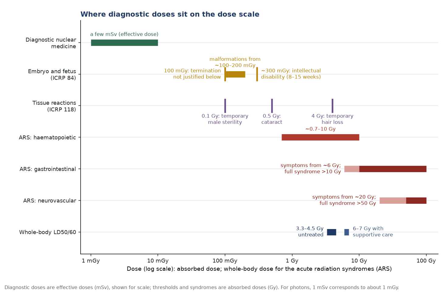
Ionising radiation harms cells mainly through DNA double-strand breaks, about two-thirds of them (for X- and γ-rays) caused indirectly by free radicals formed in water. The effects are either tissue reactions (deterministic), which need a threshold dose — skin injury, cataract, sterility, the acute radiation syndrome — or stochastic effects (cancer, heritable), whose probability, not severity, rises with dose. Diagnostic nuclear medicine doses are far below every threshold.
Radiosensitivity is greatest in dividing, undifferentiated cells (law of Bergonié and Tribondeau) and in the M and G2 phases of the cell cycle. Cell survival follows the linear-quadratic model, S = e^(−αD − βD²); LET, dose rate and oxygen all modify the response.
How radiation damages cells
- DNA is the critical target; the double-strand break is the lethal lesion.
- Direct action dominates for high-LET radiation (α, neutrons); indirect action (OH• radicals) for X- and γ-rays.
- Oxygen fixes radical damage: OER ≈ 2.5–3 for low-LET radiation, ≈ 1 for α particles.
- High-LET radiation has a high RBE (peak near 100 keV/µm) and little dose-rate effect.
Deterministic vs stochastic
- Tissue reactions: threshold dose; severity rises with dose (e.g. temporary male sterility 0.1 Gy, cataract 0.5 Gy, temporary hair loss 4 Gy).
- Stochastic effects: no threshold assumed (linear no-threshold model); probability rises with dose.
- BEIR VII: about 1 in 100 people would develop cancer from 0.1 Sv, against about 42 in 100 from other causes.
Acute radiation syndrome
- Phases: prodromal → latent → manifest illness → recovery or death.
- Haematopoietic (~0.7–10 Gy), gastrointestinal (symptoms from ~6 Gy, full syndrome >10 Gy) and neurovascular (symptoms from ~20 Gy, full syndrome >50 Gy) syndromes.
- LD50/60 about 3.3–4.5 Gy without treatment, 6–7 Gy with supportive care.
Embryo and fetus
- Pre-implantation: all-or-none. Organogenesis: malformations above about 100–200 mGy.
- 8–15 weeks: intellectual disability from about 300 mGy; IQ falls about 25 points per Gy.
- Termination of pregnancy is not justified on radiation grounds below 100 mGy (ICRP 84).
In nuclear medicine
- Diagnostic effective doses are a few mSv — only a small stochastic risk applies.
- Radionuclide therapy is planned in absorbed dose (Gy); effective dose (Sv) is for radiation protection.
- α emitters (²²³Ra, ²²⁵Ac): high LET, range of a few cell diameters; β⁻ emitters (¹³¹I, ¹⁷⁷Lu, ⁹⁰Y): low LET, millimetre ranges.
In depth
- Linear-quadratic model: α/β is about 10 Gy for early-responding tissues and many tumours and about 3 Gy for late-responding tissues; for fractionated treatment BED = nd[1 + d/(α/β)].
- Radionuclide therapy delivers a continuously falling dose rate. For single-exponential decay (constant λ) and repair rate µ (µ = ln2/T_repair), Dale's formulation gives BED = D[1 + Dλ/((µ + λ)(α/β))], so slow delivery spares late-responding tissue.
- ICRP 103/118 thresholds for about 1% incidence after acute exposure: temporary male sterility 0.1 Gy, permanent male sterility about 6 Gy, permanent female sterility about 3 Gy, depressed haematopoiesis about 0.5 Gy, cataract 0.5 Gy, circulatory disease 0.5 Gy.
- ICRP 118 applies the 0.5 Gy cataract and circulatory thresholds to acute and protracted exposure alike, which is why the occupational lens limit fell to 20 mSv per year.
- RBE peaks at an LET of about 100 keV/µm, where the spacing of ionisations matches the 2 nm diameter of the DNA double helix; higher LET wastes energy (overkill) and RBE falls.
- The fetal thyroid begins to concentrate iodine at about 10–12 weeks of gestation, so ¹³¹I given after that can ablate it; pregnancy must be excluded before radioiodine.
- ICRP 103 keeps the linear no-threshold model with a dose and dose-rate effectiveness factor of 2; BEIR VII used 1.5. Both are judgements for protection, not measured thresholds.
Sources: ICRP 103 (2007) · ICRP 118 (2012) · ICRP 84 (2000) · BEIR VII (2006) · Dale, Br J Radiol 1985 (PMID 4063711) · Hall & Giaccia, 8th ed. (2018)
Sources
- ICRP. The 2007 Recommendations of the International Commission on Radiological Protection. ICRP Publication 103. Ann ICRP. 2007;37(2-4).
- Stewart FA, Akleyev AV, Hauer-Jensen M, et al. ICRP Publication 118: ICRP statement on tissue reactions and early and late effects of radiation in normal tissues and organs – threshold doses for tissue reactions in a radiation protection context. Ann ICRP. 2012;41(1-2):1-322.
- ICRP. Pregnancy and medical radiation. ICRP Publication 84. Ann ICRP. 2000;30(1).
- National Research Council. Health Risks from Exposure to Low Levels of Ionizing Radiation: BEIR VII Phase 2. Washington, DC: National Academies Press; 2006.
- Hall EJ, Giaccia AJ. Radiobiology for the Radiologist. 8th ed. Philadelphia: Wolters Kluwer; 2018.
- Dale RG. The application of the linear-quadratic dose-effect equation to fractionated and protracted radiotherapy. Br J Radiol. 1985;58:515-28.