Radiation Protection & Dosimetry
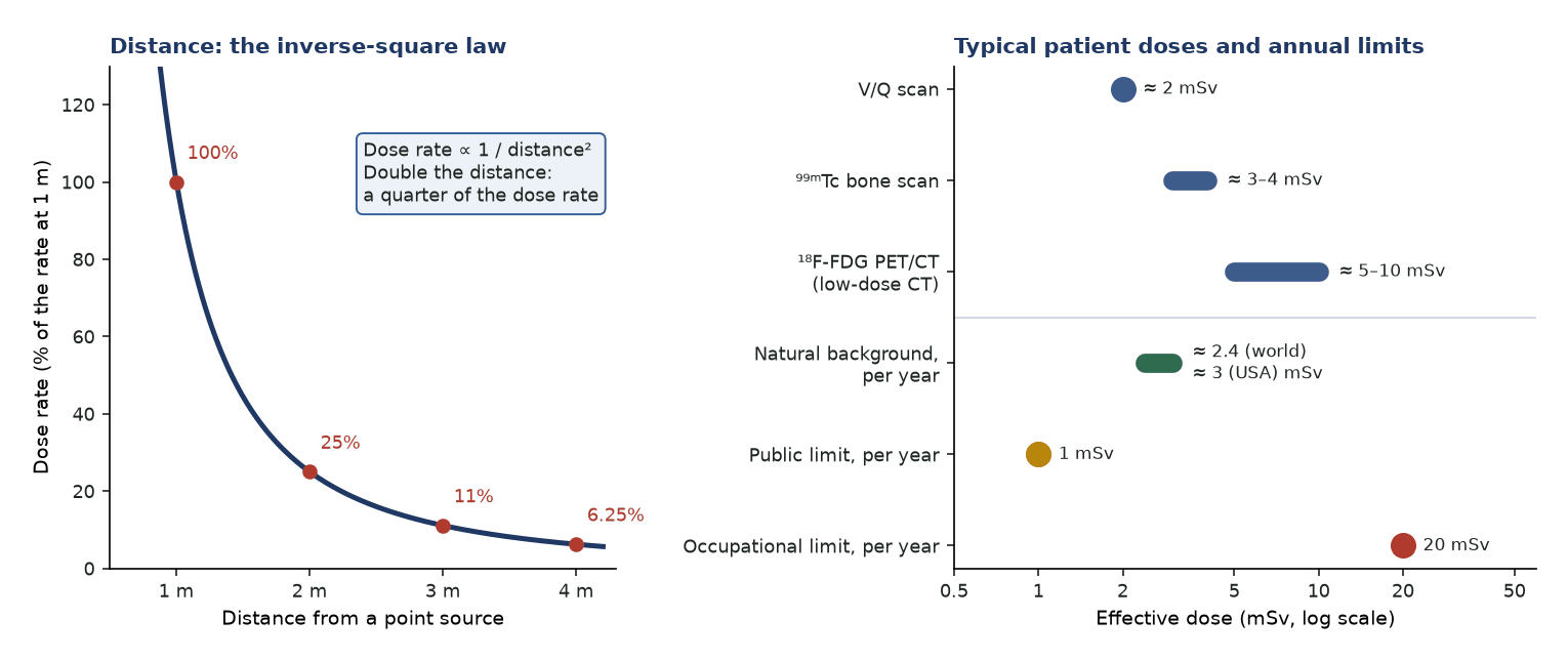
Radiation protection rests on justification, optimisation (ALARA) and dose limitation, applied through time, distance and shielding. ICRP, EU and UK limits are 20 mSv/year for workers and 1 mSv/year for the public; the US NRC worker limit is 50 mSv/year. Patient effective doses are modest (bone scan about 3–4 mSv, FDG PET/CT about 5–10 mSv with low-dose CT, V/Q about 2 mSv), and therapy patients need release criteria and waste management.
Justification, optimisation and dose limitation are the core principles. Internal dosimetry (MIRD schema) estimates absorbed dose to organs from administered activity, residence times and S-values.
- Effective dose E = Σ wT·HT, with HT = Σ wR·DT,R (ICRP 103).
- wR (ICRP 103): photons and electrons 1; protons 2; α particles 20; neutrons a continuous function of energy.
- wT (ICRP 103): red marrow, colon, lung, stomach, breast, remainder 0.12; gonads 0.08; bladder, oesophagus, liver, thyroid 0.04; bone surface, brain, salivary glands, skin 0.01.
- Nominal risk (ICRP 103, whole population): about 5.5% per Sv for cancer and 0.2% per Sv heritable.
- MIRD: D = ÷S; for uptake fraction f and single-exponential clearance, à = 1.443·f·A₀·T_eff.
- ¹⁸F dose-rate constant 0.143 µSv·m²/(MBq·h); from a patient just after injection 0.092 µSv·m²/(MBq·h) (AAPM TG-108).
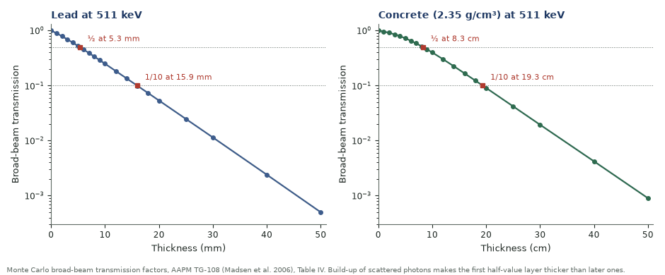
- Lead at 511 keV (broad beam): half at about 5 mm, one-tenth at about 16 mm (AAPM TG-108).
Lung dose from ⁹⁹ᵐTc-MAA (MIRD)
Given. 148 MBq (4 mCi), 99% trapped in lung; S(lung←lung) = 5.2×10⁻⁵ rad/(µCi·h); 45% clears with T_b = 3 h and 55% with T_b = 7 h; T_p = 6 h.
- T_eff1 = 3 × 6/9 = 2.0 h; T_eff2 = 7 × 6/13 = 3.2 h.
- Ã = 1.443 × 4 000 µCi × 0.99 × (0.45 × 2.0 + 0.55 × 3.2) ≈ 15 200 µCi·h.
- D = ÷S = 15 200 × 5.2×10⁻⁵ rad.
Answer. ≈ 0.79 rad = 7.9 mGy to the lungs from lung self-irradiation.
Dose rate from an FDG patient
Given. 370 MBq FDG just injected; patient dose-rate constant 0.092 µSv·m²/(MBq·h); 10 mm lead transmits 0.25 at 511 keV (TG-108).
- At 1 m: 0.092 × 370 = 34 µSv/h.
- At 2 m: 34/4 = 8.5 µSv/h; behind 10 mm lead at 1 m: 34 × 0.25 = 8.5 µSv/h.
- Averaged over the first hour, decay (T½ 110 min) reduces the rate by a factor of 0.83: 1 h at 1 m ≈ 28 µSv.
Answer. 34 µSv/h at 1 m; doubling the distance does as much as 10 mm of lead.
Principles
- Justification (net benefit), optimisation (ALARA) and dose limitation; limits apply to workers and the public, not to patients.
- Time (minimise exposure), distance (inverse-square law) and shielding (lead for γ, low-Z plastic first for β).
- Diagnostic reference levels benchmark administered activities; they are not limits.
Dose limits by jurisdiction
| Category | ICRP 103/118 · EU BSS · UK IRR17 | US NRC (10 CFR 20) |
|---|---|---|
| Worker, effective dose | 20 mSv/year (ICRP: may average over 5 years, no year above 50 mSv) | 50 mSv/year |
| Worker, lens of eye | 20 mSv/year | 150 mSv/year |
| Worker, skin and extremities | 500 mSv/year | 500 mSv/year |
| Embryo/fetus of a pregnant worker | 1 mSv for the rest of the pregnancy | 5 mSv over the pregnancy (once declared) |
| Member of the public | 1 mSv/year | 1 mSv/year |
Typical effective doses
- ⁹⁹ᵐTc bone scan about 3–4 mSv; V/Q about 2 mSv; ⁹⁹ᵐTc MPI varies with protocol.
- ¹⁸F-FDG PET/CT about 5–10 mSv with low-dose CT (FDG 0.019 mSv/MBq, ICRP 128); more with diagnostic-quality CT.
- Compare with the worldwide average natural background of 2.4 mSv/year (UNSCEAR 2008) and 3.1 mSv/year in the USA (NCRP 160).
Special situations
- Pregnancy: justify and minimise; estimate fetal dose; defer where possible.
- Breastfeeding: interrupt or cease according to the agent and the national rules, which differ widely: ICRP advises none for most ⁹⁹ᵐTc agents and 12 h for pertechnetate and MAA, UK ARSAC gives activity-based times that can be much longer, and the US NRC advises 24 h for any ⁹⁹ᵐTc agent; breastfeeding stops for good after ¹³¹I; see pregnancy and breastfeeding.
- Therapy release. US: 10 CFR 35.75 allows release if no other person is likely to receive more than 5 mSv (e.g. ¹³¹I retained activity ≤1.22 GBq or ≤70 µSv/h (7 mrem/h) at 1 m, NUREG-1556 Vol. 9 Table U.1). EU/UK: release is based on national dose constraints for carers, comforters and the public, with written instructions.
Practical safety
- Wear personal dosimeters; monitor fingers in radiopharmacy and during injection.
- Contamination monitoring and spill procedures.
- Segregate radioactive waste and store it for decay.
In the clinic — why the physics matters
- Time, distance, shielding and activity: distance is the cheapest, because dose rate falls with the square of distance.
- Tissue reactions have thresholds; stochastic risk is assumed to rise linearly without threshold, which is the basis of ALARA.
- Shield β emitters (³²P, ⁹⁰Y) with low-Z plastic first and lead outside it, to limit bremsstrahlung.
- US NRC model ALARA programmes set investigation levels at 10% (Level I) and 30% (Level II) of the occupational limits.
Radiation protection quantities
| Quantity | Unit | Definition | Use |
|---|---|---|---|
| Absorbed dose D | Gy (J/kg) | Energy per unit mass | Tissue reactions; therapy dosimetry |
| Equivalent dose HT | Sv | Σ wR·DT,R | Organ limits (lens, skin) |
| Effective dose E | Sv | Σ wT·HT | Limits, optimisation, comparing procedures |
| Operational quantities H*(10), Hp(10), Hp(0.07) | Sv | Measured estimates | Area and personal monitoring |
Common pitfalls & misconceptions
- Absorbed dose (Gy), equivalent dose (Sv = Gy·W_R) and effective dose (ΣW_T·H_T) are distinct — don't interchange.
- 1 R ≠ 1 rad: 1 R = 0.96 rad in soft tissue, defined only for air and photons <3 MeV.
- Use the average β energy (⅓E_max), not E_max, in dose calculations.
- S depends on photon energy, target mass and shape — but not on the amount of activity in the source organ.
In depth
- ICRP 147 (2021): effective dose is for planning and optimisation and for comparing procedures, and can be used as a rough indicator of possible risk; individual risk estimates should use organ absorbed doses with age- and sex-specific risk models.
- ICRP 103 detriment-adjusted nominal risk is about 5.7% per Sv for the whole population (5.5% cancer, 0.2% heritable) and about 4.2% per Sv for adult workers, using a dose and dose-rate effectiveness factor of 2.
- Lens of the eye: the 2011 ICRP statement (published in ICRP 118) cut the occupational limit from 150 to 20 mSv/year averaged over 5 years; the EU BSS and UK IRR17 adopted it, the US NRC has not.
- Pregnant workers: ICRP, the EU and the UK limit the embryo or fetus to about 1 mSv for the rest of the pregnancy once declared; the US NRC allows 5 mSv over the whole pregnancy (10 CFR 20.1208).
- EU guidance on ¹³¹I patients (EC Radiation Protection 97) proposed dose constraints of 1 mSv for children, 3 mSv for adults up to about 60 and 15 mSv for older carers; the US uses a single 5 mSv criterion for release.
- Diagnostic reference levels (ICRP 135) are set at about the 75th percentile of the median values from facilities; in nuclear medicine they are expressed as administered activity or activity per kilogram.
- MIRD Pamphlet 21 nomenclature: absorbed dose D(rT) = Σ Ã(rS)·S(rT←rS), with S in Gy/(Bq·s) and the time-integrated activity coefficient in hours.
- TG-108 patient dose rate assumes 36% body absorption; voiding before imaging removes about 15% of the activity; barrier design uses broad-beam transmission, which at 511 keV needs centimetres of lead or tens of centimetres of concrete.
Sources: ICRP 103 (2007), 118 (2012), 128 (2015), 135 (2017), 147 (2021) · EU BSS 2013/59/Euratom; UK IRR17 · 10 CFR 20 and 35; NUREG-1556 Vol. 9 · EC Radiation Protection 97 (1998) · AAPM TG-108 (PMID 16485403) · MIRD 21 (PMID 19258258)
Sources
- ICRP. The 2007 Recommendations of the International Commission on Radiological Protection. ICRP Publication 103. Ann ICRP. 2007;37(2-4).
- Stewart FA, Akleyev AV, Hauer-Jensen M, et al. ICRP Publication 118: ICRP statement on tissue reactions and early and late effects of radiation in normal tissues and organs. Ann ICRP. 2012;41(1-2):1-322.
- ICRP. Use of dose quantities in radiological protection. ICRP Publication 147. Ann ICRP. 2021;50(1).
- ICRP. Radiation dose to patients from radiopharmaceuticals: a compendium of current information related to frequently used substances. ICRP Publication 128. Ann ICRP. 2015;44(2S).
- ICRP. Diagnostic reference levels in medical imaging. ICRP Publication 135. Ann ICRP. 2017;46(1).
- Council Directive 2013/59/Euratom laying down basic safety standards for protection against the dangers arising from exposure to ionising radiation; and the UK Ionising Radiations Regulations 2017 (SI 2017/1075).
- US Nuclear Regulatory Commission. 10 CFR Part 20 (Standards for protection against radiation) and Part 35 (§35.75); NUREG-1556 Vol. 9, consolidated guidance for medical use licences.
- Madsen MT, Anderson JA, Halama JR, et al. AAPM Task Group 108: PET and PET/CT shielding requirements. Med Phys. 2006;33:4-15.
- Bolch WE, Eckerman KF, Sgouros G, Thomas SR. MIRD pamphlet No. 21: a generalized schema for radiopharmaceutical dosimetry. J Nucl Med. 2009;50:477-84.
- UNSCEAR. Sources and Effects of Ionizing Radiation. UNSCEAR 2008 Report, Vol. I, Annex B. New York: United Nations; 2010.