CTEPH and Chronic Pulmonary Embolism
Snapshot
Chronic thromboembolic pulmonary hypertension (CTEPH) is the one form of pulmonary hypertension that surgery or angioplasty can cure, and it is under-diagnosed. V/Q scintigraphy is the screening test of choice: sensitivity 96–97% and specificity 90–95%, against 51% sensitivity for CTPA in the landmark comparison. A normal perfusion scan effectively excludes CTEPH; mismatched segmental defects after at least 3 months of anticoagulation send the patient to a PH/CTEPH centre. Not every mismatch is thromboembolic, and the mimics matter because the treatments differ.
Read the full article →In-depth, fully referenced version
96–97%V/Q sensitivity for CTEPH
51%CTPA sensitivity, same series
≥3 monthsAnticoagulation before the diagnosis
Reference values
- PH: mean pulmonary artery pressure (mPAP) >20 mmHg at rest (ESC/ERS 2022)
- Pre-capillary PH: pulmonary arterial wedge pressure ≤15 mmHg and PVR >2 Wood units
- CTEPD: symptoms, mismatched perfusion defects and organised clot on imaging after ≥3 months of therapeutic anticoagulation, with or without PH
- CTEPH after acute PE: 0.56% of all comers, about 3% of survivors (meta-analysis); 2.3% at 2 years (FOCUS)
- Post-PE impairment (persistent clinical, functional or imaging abnormality): 16% at 2 years (FOCUS)
- Residual perfusion defects: 57% at 6 months and 52% at 11 months after PE (systematic review)
Definitions
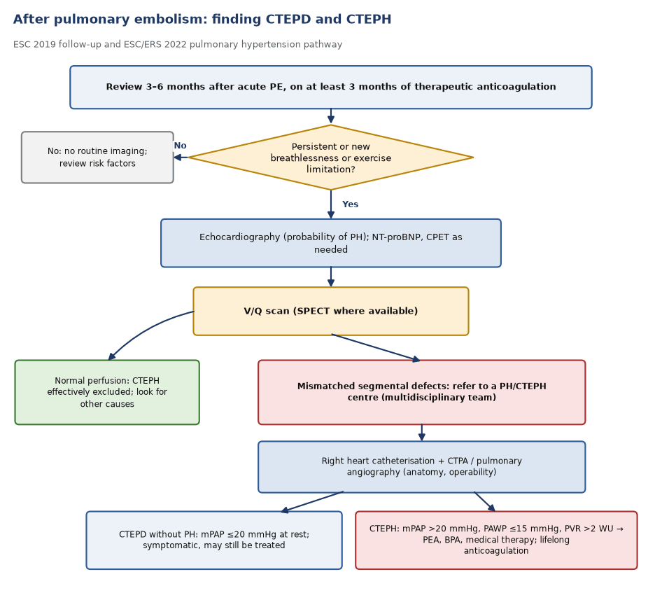
- ESC/ERS 2022 lowered the PH threshold to mPAP above 20 mmHg at rest. Pre-capillary PH also needs a wedge pressure of 15 mmHg or less and PVR above 2 Wood units, so right heart catheterisation is required.
- CTEPH is pre-capillary PH with mismatched perfusion defects and signs of organised clot (ring-like stenoses, webs or slits, chronic total occlusions) after at least 3 months of therapeutic anticoagulation.
- Chronic thromboembolic pulmonary disease (CTEPD) covers symptomatic patients with the same vascular findings, with or without PH at rest. CTEPD without PH may still be treated.
- Chronic PE simply means perfusion defects that have not resolved. It is common after PE and is not by itself CTEPH.
Why V/Q is the screening test
- In 227 patients referred to a PH service (78 with CTEPH), V/Q had sensitivity 96–97.4% and specificity 90–95%. CTPA had sensitivity 51% and specificity 99% (Tunariu 2007).
- EANM 2019 calls V/Q the imaging test of choice to exclude CTEPH, and reports agreement between CTPA and scintigraphy as only slight to fair.
- ESC/ERS 2022 places a V/Q or perfusion lung scan, planar or SPECT, in the work-up of unexplained PH to look for CTEPH. SPECT is less validated for CTEPH than for acute PE.
- Perfusion shows the consequence of obstruction anywhere from the main to the subsegmental arteries, including disease that CTPA misses. Recanalised vessels may reperfuse, so the scan can understate the extent of obstruction.
Patterns
- CTEPH: one or, more often, several mismatched segmental or larger defects, pleural-based and following vascular anatomy. Defects may be partial where vessels have recanalised.
- Pulmonary arterial hypertension and other small-vessel disease: normal perfusion or a patchy, mottled non-segmental pattern.
- Pulmonary veno-occlusive disease is usually normal or mottled, but mismatched segmental defects occur (4 of 56 in one series).
- Unilateral absent perfusion: consider central tumour, fibrosing mediastinitis, proximal interruption of a pulmonary artery or sarcoma before chronic clot.
- Compare with any scan from the acute episode. A predischarge baseline V/Q (EANM) makes later residual and new defects much easier to separate.
Follow-up after PE and referral
- ESC 2019: review patients 3–6 months after acute PE. If breathlessness or exercise limitation persists or appears, assess with echocardiography and a V/Q scan.
- Symptomatic patients with mismatched perfusion defects beyond 3 months of anticoagulation should be referred to a PH/CTEPH centre (ESC/ERS 2022).
- The centre confirms the diagnosis with right heart catheterisation and defines anatomy with CTPA and pulmonary angiography.
- A multidisciplinary team chooses pulmonary endarterectomy for surgically accessible disease, balloon pulmonary angioplasty for distal or inoperable disease, and medical therapy, often in combination. Anticoagulation is lifelong.
- Recurrent VTE and unprovoked PE raise the risk of CTEPH (odds ratios 3.2 and 4.1).
Mimics of CTEPH
- Pulmonary artery sarcoma: mismatched defects like CTEPH, but a large, often unilateral, expansile filling defect that may enlarge despite anticoagulation. It is FDG-avid, whereas bland chronic clot is not. About 11% of 391 reported cases were first labelled CTEPH.
- Fibrosing mediastinitis: large mismatched defects from extrinsic compression. CT shows infiltrative, often calcified mediastinal soft tissue compressing airways and veins as well as arteries.
- Large-vessel vasculitis (Takayasu, Behçet): stenoses and occlusions with mural thickening, systemic features and aortic involvement; FDG shows vessel-wall inflammation.
- Pulmonary vein stenosis after atrial fibrillation ablation: mismatched defects in up to 80%; look for venous congestion and the ablation history.
- Extrinsic compression by tumour or nodes, sarcoidosis, tumour emboli and in situ thrombosis also mimic CTEPH. In situ thrombosis is non-obstructive and the V/Q scan is usually normal or mottled.
In depth
- Tunariu 2007 read V/Q by modified PIOPED criteria: 75 of 78 CTEPH patients had high-probability scans; 8 of 149 non-CTEPH patients also did.
- The CTEPH incidence depends on who is counted: 0.56% in all comers, 3.2% in survivors and 2.8% in survivors without major comorbidity. Studies without right heart catheterisation overestimate it (6.3%).
- In FOCUS (1017 patients), CTEPH was diagnosed a median 129 days after PE, and post-PE impairment identified 15 of the 16 patients who developed CTEPH.
- Residual defects are common: 29% of patients had defects of at least two segments at a median of 12 months in one cohort. A residual defect alone does not make CTEPH; symptoms and haemodynamics decide.
- In sarcoma, one study found an FDG SUV cut-off of 3.5 separated sarcoma from CTEPH, but low-uptake sarcomas are reported and infarcts in CTEPH can take up FDG.
- Large-vessel vasculitis involves the pulmonary arteries in roughly half of patients with Takayasu arteritis, though symptomatic pulmonary disease is less common.
Sources: Tunariu 2007 (PMID 17475953) · Ende-Verhaar 2017 (PMID 28232411) · Valerio 2022 (PMID 35484821) · Sanchez 2010 (PMID 20236393) · Narechania 2020 (PMID 32257112)
Sources
- Humbert M, et al. 2022 ESC/ERS Guidelines for the diagnosis and treatment of pulmonary hypertension. Eur Heart J. 2022;43:3618–731.
- Konstantinides SV, et al. 2019 ESC Guidelines for the diagnosis and management of acute pulmonary embolism. Eur Heart J. 2020;41:543–603.
- Bajc M, et al. EANM guideline for ventilation/perfusion SPECT for diagnosis of pulmonary embolism and beyond. Eur J Nucl Med Mol Imaging. 2019;46:2429–51.
- Tunariu N, et al. Ventilation-perfusion scintigraphy is more sensitive than multidetector CTPA in detecting chronic thromboembolic pulmonary disease as a treatable cause of pulmonary hypertension. J Nucl Med. 2007;48:680–4.
- Ende-Verhaar YM, et al. Incidence of chronic thromboembolic pulmonary hypertension after acute pulmonary embolism: a contemporary view of the published literature. Eur Respir J. 2017;49:1601792.
- Valerio L, et al. Chronic thromboembolic pulmonary hypertension and impairment after pulmonary embolism: the FOCUS study. Eur Heart J. 2022;43:3387–98.
- Nijkeuter M, et al. Resolution of thromboemboli in patients with acute pulmonary embolism: a systematic review. Chest. 2006;129:192–7.
- Sanchez O, et al. Perfusion defects after pulmonary embolism: risk factors and clinical significance. J Thromb Haemost. 2010;8:1248–55.
- Narechania S, et al. Mimickers of chronic thromboembolic pulmonary hypertension on imaging tests: a review. Pulm Circ. 2020;10:2045894019882620.
Practise with a case
- Pleuritic pain after a long-haul flight in a 54-year-old · Intermediate