Suspected Pulmonary Embolism in Pregnancy
Snapshot
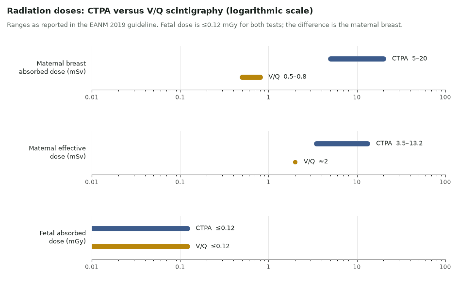
Pregnancy-adapted clinical algorithms now exclude PE without imaging in a third or more of pregnant women. When imaging is needed, a chest radiograph comes first; if it is normal, a perfusion-first scan is the preferred test in most guidelines. The fetal dose is tiny and similar for V/Q and CTPA (≤0.12 mGy); the real difference is the maternal breast, which receives 5–20 mSv from CTPA and 0.5–0.8 mSv from V/Q.
Read the full article →In-depth, fully referenced version
≤0.12 mGyFetal dose, V/Q or CTPA (EANM)
0.5–0.8 vs 5–20 mSvMaternal breast: V/Q vs CTPA
50 MBqDay-1 perfusion-only ⁹⁹ᵐTc-MAA
Reference values
- Day 1: perfusion-only SPECT, ⁹⁹ᵐTc-MAA 50 MBq (EANM 2019, first 3 months of pregnancy)
- Day 2, only if perfusion is abnormal: ventilation SPECT, 20–30 MBq deposited in the lungs
- After the first trimester the standard one-day V/Q protocol may be considered
- Pregnancy-adapted YEARS: D-dimer <1000 ng/mL with no item, <500 ng/mL with ≥1 item
- ICRP 84: fetal doses below 100 mGy are not a reason to terminate a pregnancy
- Breastfeeding after MAA: UK ARSAC 13 h (100 MBq) or 20 h (200 MBq); ICRP 106 12 h; US NRC 24 h
Clinical algorithms first
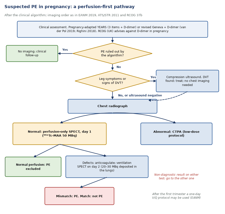
- Pregnancy-adapted YEARS (van der Pol 2019): three YEARS items plus D-dimer, and compression ultrasound first if there are leg symptoms. PE was excluded without CTPA in 39% of women, with a 3-month VTE rate of 0.21%. The gain was largest in the first trimester (65%) and smallest in the third (32%).
- The Geneva-based strategy (Righini 2018, CT-PE-Pregnancy): revised Geneva probability and high-sensitivity D-dimer; if not excluded, bilateral leg ultrasound, then CTPA, with V/Q only when CTPA was inconclusive. Among 395 women, no untreated woman had VTE at 3 months (0.0%; 95% CI 0.0–1.0%).
- D-dimer rises through normal pregnancy, so its yield falls with gestation. The UK RCOG guideline advises against D-dimer testing in pregnancy; ESC 2019 keeps clinical probability and D-dimer in its pregnancy algorithm.
- If leg ultrasound confirms DVT, treat. No chest imaging is needed (RCOG, ATS/STR).
Which imaging test
- ATS/STR 2011 (strong recommendations): chest radiograph as the first radiation-associated test; lung scintigraphy as the preferred test when the radiograph is normal; CTPA rather than catheter angiography after a non-diagnostic V/Q.
- ESC 2019: perfusion scintigraphy or low-dose CTPA should be considered to rule out PE; CTPA first if the chest radiograph is abnormal.
- RCOG Green-top 37b (2015): V/Q or CTPA; CTPA in preference when the radiograph is abnormal. The ventilation component can often be omitted, and perfusion scans are the first choice for young women, especially with a family history of breast cancer.
- CTPA is harder in pregnancy: higher cardiac output and unopacified inferior vena caval blood degrade enhancement, and 6–36% of pregnancy CTPAs are non-diagnostic (EANM).
The perfusion-first protocol
- Day 1: perfusion-only SPECT with 50 MBq ⁹⁹ᵐTc-MAA. Pulmonary disease is uncommon in pregnant women, so a normal perfusion pattern usually excludes PE.
- If perfusion is abnormal, start anticoagulation and add ventilation SPECT on day 2 (20–30 MBq in the lungs). A segmental mismatch confirms PE; matched defects do not.
- After the first trimester, EANM allows the standard one-day V/Q protocol.
- Performance: in one series, 225 low-dose perfusion scans and 97 CTPAs were each non-diagnostic in 9.3%; the negative predictive value was 100% for perfusion and 97.5% for CTPA. A meta-analysis found a pooled false-negative rate of 0% for both tests.
Radiation and counselling
- Fetal absorbed dose is ≤0.12 mGy for both CTPA and perfusion SPECT, far below the 100 mGy that ICRP 84 treats as a threshold for considering termination. Fetal dose should not drive the choice.
- Maternal breast dose differs by about an order of magnitude: 5–20 mSv for CTPA against 0.5–0.8 mSv for V/Q. EANM calls this difference critical in pregnancy.
- RCOG wording for counselling: compared with CTPA, V/Q may carry a slightly increased risk of childhood cancer but a lower risk of maternal breast cancer; in both cases the absolute risk is very small.
- Prenatal exposure to iodinated contrast is not a risk factor for neonatal thyroid dysfunction (EANM).
- General pregnancy and breastfeeding rules for nuclear medicine: pregnancy and breastfeeding.
Breastfeeding after ⁹⁹ᵐTc-MAA
Advice differs by region. Ask the mother to express and store milk beforehand, and to express and discard during the interruption.
| Source | Interruption after ⁹⁹ᵐTc-MAA |
|---|---|
| UK: ARSAC Notes for Guidance (Table 7.2) | 13 h after 100 MBq; 20 h after 200 MBq (infant dose under 1 mSv) |
| ICRP Publication 106 (Annex D) | 12 h |
| USA: NRC Regulatory Guide 8.39 Rev. 1 (2020) | 24 h (single interruption period advised for all ⁹⁹ᵐTc agents) |
In depth
- ICRP 106 groups MAA with pertechnetate, in vivo labelled red cells and white cells at 12 h; most other ⁹⁹ᵐTc agents need no interruption (a 4-h pause with one feed discarded is offered as a precaution).
- Non-diagnostic rates depend on definitions. The Haematologica meta-analysis counted PIOPED low and intermediate probability as non-diagnostic and reported 12% for CTPA and 14% for V/Q; binary SPECT reporting gives far fewer.
- PE prevalence among pregnant women imaged for suspected PE is low (3.1% in the Sheen series; 4.0% and 7.1% in the two algorithm studies), which is why a normal perfusion study carries such a high negative predictive value.
- EANM (2019) states that fetal doses for CTPA and perfusion SPECT are ≤0.12 mGy and maternal (mainly breast) doses are 5–20 mSv for CTPA and 0.5–0.8 mSv for V/Q, which is the basis of its preference for V/Q when both are available.
- The pregnancy-adapted YEARS study was a management study: CTPA was the only imaging test used, so its safety data apply to the clinical rule, not to V/Q.
- The ICRP 84 wording is specific: fetal doses below 100 mGy should not be considered a reason for terminating a pregnancy; above this, decisions are individual.
Sources: ICRP 106 (2008) Annex D · Tromeur 2019 (PMID 30115658) · Sheen 2018 (PMID 28823756) · van der Pol 2019 (PMID 30893534) · Righini 2018 (PMID 30357273) · EANM 2019 (PMID 31410539) · ICRP 84 (2000)
Sources
- Bajc M, et al. EANM guideline for ventilation/perfusion SPECT for diagnosis of pulmonary embolism and beyond. Eur J Nucl Med Mol Imaging. 2019;46:2429–51.
- Leung AN, et al. An official ATS/STR clinical practice guideline: evaluation of suspected pulmonary embolism in pregnancy. Am J Respir Crit Care Med. 2011;184:1200–8.
- Royal College of Obstetricians and Gynaecologists. Thromboembolic disease in pregnancy and the puerperium: acute management. Green-top Guideline No. 37b. 2015.
- Konstantinides SV, et al. 2019 ESC Guidelines for the diagnosis and management of acute pulmonary embolism. Eur Heart J. 2020;41:543–603.
- van der Pol LM, et al. Pregnancy-adapted YEARS algorithm for diagnosis of suspected pulmonary embolism. N Engl J Med. 2019;380:1139–49.
- Righini M, et al. Diagnosis of pulmonary embolism during pregnancy: a multicenter prospective management outcome study. Ann Intern Med. 2018;169:766–73.
- Sheen JJ, et al. Performance of low-dose perfusion scintigraphy and CT pulmonary angiography for pulmonary embolism in pregnancy. Chest. 2018;153:152–60.
- Tromeur C, et al. Computed tomography pulmonary angiography versus ventilation-perfusion lung scanning for diagnosing pulmonary embolism during pregnancy: a systematic review and meta-analysis. Haematologica. 2019;104:176–88.
- ICRP Publication 84. Pregnancy and medical radiation. Ann ICRP. 2000;30(1).
- ICRP Publication 106. Radiation dose to patients from radiopharmaceuticals. Ann ICRP. 2008;38(1–2).
- ARSAC. Notes for Guidance on the Clinical Administration of Radiopharmaceuticals and Use of Sealed Radioactive Sources. 2026.
- US Nuclear Regulatory Commission. Regulatory Guide 8.39, Revision 1: Release of patients administered radioactive material. 2020.
Practise with a case
- Breathlessness at 11 weeks of pregnancy · Intermediate