Acute Pulmonary Embolism and V/Q SPECT
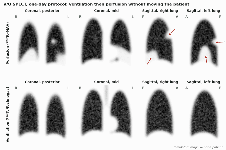
V/Q scintigraphy shows what an embolus does: it removes perfusion from a segment whose airways stay open. EANM (2019) recommends one-day V/Q SPECT, read holistically and reported as PE present or absent; PE is diagnosed with a mismatch of at least one segment or two subsegments. With these criteria, series of over 5000 patients report sensitivity 96–99%, specificity 96–98% and only 1–4% non-diagnostic reports. The test has no contraindications, but it answers the question only after clinical probability and D-dimer have selected the patient.
- Perfusion: ⁹⁹ᵐTc-MAA 140–160 MBq for one-day SPECT (EANM 2019); SNM 4.0 adult range 40–150 MBq
- Particles: about 400 000 usually; 60 000 suffice for imaging; 100 000–200 000 in pulmonary hypertension, right-to-left shunt, pneumonectomy or single-lung transplant
- Ventilation: 25–30 MBq in the lungs (Technegas or ⁹⁹ᵐTc-DTPA aerosol), acquired first
- SPECT: general-purpose collimator, 64 × 64, 120–128 projections; 10 s per projection (V), 5 s (Q); iterative reconstruction
- Effective dose about 2 mSv; low-dose CT adds 1–2 mSv; breast dose under 1 mSv
- Three-month VTE rate after a negative V/Q SPECT without anticoagulation: 0.38% (1/262)
Who needs imaging
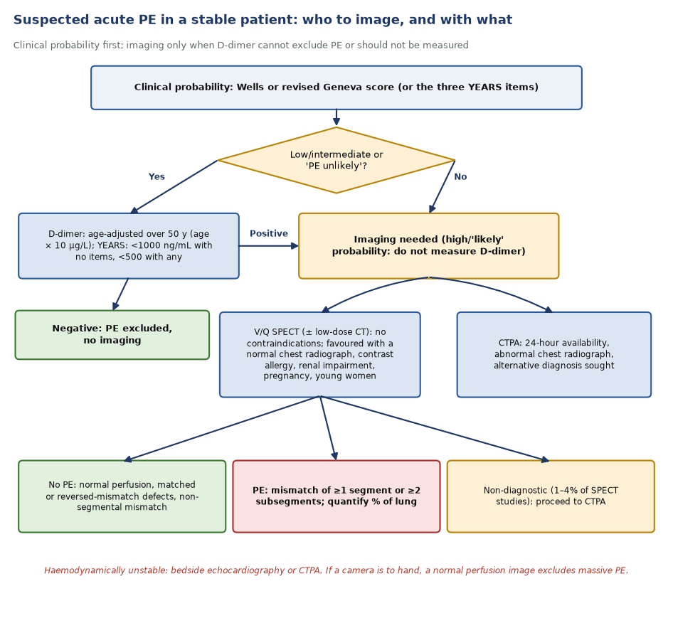
- Start with clinical probability: Wells or revised Geneva score, or clinical judgement.
- Measure D-dimer only when probability is low, intermediate or 'PE unlikely'. A negative result excludes PE; an age-adjusted cut-off (age × 10 µg/L over 50 years) had a 3-month failure rate of 0.3% in ADJUST-PE.
- YEARS uses three items (signs of DVT, haemoptysis, PE the most likely diagnosis) with a D-dimer cut-off of 1000 ng/mL when no item is present and 500 ng/mL otherwise. It avoided CTPA in 48% of patients (34% with Wells); 3-month VTE 0.61%.
- With a high or 'likely' probability, go straight to imaging. D-dimer has no discriminating value there.
- Image early: perfusion can recover within days.
V/Q or CTPA?
- Prefer V/Q when the chest radiograph is normal or near-normal, when there is contrast allergy, renal impairment or thyroid disease, and in pregnancy and young women, where breast dose matters. See suspected PE in pregnancy.
- Prefer CTPA when you need 24-hour availability, when the radiograph is abnormal, or when an alternative diagnosis or right ventricular strain must be shown.
- In a randomised trial of 1417 patients, CTPA was non-inferior to V/Q (3-month VTE after a negative test 0.4% vs 1.0%). It diagnosed more PE (19.2% vs 14.2%), and the clinical meaning of the extra small emboli is uncertain.
- In PIOPED II, CTPA had sensitivity 83% and specificity 96%, and 6.2% of studies were inconclusive on image quality. In a head-to-head series, V/Q SPECT with low-dose CT had sensitivity 97% and specificity 100%, against 68% and 100% for CTPA.
- In an unstable patient, CTPA or bedside echocardiography is usual; a normal perfusion image, even one planar view, excludes massive PE.
Radiopharmaceuticals and acquisition
- ⁹⁹ᵐTc-MAA particles (15–100 µm) lodge in precapillary arterioles in proportion to flow. Shake the vial, never draw blood into the syringe (clumps make hot spots), and inject slowly over about 30 s with the patient supine.
- For ventilation, Technegas (0.005–0.2 µm carbon particles, used within 10 min of generation) or ⁸¹ᵐKr is preferred in COPD. ⁹⁹ᵐTc-DTPA droplets (1.2–2 µm) deposit centrally in obstructed airways.
- Acquire ventilation SPECT, then perfusion SPECT, without moving the patient. The perfusion study has about five times the ventilation activity, so ventilation counts barely affect the perfusion images.
- Low-dose CT (V/Q SPECT/CT) adds specificity by showing emphysema, consolidation, effusion or vascular compression.
Reading the study
EANM reads V/Q SPECT holistically. Every defect is judged against pulmonary vascular anatomy and against ventilation.
| Pattern | What it means |
|---|---|
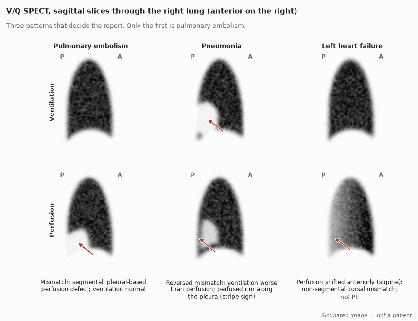
| Mismatch: pleural-based, segmental or subsegmental perfusion defect with normal ventilation | PE, if ≥1 segment or ≥2 subsegments |
| Matched defect | Airway or parenchymal disease; not PE |
| Reversed mismatch: ventilation worse than perfusion | Pneumonia, COPD, mucus plugging |
| Stripe sign: perfused rim at the pleura beyond a defect | Argues against PE in that zone; typical of pneumonia |
| Antigravitational perfusion (anterior more than posterior, supine) with a dorsal, non-segmental mismatch | Left heart failure with congestion; not PE |
| Uneven, patchy ventilation with milder perfusion change | COPD; grade severity from the ventilation images |
| Normal perfusion | PE excluded |
| Widespread abnormalities typical of no single disease | Non-diagnostic (1–4%): proceed to CTPA |
Reporting and pitfalls
- Report a binary conclusion: PE present, PE absent, or (rarely) non-diagnostic. Do not use PIOPED probability categories for SPECT; they generated large numbers of non-diagnostic reports.
- Record a single subsegmental mismatch, but it does not meet the criteria for PE. Its clinical significance is unproven.
- Quantify embolic extent as the number of mismatched segments and subsegments, expressed as a percentage of lung. Extent predicts recurrence and supports outpatient treatment decisions.
- Compare with any previous study. Without both, old and new emboli are hard to separate.
- Technical pitfalls: MAA hot spots, free pertechnetate (thyroid, stomach, kidneys), central DTPA deposition, and patient movement between ventilation and perfusion, which creates false mismatch.
- Non-segmental defects from cardiomegaly, hilar nodes, tumour or effusion are not PE. More in the full article: V/Q imaging.
In depth
- PIOPED (1990) explains why probabilistic planar reporting failed: a high-probability scan had sensitivity 41% and specificity 97%; 33% of intermediate-probability scans had PE, as did about 12% of low-probability scans.
- In a head-to-head study, V/Q SPECT alone had specificity 88%. Adding low-dose CT raised it to 100%, while perfusion SPECT plus low-dose CT without ventilation fell to 51%. CT does not replace the ventilation study.
- A meta-analysis of 9 studies (3454 patients) gave pooled sensitivity 96% and specificity 97% for V/Q SPECT. A later systematic review found a high risk of bias, so the exact accuracy is still uncertain; management outcome data (3-month VTE 0.38% after a negative study) are the stronger evidence.
- The stripe sign was validated in PIOPED: in 93% of lung zones with the sign (79 of 85), there was no PE in that zone.
- When ventilation was dropped during the COVID-19 pandemic to avoid aerosols, perfusion alone confidently excluded PE in only 57% of patients (French registry).
- Isolated subsegmental PE managed without anticoagulation (after negative leg ultrasound) had a 90-day recurrent VTE rate of 3.1%: 2.1% for a single subsegmental clot and 5.7% for several.
Sources: PIOPED, JAMA 1990 (PMID 2332918) · Gutte 2009 (PMID 19910421) · Kan 2015 (PMID 24917606) · Le Roux 2020 (PMID 33433051) · Le Roux 2014 (PMID 24719158) · Sostman 1992 (PMID 1620847) · EANM 2019 (PMID 31410539) · Le Roux 2022 (PMID 34649944) · Le Gal 2022 (PMID 34807722)
Sources
- Bajc M, et al. EANM guideline for ventilation/perfusion single-photon emission computed tomography (SPECT) for diagnosis of pulmonary embolism and beyond. Eur J Nucl Med Mol Imaging. 2019;46:2429–51.
- Parker JA, et al. SNM practice guideline for lung scintigraphy 4.0. J Nucl Med Technol. 2012;40:57–65.
- PIOPED Investigators. Value of the ventilation/perfusion scan in acute pulmonary embolism. JAMA. 1990;263:2753–9.
- Stein PD, et al. Multidetector computed tomography for acute pulmonary embolism. N Engl J Med. 2006;354:2317–27.
- Anderson DR, et al. Computed tomographic pulmonary angiography vs ventilation-perfusion lung scanning in patients with suspected pulmonary embolism: a randomized controlled trial. JAMA. 2007;298:2743–53.
- Gutte H, et al. Detection of pulmonary embolism with combined ventilation-perfusion SPECT and low-dose CT: head-to-head comparison with multidetector CT angiography. J Nucl Med. 2009;50:1987–92.
- Righini M, et al. Age-adjusted D-dimer cutoff levels to rule out pulmonary embolism: the ADJUST-PE study. JAMA. 2014;311:1117–24.
- van der Hulle T, et al. Simplified diagnostic management of suspected pulmonary embolism (the YEARS study). Lancet. 2017;390:289–97.
- Le Roux PY, et al. Safety of ventilation/perfusion single photon emission computed tomography for pulmonary embolism diagnosis. Eur J Nucl Med Mol Imaging. 2014;41:1957–64.
- Le Gal G, et al. Risk for recurrent venous thromboembolism in patients with subsegmental pulmonary embolism managed without anticoagulation. Ann Intern Med. 2022;175:29–35.
- Pleuritic pain after a long-haul flight in a 54-year-old · Intermediate