Thyroid Cancer Imaging (Radioiodine)
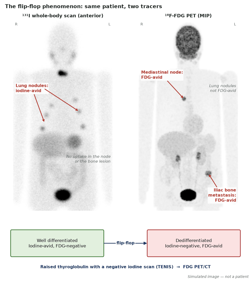
Differentiated thyroid cancer concentrates iodine, so ¹²³I/¹³¹I whole-body scintigraphy detects iodine-avid remnant and metastatic disease and underpins ¹³¹I therapy — the classic theranostic. FDG PET is reserved for dedifferentiated, iodine-negative disease (the “TENIS” scenario: raised thyroglobulin, negative iodine scan). (See also PET/CT → Thyroid cancer.)
Radioiodine imaging requires TSH stimulation (thyroid-hormone withdrawal or recombinant TSH) and a low-iodine diet to maximise uptake. Post-therapy scans are more sensitive than diagnostic scans.
When to image
- Around ¹³¹I therapy: optional pre-therapy ¹²³I/low-activity ¹³¹I scan to guide planning, and a post-therapy scan after every treatment. Remnant ablation is not routinely recommended for ATA low-risk DTC.
- Suspected iodine-avid recurrence in intermediate-high/high-risk patients; routine surveillance scans are not needed in low-risk patients with an excellent response or in those never given RAI.
- High-risk DTC with elevated thyroglobulin (generally >10 ng/mL), especially with negative iodine imaging or oncocytic/aggressive histology → FDG PET/CT.
How to read it
- Thyroid-bed uptake is usually remnant, but nodal disease can mimic it — SPECT/CT helps localise; uptake in lung or bone indicates metastasis.
- Physiological uptake in salivary glands, stomach, bowel and bladder must not be mistaken for disease.
- Post-therapy scans reveal disease not seen on diagnostic scans.
Protocol
- TSH stimulation to TSH >30 mIU/L (rhTSH preferred for ablation/adjuvant therapy, or LT4 withdrawal for 3–4 weeks) and a low-iodine diet (<50 µg/day) for ~1–2 weeks.
- ¹²³I (or low-dose ¹³¹I) diagnostic scan; post-therapy ¹³¹I scan.
- Avoid recent iodinated contrast (urinary iodine usually normalises within ~1 month; a spot urinary iodine can be checked if in doubt).
Pitfalls
- Iodine contamination (contrast) blocks uptake.
- Physiological activity mimics metastases.
- Dedifferentiated disease becomes iodine-negative and FDG-avid.
Evidence & guidelines
- 2025 ATA adult DTC guidelines (Ringel et al., Thyroid 2025;35:841–985), superseding ATA 2015.
- FDG PET/CT may be performed in high-risk DTC with elevated Tg (particularly RAI-negative, oncocytic or aggressive histology) and for prognosis/response in advanced disease; not routinely before initial treatment.
In depth
- ¹³¹I emits a 364 keV gamma photon (about 81%) and beta particles (maximum 606 keV) with a short tissue range of about 2 mm at most; its physical half-life is 8.0 days. It is actively concentrated into thyroid follicular cells well above plasma levels by the sodium–iodide symporter (NIS).
- ATA 2025 replaced the three-tier 2015 system (low, intermediate, high) with four recurrence-risk tiers: low, low-intermediate, intermediate-high and high. High risk still includes gross extrathyroidal extension, incomplete resection, distant metastases and nodal metastases ≥3 cm; N1a is now ranked below N1b, and limited vascular invasion in follicular or oncocytic cancer moves from low to low-intermediate risk.
- ATA 2025 does not recommend routine remnant ablation in low-risk DTC (30–50 mCi if given); it suggests considering 30–100 mCi for low-intermediate and intermediate-high risk, 100–150 mCi for high risk, and 100–200 mCi or dosimetry for distant metastases (100–150 mCi in patients over 70).
- Maximum-tolerated-activity dosimetry limits the blood (bone-marrow surrogate) dose to 2 Gy and whole-body retention at 48 h to 4.44 GBq (120 mCi), or 2.96 GBq (80 mCi) with diffuse lung metastases; it is favoured in renal insufficiency, children, older patients and diffuse lung metastases.
- Preparation: a low-iodine diet for about 1–2 weeks and TSH >30 mU/L by thyroid hormone withdrawal or rhTSH; exclude recent iodine loads such as IV contrast in the previous 3 months (urinary iodine can confirm clearance, often within about 1 month) or amiodarone.
- ATA 2025 no longer gives numerical TSH targets: TSH is kept within the reference range for an excellent or indeterminate response and below the reference range for biochemical or structural incomplete response, individualised to risk and side effects.
- Measure thyroglobulin with an assay calibrated to the BCR457 (CRM-457) standard, with quantitative anti-Tg antibodies on every sample. FDG PET/CT is used mainly in high-risk patients with elevated Tg (generally >10 ng/mL), particularly when radioiodine imaging is negative or histology is aggressive or oncocytic.
Sources: NNDC/LNHB nuclear decay data for I-131 · Dohán et al. Endocr Rev 2003 (PMID 12588808) · ATA 2025 DTC guidelines, Thyroid 2025 · 35:841-985 (PMID 40844370) · ATA 2025 DTC guidelines, Table 10 and Recommendations 32, 55–56 (PMID 40844370) · ATA 2025 DTC guidelines, Recommendations 55–56 (PMID 40844370) · ATA 2025 DTC guidelines, Recommendations 34–35 (PMID 40844370) · Padovani et al. Thyroid 2012
- Chest uptake after thyroid remnant ablation · Intermediate