PET/MR
PET/MR combines PET with the soft-tissue contrast, diffusion and functional sequences of MRI and removes the CT radiation dose. Integrated scanners acquire both at once using photodetectors that work in a 3-T field (avalanche photodiodes or silicon photomultipliers). Attenuation correction must be derived from MR images, so bone, metal and truncated arms need special handling. Evidence is strongest in paediatric oncology, prostate and pelvic cancer, liver and brain metastases, neuro-oncology, epilepsy and cardiac sarcoidosis.
MR signal reflects proton density and relaxation, not electron density, and cortical bone, air and metal all appear dark. A PET/MR attenuation (µ) map is therefore built by segmenting MR images into tissue classes, adding bone from a model or special sequences, or predicting a pseudo-CT.
- Photomultiplier tubes fail in strong magnetic fields; integrated PET/MR uses APDs (Biograph mMR, no TOF) or SiPMs (SIGNA PET/MR, TOF below 400 ps).
- NEMA: Biograph mMR 4.3 mm FWHM, 15.0 kcps/MBq; SIGNA PET/MR 4.1–5.3 mm, 23.3 kcps/MBq.
- Standard whole-body µ-map: Dixon segmentation into air, lung, fat and soft tissue; bone treated as soft tissue.
- Ignoring bone underestimates SUV in bone lesions by 11% on average (up to 31%).
- Model-based bone reduced the bias in bone from −25% to −5%.
- Modern brain MR-AC methods agree with CT-AC within ±5% on average.
- Paediatric FDG PET/MR: same lesion detection as PET/CT with 73% lower radiation dose.
- Cost per examination about 50% higher than PET/CT.
Hardware
- Integrated systems place the PET ring inside the MR bore and acquire simultaneously; the Biograph mMR (LSO with avalanche photodiodes) was the first commercial whole-body system, and SiPM systems add time-of-flight.
- Sequential systems (Philips Ingenuity TF) pair a TOF PET scanner with shielded photomultipliers and a 3-T MR on a shared bed; there is no simultaneity and the patient may move between scans.
- Mutual interference has been solved: PET performance matches PET/CT and MR quality stays within ACR tolerances.
- Rigid hardware (bed, head coil) is added to the µ-map as a template; flexible surface coils usually are not.
MR-based attenuation correction
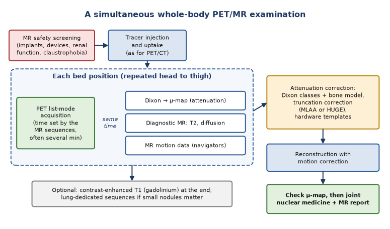
- Dixon water–fat imaging is segmented into four classes with fixed µ values: fast and robust, but it ignores bone and assumes one lung density.
- Bone can come from a registered bone model (whole body), ultrashort- or zero-echo-time sequences (mainly the head) or deep-learning pseudo-CT prediction.
- Truncation: the MR field of view is smaller than PET's, so arms are cut off; MLAA (activity and attenuation estimated from PET data) or HUGE (MR field-of-view extension) restores them.
- Metal implants give signal voids segmented as air, so uptake near prostheses and spinal hardware is underestimated — always check the µ-map (see quantification).
Motion and workflow
- Simultaneous MR data (navigators, self-gating) can correct respiratory and cardiac motion in PET without extra radiation.
- MR safety screening comes before injection; after uptake, PET is acquired at each bed position while the Dixon µ-map and diagnostic sequences (T2, diffusion, contrast T1) run.
- PET time per bed is set by the MR sequences, which improves counts or allows lower activity; full diagnostic protocols take much longer than PET/CT.
Clinical indications and evidence
- Paediatric oncology: equivalent lesion detection to PET/CT with 73% dose reduction (see paediatric practice).
- Prostate: simultaneous ⁶⁸Ga-PSMA PET/MRI localised primary cancer better than mpMRI or PET alone (AUC 0.88 vs 0.73 and 0.83).
- Rectal, cervical and head and neck cancer: MRI is already the local-staging reference, so PET/MR stages locally and distantly in one visit.
- In a mixed oncology series, extra PET/MR findings were mainly liver and brain metastases, changing management in 8%.
- Neurology: FDG or amyloid PET with volumetric MRI for dementia; drug-resistant epilepsy; amino-acid PET for gliomas (see epilepsy, brain tumours).
- Cardiac: FDG with late gadolinium enhancement for active sarcoidosis and viability (see cardiac sarcoidosis).
Safety, quantification and limitations
- Full MR safety rules apply: pacemakers and defibrillators only if MR-conditional and scanned under their conditions; screen for cochlear implants, neurostimulators, ferromagnetic foreign bodies and claustrophobia; observe gadolinium precautions in renal impairment.
- SUVs correlate closely with PET/CT (lesion ρ = 0.93) but are not interchangeable; use one modality for serial response assessment.
- MR misses small lung nodules: of CT nodules under 1 cm, Dixon showed 9 of 33 and contrast-enhanced VIBE 15 of 33, although FDG-avid lesions were detected equally.
- Longer slots, cost, limited availability and the need for dual expertise confine PET/MR to indications where MRI is needed anyway or dose matters most.
In depth
- APDs have low gain and slow timing, so the mMR has no time-of-flight; SiPMs combine high gain with fast timing (SIGNA coincidence timing below 400 ps). The sequential Ingenuity TF kept shielded photomultipliers (525 ps).
- In 11 brain MR-AC methods tested in 359 patients, vendor Dixon-based correction underestimated FDG uptake by 11.3%, while all newer atlas, segmentation and reconstruction methods were within ±5% of CT-AC.
- Replacing bone by soft tissue in CT maps gave SUV errors of −15.9% in sclerotic and −7.2% in lytic spinal lesions and −3.2% in soft-tissue lesions next to bone.
- Zero-echo-time AC improved brain accuracy by about 25% over the atlas method but misclassified air and bone in the mastoids and sinuses, overestimating temporal and cerebellar uptake by 2–3%.
- Truncation correction (HUGE or MLAA) raised lesion SUVs by 4–5% on average and changed values in truncated regions by up to 40%; MLAA failed with a non-FDG tracer, whereas HUGE is tracer-independent.
- In 330 same-day examinations, PET/MRI was accurate in 97.3% and PET/CT in 83.9%, at 597 vs 406 EUR per examination; the authors propose triage by tumour type.
Sources: Delso 2011; Grant 2016; Zaidi 2011 · Ladefoged 2017 (PMID 27988322) · Samarin 2012 (PMID 22526955) · Sekine 2016 (PMID 27339875) · Lindemann 2017 (PMID 28675598) · Mayerhoefer 2020 (PMID 31410538)
Sources
- Delso G, et al. Performance measurements of the Siemens mMR integrated whole-body PET/MR scanner. J Nucl Med. 2011;52:1914–22.
- Grant AM, et al. NEMA NU 2-2012 performance studies for the SiPM-based ToF-PET component of the GE SIGNA PET/MR system. Med Phys. 2016;43:2334.
- Samarin A, et al. PET/MR imaging of bone lesions — implications for PET quantification from imperfect attenuation correction. Eur J Nucl Med Mol Imaging. 2012;39:1154–60.
- Paulus DH, et al. Whole-body PET/MR imaging: a novel model-based MR attenuation correction method including bone. J Nucl Med. 2015;56:1061–6.
- Ladefoged CN, et al. A multi-centre evaluation of eleven clinically feasible brain PET/MRI attenuation correction techniques. NeuroImage. 2017;147:346–59.
- Schäfer JF, et al. Simultaneous whole-body PET/MR imaging in comparison to PET/CT in pediatric oncology. Radiology. 2014;273:220–31.
- Eiber M, et al. Simultaneous ⁶⁸Ga-PSMA HBED-CC PET/MRI improves the localization of primary prostate cancer. Eur Urol. 2016;70:829–36.
- Rauscher I, et al. PET/MR imaging in the detection and characterization of pulmonary lesions. J Nucl Med. 2014;55:724–9.
- Mayerhoefer ME, et al. PET/MRI versus PET/CT in oncology: a prospective study of 330 examinations. Eur J Nucl Med Mol Imaging. 2020;47:51–60.
- Dweck MR, et al. Hybrid MRI and PET with FDG to diagnose active cardiac sarcoidosis. JACC Cardiovasc Imaging. 2018;11:94–107.