Neuro-Oncology PET (Amino-Acid Tracers)
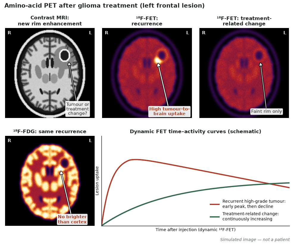
For brain tumours, amino-acid PET (¹⁸F-FET, ¹⁸F-FDOPA, ¹¹C-methionine) outperforms FDG because normal cortex has low amino-acid uptake, giving high tumour-to-background contrast. It aids glioma grading and delineation, biopsy and radiotherapy planning, and — most usefully — distinguishing recurrent/residual tumour from treatment-related change (pseudoprogression, radionecrosis). FDG PET is limited by high cortical background.
Gliomas over-express L-type amino-acid transporters (LAT1), so amino-acid analogues accumulate in tumour with little uptake in surrounding brain. This contrast underpins their advantage over FDG for delineation and recurrence assessment.
When to image
- Distinguishing tumour recurrence/residual disease from radionecrosis or pseudoprogression.
- Delineating tumour extent for biopsy and radiotherapy planning.
- Supporting glioma grading and prognostication.
- Assessing treatment response.
How to read it
- Increased amino-acid uptake with high tumour-to-brain ratio suggests active tumour.
- Dynamic ¹⁸F-FET time–activity curves help separate high-grade tumour from benign change.
- Treatment-related change tends to show lower tumour-to-brain ratios and continuously increasing FET time–activity curves (an early peak with a declining curve suggests high-grade tumour) — correlate with MRI.
Protocol
- Static ± dynamic amino-acid PET (FET/FDOPA/MET) with tumour-to-background ratios.
- Co-register with contrast-enhanced MRI for anatomical correlation.
Diagnostic performance
- Amino-acid PET separates recurrence from treatment change more accurately than FDG.
- High tumour-to-background contrast improves delineation for planning.
- Some uptake occurs in inflammation — interpret with MRI.
Pitfalls
- FDG’s high cortical uptake obscures many gliomas — prefer amino-acid tracers.
- Inflammatory and post-treatment changes can take up amino-acid tracers.
- Availability of amino-acid tracers is limited in some centres.
Evidence & guidelines
- PET-RANO/EANO recommendations (Albert et al., 2016) and the joint EANM/EANO/RANO/SNMMI guideline (Law et al., 2019) support amino-acid PET in glioma management.
- Amino-acid PET is complementary to advanced MRI (perfusion/spectroscopy).
In depth
- Amino-acid and receptor tracers exploit high tumour-to-brain contrast; NK-1 receptor (substance P) is overexpressed in 55/58 gliomas (WHO II–IV) and on tumour neovasculature.
- Targeted alpha therapy delivers ²¹³Bi- or ²²⁵Ac-DOTA-substance P locoregionally through stereotactically placed intratumoral or intracavitary catheters, with co-injected ⁶⁸Ga-DOTA-substance P PET/CT used to check the distribution.
- Alpha particles have a very short tissue range (<100 µm) and high LET (roughly 100 keV/µm), causing complex DNA double-strand breaks whose effect depends little on oxygenation or cell-cycle phase, which may help overcome radioresistance.
- ²¹³Bi (half-life 46 min) decays mainly via the alpha emitter ²¹³Po; ²²⁵Ac (half-life 9.9 days) releases four alpha particles through its decay chain, and its longer half-life was introduced to improve dose distribution within the tumour.
- The ²¹³Bi alpha emission has a mean tissue range of about 81 µm, far shorter than the several-millimetre range of the beta emitter ⁹⁰Y, favouring treatment of gliomas in eloquent (critically located) brain.
- ⁹⁰Y-DOTATOC targets somatostatin receptor subtype 2, which is expressed mainly in lower-grade (WHO grade II–III) gliomas; somatostatin-receptor expression is inversely related to substance P receptor expression in glioblastoma, limiting its use there.
- In a 2008 pilot study, resection-cavity ¹³¹I-81C6 (anti-tenascin) plus radiotherapy and temozolomide gave a median overall survival of about 21 months in glioblastoma, and ²¹¹At-ch81C6 in the resection cavity was feasible and safe in recurrent brain tumours.
Sources: Kneifel et al. Clin Cancer Res 2006 · 12:3843-50 (PMID 16778112) · Hennig et al. Int J Cancer 1995 (PMID 7790112) · Królicki et al. EJNMMI 2021 · 48:3595-605 (PMID 33860346) · Królicki et al. EJNMMI 2021 (PMID 33860346) · Królicki et al. Clin Nucl Med 2023 (PMID 36854309) · Cordier et al. EJNMMI 2010