Nuclear Medicine After Organ Transplantation
At a glance
- Function, not just structure. Scintigraphy shows graft perfusion and function when ultrasound is normal or equivocal.
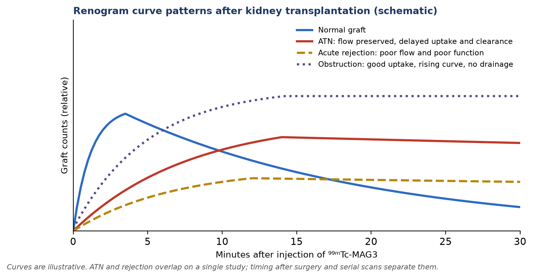
- Kidney. Get a baseline MAG3 scan early; ATN preserves perfusion with poor function, rejection reduces both, and serial scans separate them.
- FDG for rejection. A low renal FDG uptake (mean SUVmean <1.6) makes acute kidney rejection very unlikely (sensitivity 100%, specificity 30%).
- Liver. HIDA is sensitive for bile leaks but misses many anastomotic strictures.
- Heart and lung. PET flow reserve for allograft vasculopathy; V/Q and gastric emptying after lung transplantation.
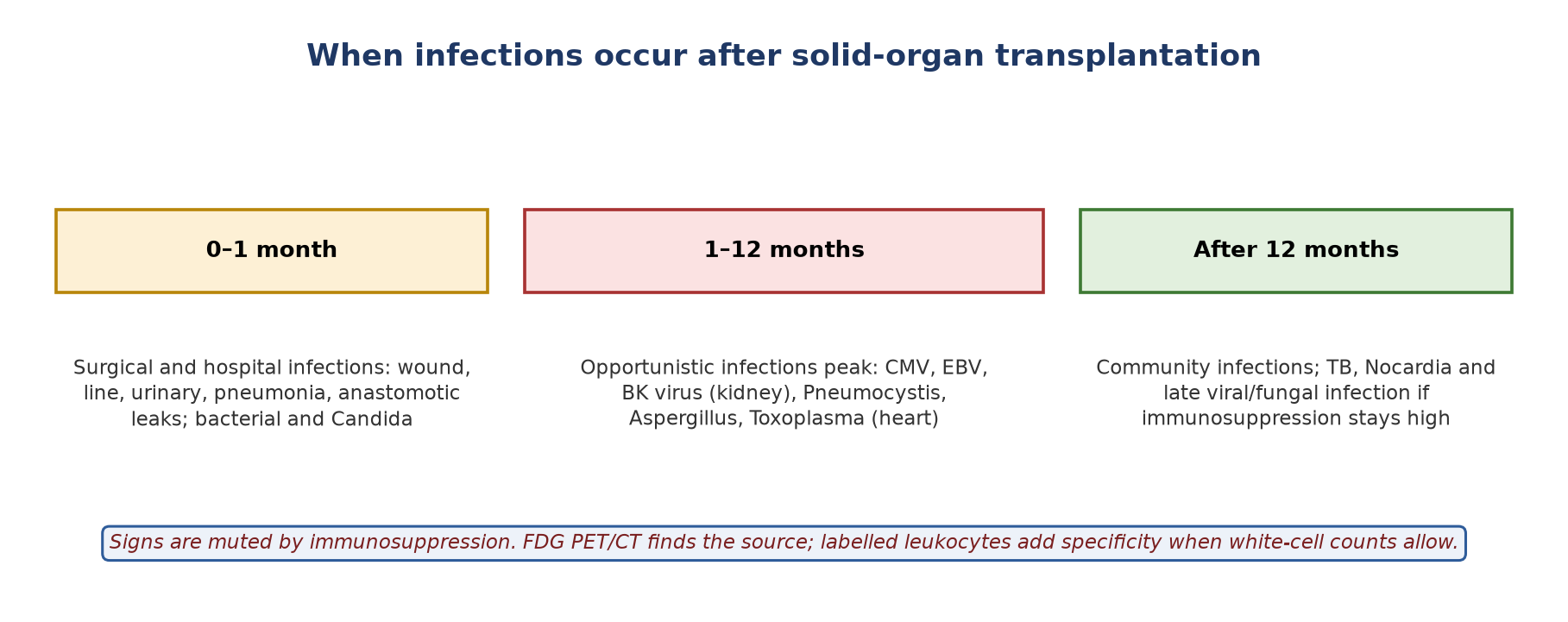
- Infection and PTLD. FDG PET/CT localises occult infection and stages PTLD (pooled sensitivity ~90%, specificity ~91%).
1. Kidney transplant: the renogram
- Biopsy is the reference for graft dysfunction but is invasive; ultrasound shows anatomy but not function. ⁹⁹ᵐTc-MAG3 renography adds a non-invasive functional assessment.
- Tracer: MAG3 is preferred over DTPA because its high renal extraction gives good images even with poor function. Typical adult activity is 37–185 MBq; higher activity improves the perfusion (flow) phase.
- Acquisition: supine, camera anterior over the iliac fossa; a fast flow phase, then dynamic images for 20–30 min. Add delayed static images (up to ~2 h) if a urine leak is suspected. Diuretic is less useful early after surgery.
- Baseline scan (ideally within 24–48 h): confirms perfusion, gives the reference for every later study and has prognostic value.
Complications and their scan patterns
| Problem | Timing | Perfusion | Uptake / function | Drainage | Clue |
|---|---|---|---|---|---|
| Normal graft | — | Prompt | Prompt | Prompt | Normal curve |
| Acute tubular necrosis (ATN) | From day 0; improves over days to weeks | Preserved or mildly reduced | Poor, cortical retention | Delayed | Perfusion–function mismatch; improves on serial scans |
| Acute rejection | Usually after ~5 days | Reduced | Reduced | Delayed | New or progressive worsening from baseline |
| Calcineurin-inhibitor toxicity | Variable | Usually preserved | Delayed clearance | Delayed | Resembles ATN; hard to separate from rejection |
| Chronic allograft nephropathy | Months to years | Gradually reduced | Patchy, falling | Delayed | Small, scarred graft over serial scans |
| Urine leak | Early | Normal | Normal | Tracer outside the urinary tract | Growing extra-urinary collection |
| Obstruction | Any | Normal | Normal or delayed | Absent; rising curve | Tracer retained in the collecting system |
| Arterial or venous thrombosis | Early | Absent | Absent | Absent | Photopenic graft |
Numbers that help
- Hilson perfusion index: area under the iliac artery curve divided by area under the graft curve (to peak) × 100. Normal is below 150; it rises when graft perfusion falls (rejection, arterial stenosis).
- Time to peak and clearance: delayed peak and poor clearance are sensitive but non-specific signs of dysfunction (ATN, rejection, toxicity or obstruction); the trend from baseline matters more than one value.
- Measured function: MAG3 clearance (ERPF) or DTPA/⁵¹Cr-EDTA GFR quantify function; a fall from the patient's own baseline is the most useful signal.
- Infection: ⁹⁹ᵐTc-DMSA is more sensitive than ultrasound for graft pyelonephritis.
FDG PET/CT for suspected acute rejection
- Inflammatory cells in rejection take up FDG. In a validation cohort of 79 recipients (86 scans) with biopsy, a mean renal SUVmean threshold of 1.6 gave sensitivity 100% and specificity 30% (AUC 0.86).
- So a low value argues strongly against acute rejection and may avoid a biopsy; a high value is non-specific (pyelonephritis, BK nephropathy, borderline changes).
2. Liver transplant
- Complications include rejection, bile leak, anastomotic stricture or obstruction, and vascular occlusion.
- HIDA (⁹⁹ᵐTc-mebrofenin): shows hepatocyte uptake, excretion and biliary drainage in one study.
| Finding | Uptake | Biliary/bowel activity | Clue |
|---|---|---|---|
| Normal | Prompt | Prompt bowel activity | Normal uptake and excretion |
| Bile leak | Normal | May be seen | Growing collection outside the biliary tree (perihepatic, peritoneal) |
| Obstruction / stricture | Normal or slightly reduced | Delayed or absent | Uptake–excretion mismatch; retained ductal activity |
| Hepatocyte dysfunction (rejection, preservation injury) | Reduced, slow blood-pool clearance | Delayed | Poor extraction from blood |
Know the limits
- In 104 living-donor recipients, HIDA detected anastomotic strictures with sensitivity 56% and specificity 73% (PPV 93%, NPV 22%): a scan without obstruction does not exclude a stricture.
- HIDA is not a reliable test for vascular occlusion; use Doppler ultrasound or CT angiography.
3. Heart transplant
- Endomyocardial biopsy remains the reference for rejection but is invasive and prone to sampling error.
- Graft function: radionuclide ventriculography (MUGA) gives reproducible LVEF; a fall from baseline suggests rejection, ischaemia or infection.
- Innervation: the transplanted heart is denervated, so ¹²³I-MIBG uptake is initially absent; partial sympathetic re-innervation over years can be tracked and has prognostic value.
- Acute cellular rejection: FDG uptake from inflammatory infiltrates has been described, but specificity is low and it remains investigational.
- Cardiac allograft vasculopathy (CAV): a diffuse, largely microvascular coronary disease affecting about half of recipients by 10 years. Relative SPECT perfusion can look normal when disease is uniform; PET absolute myocardial blood flow and flow reserve detect the global reduction and predict outcome.
4. Lung transplant
- V/Q scintigraphy: split lung function, suspected embolism, and surgical complications such as bronchial or arterial anastomotic stenosis.
- Chronic lung allograft dysfunction (CLAD): affects about half of recipients by 5 years; bronchiolitis obliterans syndrome is the commonest form. Small-airway obstruction causes air trapping and reflex vasoconstriction, giving matched patchy V/Q defects.
- Gastric emptying: vagal injury at surgery commonly causes gastroparesis; delayed emptying promotes reflux and micro-aspiration, a recognised risk factor for CLAD.
5. Infection and fever of unknown origin
| ¹⁸F-FDG PET/CT | Labelled leukocytes (¹¹¹In-oxine or ⁹⁹ᵐTc-HMPAO) | |
|---|---|---|
| Principle | Glucose metabolism of inflammatory cells | Migration of the patient's own labelled white cells |
| Best use | Whole-body search in FUO; monitoring treatment | Problem-solving when high specificity is needed |
| Sensitivity / specificity | High / low (also positive in rejection, PTLD, tumour) | Good / high for bacterial infection |
| Practical | Single visit, ~1–2 h | Needs blood handling; ⁹⁹ᵐTc-HMPAO images at 3–4 h and 20–24 h; not possible in marked leukopenia; poor for spine, fungal and viral infection |
6. Post-transplant lymphoproliferative disorder (PTLD)
- Lymphoid proliferations after solid-organ or stem-cell transplantation, linked to immunosuppression and Epstein–Barr virus.
- Risk by organ: lowest after kidney transplantation (about 1–2%), higher after heart and lung, highest after intestinal/multivisceral transplantation (up to ~20%).
- Timing: bimodal — early (first year, mostly EBV-positive) and late (often EBV-negative).
- WHO 2017 categories: non-destructive (plasmacytic hyperplasia, infectious mononucleosis-like, florid follicular hyperplasia); polymorphic; monomorphic (B-cell, most often DLBCL; T/NK-cell); classic Hodgkin lymphoma-type.
- Extranodal disease is common (gut, lung, CNS, skin, the graft itself) and can mimic infection or rejection.
Role of FDG PET/CT
- Diagnosis and staging: pooled sensitivity 89.7% and specificity 90.9% (bivariate meta-analysis, 336 recipients); FDG found lesions missed by CT/MRI in about 28% of patients.
- Response: Deauville/Lugano scoring as in lymphoma; FDG findings altered or guided treatment in about 29%. Criteria are validated for DLBCL- and Hodgkin-type disease, less so for polymorphic and non-destructive lesions.
- Pitfalls: false negatives in early lesions and high-background organs; false positives from inflammation.
7. What is coming
- CXCR4 PET (⁶⁸Ga-pentixafor): images leukocyte infiltration; in 13 kidney recipients with complicated urinary infection, PET/MRI identified allograft infection. Investigational.
- Other targets of alloimmune activation (e.g. activated T-cell markers) are being studied in animal models only.
Summary
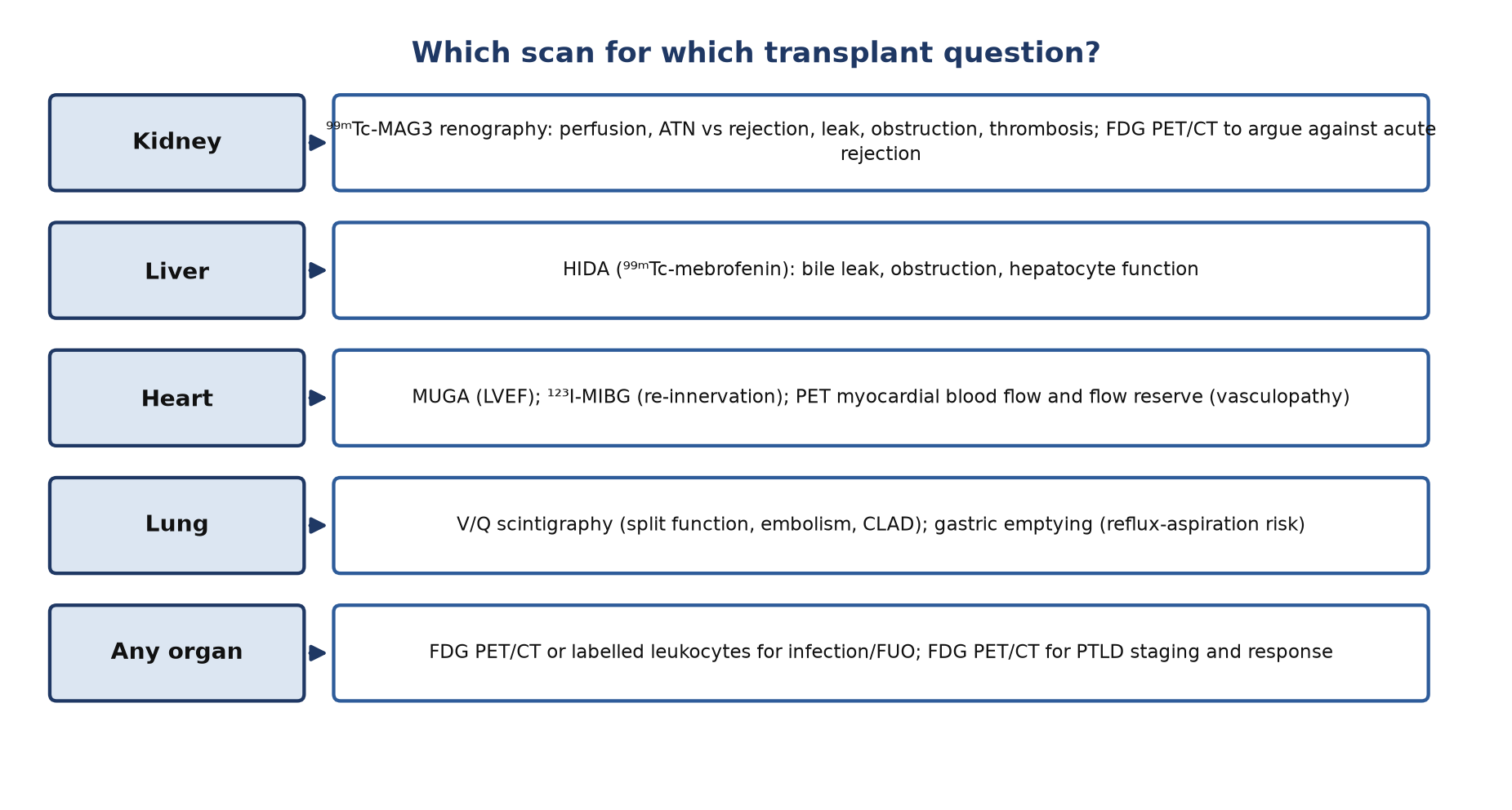
- Do a baseline MAG3 scan early after kidney transplantation and compare every later scan with it.
- ATN: preserved perfusion with poor function that improves; rejection: reduced perfusion and function, usually after day 5.
- A low renal FDG uptake argues strongly against acute rejection.
- HIDA is good for bile leaks but can miss strictures.
- Use PET flow reserve for heart allograft vasculopathy, V/Q for lung grafts, and FDG PET/CT for infection and PTLD.
Test yourself
5 quick questions. Pick an answer to see the explanation.
1. Day 1 after deceased-donor kidney transplantation, MAG3 shows good perfusion but poor uptake with cortical retention and little excretion. Most likely:
2. A Hilson perfusion index of 220 indicates:
3. A kidney recipient with rising creatinine has a renal FDG mean SUVmean of 1.4. This suggests:
4. Which statement about HIDA after liver transplantation is correct?
5. Why can relative SPECT perfusion look normal in cardiac allograft vasculopathy?
References
- Lovinfosse P, Weekers L, Pottel H, et al. [18F]FDG PET/CT imaging disproves renal allograft acute rejection in kidney transplant recipients with acute kidney dysfunction: a validation cohort. Eur J Nucl Med Mol Imaging. 2022;49(1):331-5.
- Kim YJ, Lee KT, Jo YC, et al. Hepatobiliary scintigraphy for detecting biliary strictures after living donor liver transplantation. World J Gastroenterol. 2011;17(21):2626-31.
- Fishman JA. Infection in organ transplantation. Am J Transplant. 2017;17(4):856-79.
- Erba PA, Glaudemans AWJM, Veltman NC, et al. Image acquisition and interpretation criteria for 99mTc-HMPAO-labelled white blood cell scintigraphy: results of a multicentre study. Eur J Nucl Med Mol Imaging. 2014;41(4):615-23.
- Ballova V, Muoio B, Albano D, et al. Diagnostic performance of 18F-FDG PET or PET/CT for detection of post-transplant lymphoproliferative disorder: a systematic review and a bivariate meta-analysis. Diagnostics (Basel). 2020;10(2):101.
- Montes de Jesus FM, Kwee TC, Nijland M, et al. Performance of advanced imaging modalities at diagnosis and treatment response evaluation of patients with post-transplant lymphoproliferative disorder: a systematic review and meta-analysis. Crit Rev Oncol Hematol. 2018;132:27-38.
- Swerdlow SH, Campo E, Pileri SA, et al. The 2016 revision of the World Health Organization classification of lymphoid neoplasms. Blood. 2016;127(20):2375-90.
- Derlin T, Gueler F, Bräsen JH, et al. Integrating MRI and chemokine receptor CXCR4-targeted PET for detection of leukocyte infiltration in complicated urinary tract infections after kidney transplantation. J Nucl Med. 2017;58(11):1831-7.
Practise with a case
- Oliguria two days after a kidney transplant · Intermediate