Myocardial Perfusion PET & Flow Reserve
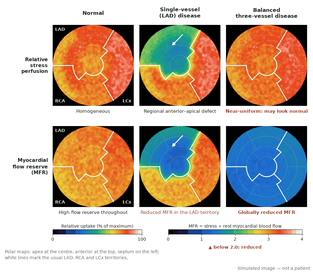
Myocardial perfusion PET uses ⁸²Rb (generator), ¹³N-ammonia or ¹⁵O-water (on-site cyclotron) and, in the US since September 2024, ¹⁸F-flurpiridaz. Pooled ⁸²Rb PET studies gave sensitivity 90% and specificity 88% against angiography, better than attenuation-corrected SPECT, and ¹⁵O-water PET was the most accurate test against FFR in the PACIFIC trial. Its unique strength is absolute myocardial blood flow (MBF, mL/min/g) and myocardial flow reserve (MFR = stress ÷ rest MBF), which expose balanced multivessel disease and microvascular dysfunction that relative imaging can miss.
A kinetic model applied to dynamic list-mode data estimates MBF, alongside relative perfusion and gated function. ⁸²Rb and ¹⁵O-water must be imaged during hyperaemia, so they need vasodilator stress; ¹⁸F-flurpiridaz also allows exercise.
- Half-lives: ⁸²Rb 75 s (parent ⁸²Sr 25.5 days); ¹⁵O 2 min; ¹³N 10 min; ¹⁸F 110 min.
- ⁸²Rb per injection (ASNC/SNMMI 2016): 2D 1480–2220 MBq; 3D BGO 370–740 MBq; 3D LSO/LYSO 1110–1480 MBq. Image 70–90 s after injection if LVEF > 50%, 90–130 s if lower.
- ¹³N-ammonia 370–740 MBq; images 1.5–3 min after infusion; 740 MBq ≈ 1.5 mSv.
- ¹⁸F-flurpiridaz one-day protocol (US label): rest 93–111 MBq; pharmacological stress 222–241 MBq (≥ 30 min later); exercise 333–352 MBq (≥ 60 min later).
- Healthy volunteers: ⁸²Rb stress MBF 2.86 mL/min/g, MFR 4.07; ¹³N-ammonia 2.58 mL/min/g, MFR 3.54.
- Global stress MBF > 2 mL/min/g with MFR > 2 excludes left main or three-vessel disease (NPV > 95%); MFR < 1.5 is high risk (Murthy 2018).
A low MFR driven by a high resting flow
Given. ⁸²Rb PET. Rest: heart rate 95/min, systolic BP 150 mmHg, MBF 1.10 mL/min/g. Regadenoson stress MBF 2.30 mL/min/g. Relative perfusion normal.
- Unadjusted MFR = 2.30 ÷ 1.10 = 2.09.
- Rest rate–pressure product = 95 × 150 = 14 250.
- Adjusted rest MBF = 1.10 × 8500 ÷ 14 250 = 0.66 mL/min/g (reference 8500, SNMMI/ASNC 2018).
- Adjusted MFR = 2.30 ÷ 0.66 = 3.5.
Answer. The borderline MFR reflects high resting work, not a poor hyperaemic response; stress MBF is above 2 mL/min/g. Report both values rather than calling microvascular dysfunction.
When to image
- Coronary disease where SPECT is limited: obesity, large breasts, or an equivocal previous study.
- Suspected left main or three-vessel disease, which can look 'balanced' on relative images.
- Angina without obstructive coronary disease, to look for microvascular dysfunction.
- Risk: the lowest tertile of flow reserve (MFR < 1.5) carried a 5.6-fold higher risk of cardiac death, independent of perfusion (Murthy 2011, 2783 patients).
- Viability: rest perfusion paired with ¹⁸F-FDG; see Myocardial viability.
The tracers
| Tracer | Source | Behaviour | Practical points |
|---|---|---|---|
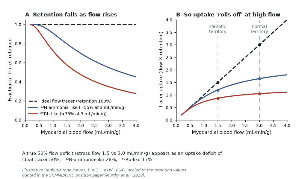
| ⁸²Rb | ⁸²Sr/⁸²Rb generator, replaced about every 6 weeks | K⁺ analogue (Na⁺/K⁺-ATPase); extraction about 35% at peak stress | No cyclotron; highest positron energy (Emax 3.38 MeV), so lowest resolution |
| ¹³N-ammonia | On-site cyclotron | Near-complete extraction, trapped as glutamine; 50–60% retained at peak stress | Good images and flow |
| ¹⁵O-water | On-site cyclotron | Freely diffusible, not retained | Reference MBF tracer but poor relative images; used in Europe, not FDA-approved |
| ¹⁸F-flurpiridaz | Regional cyclotron, unit doses | Analogue of the mitochondrial complex I inhibitor pyridaben | FDA-approved 27 September 2024; exercise possible; shortest positron range |
Protocol
- Rest then stress the same day with regadenoson, adenosine or dipyridamole (no caffeine for 12 h, no theophylline for 48 h); ⁸²Rb doses at least 10 min apart.
- List-mode acquisition from injection gives dynamic (flow), summed (perfusion) and gated (LVEF at rest and peak hyperaemia) data.
- Low-dose CT for attenuation correction at end-expiration or shallow breathing, never full inspiration; for ⁸²Rb a post-stress CT is preferred if realignment software is unavailable.
- Reduce ⁸²Rb activity on 3D scanners: detector saturation underestimates the input function and falsely raises MBF.
How to read it
- Read relative perfusion as for SPECT (17 segments, summed scores), then global and territorial stress MBF and MFR.
- Regional defect with regionally low MFR = focal stenosis. Global reduction without a regional defect = balanced multivessel disease, diffuse atheroma or microvascular dysfunction; anatomy is often needed to separate them.
- LVEF normally rises during vasodilator stress on PET; a rise above 5% had a 97% negative predictive value for left main or three-vessel disease (Dorbala 2007).
- Global MFR close to 1 with normal relative images suggests the vasodilator failed (often caffeine); the perfusion images may be invalid.
- Report coronary calcium on the attenuation CT, as for any non-contrast chest CT (SCCT/STR 2016).
Diagnostic performance
- Mc Ardle 2012 meta-analysis: ⁸²Rb PET sensitivity 90%, specificity 88%; attenuation-corrected gated SPECT 85% and 85%; AUC 0.95 v 0.90.
- PACIFIC (208 patients, FFR ≤ 0.80 in every vessel): ¹⁵O-water PET accuracy 85% (sensitivity 87%, specificity 84%), SPECT 77% (57%, 94%), CT coronary angiography 74% (90%, 60%); hybrid imaging added nothing.
- Flurpiridaz v SPECT: sensitivity 71.9% v 53.7% in the first phase 3 trial (specificity missed non-inferiority); 80.3% v 68.7% in AURORA, with non-inferior specificity.
Pitfalls
- CT–PET misregistration creates false defects in segments next to lung; check the fusion every time.
- Roll-off: ⁸²Rb uptake underestimates high flow, so mild stenoses show less contrast on relative images (see figure).
- Spillover, partial volume, motion and a poor bolus bias MBF, which also differs between tracers and software: keep both constant for serial studies.
- ⁸²Rb eluate is tested daily for ⁸²Sr/⁸⁵Sr breakthrough (US alert limits 0.002 and 0.02 µCi per mCi ⁸²Rb); eluting with the wrong solution causes high breakthrough.
In depth
- Renkin–Crone: extraction E = 1 − e−PS/MBF (PS, permeability–surface area product) and uptake K1 = E × MBF, so a low-PS tracer such as ⁸²Rb plateaus at high flow; software converts K1 back to MBF.
- ⁸²Rb emits a 777 keV gamma in about 15% of decays, adding prompt-gamma coincidences that 3D scanners must correct; it decays to stable ⁸²Kr and the generator refills in about 10 min.
- ⁸²Rb gives 1.1–3.5 mSv for 2220 MBq at each of rest and stress; the bladder is the critical organ for ¹³N-ammonia.
- Flurpiridaz: 0.019 mSv/MBq, so a one-day rest plus pharmacological study (about 330 MBq) gives about 6 mSv, matching 6.1 mSv (v 13.4 mSv for SPECT) in the first phase 3 trial.
- Thresholds: the 2016 guideline calls MFR > 2.3 favourable and < 1.5 significantly reduced; values shift a little with software.
Sources: Murthy 2018 (PMID 29242396) · Dilsizian 2016 (PMID 27392702) · Dorbala 2007 (PMID 17332611) · Flyrcado and Cardiogen-82 US labels · NNDC decay data · Maddahi 2020 (PMID 32703509)
Sources
- Dilsizian V, Bacharach SL, Beanlands RS, et al. ASNC imaging guidelines/SNMMI procedure standard for positron emission tomography (PET) nuclear cardiology procedures. J Nucl Cardiol. 2016;23:1187–226.
- Murthy VL, Bateman TM, Beanlands RS, et al. Clinical quantification of myocardial blood flow using PET: joint position paper of the SNMMI Cardiovascular Council and the ASNC. J Nucl Med. 2018;59:273–93.
- Mc Ardle BA, Dowsley TF, deKemp RA, et al. Does rubidium-82 PET have superior accuracy to SPECT perfusion imaging for the diagnosis of obstructive coronary disease? J Am Coll Cardiol. 2012;60:1828–37.
- Danad I, Raijmakers PG, Driessen RS, et al. Comparison of coronary CT angiography, SPECT, PET, and hybrid imaging for diagnosis of ischemic heart disease determined by fractional flow reserve. JAMA Cardiol. 2017;2:1100–7.
- Maddahi J, Agostini D, Bateman TM, et al. Flurpiridaz F-18 PET myocardial perfusion imaging in patients with suspected coronary artery disease. J Am Coll Cardiol. 2023;82:1598–610.
- Murthy VL, Naya M, Foster CR, et al. Improved cardiac risk assessment with noninvasive measures of coronary flow reserve. Circulation. 2011;124:2215–24.