Myocardial Perfusion SPECT
Stress/rest myocardial perfusion SPECT (⁹⁹ᵐTc-sestamibi or tetrofosmin, or ²⁰¹Tl) detects flow-limiting coronary artery disease and is a powerful risk-stratifier: a normal study carries a low annual cardiac event rate. Reported sensitivity is ≈85–90% for obstructive CAD, with more variable specificity (≈61% pooled across older studies, ≈85% for contemporary gated, attenuation-corrected SPECT); gated acquisition adds ejection fraction and wall motion. Attenuation correction and prone/upright imaging mitigate artefacts.
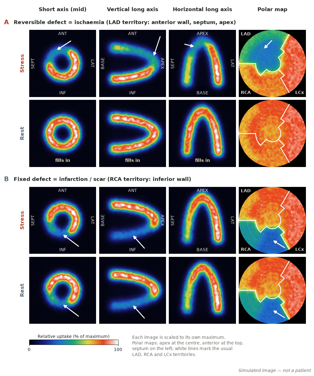
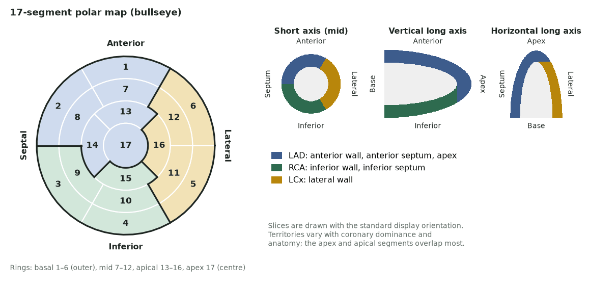
A perfusion tracer is injected at peak stress (exercise or pharmacological vasodilator/inotrope) and at rest. Regions supplied by a stenosed artery show relatively reduced stress uptake that “fills in” at rest (reversible ischaemia); fixed defects usually reflect scar.
When to image
- Symptomatic patients with intermediate pretest probability of CAD.
- Risk stratification in known CAD and before major surgery in selected cases.
- Assessing functional significance of known stenoses.
- Evaluating chest pain when the ECG is uninterpretable or exercise ECG is equivocal.
Protocol
- Stress with exercise or pharmacological agents (adenosine, regadenoson, dipyridamole; dobutamine if vasodilators contraindicated).
- ⁹⁹ᵐTc agents (gated) or ²⁰¹Tl; one- or two-day protocols.
- Attenuation correction and/or prone/upright imaging to reduce artefacts.
- Gated SPECT for EF and regional wall motion.
How to read it
- Reversible defect (stress-only) → ischaemia; fixed defect → scar; partial reversibility → mixed.
- Assess extent/severity, transient ischaemic dilatation, and post-stress EF drop as high-risk markers.
- Correlate with symptoms, ECG and coronary anatomy.
Diagnostic performance
Per-patient sensitivity for obstructive CAD is ≈85–90%; specificity varies widely (pooled ≈61% in a meta-analysis of 1990–2010 studies, ≈85% with contemporary gated, attenuation-corrected SPECT); a normal perfusion study confers a low annual rate of cardiac death/MI. PET generally out-performs SPECT (see Perfusion PET).
Pitfalls
- Attenuation (breast, diaphragm) mimics defects — use correction/positioning.
- Balanced multivessel disease can look near-normal on relative imaging.
- Sub-maximal stress reduces sensitivity.
- Left bundle branch block causes septal artefacts (prefer vasodilator stress).
Evidence & guidelines
- ASNC imaging guidelines (e.g. Henzlova et al., 2016, on stress, protocols and tracers) cover acquisition, stress protocols and reporting.
- Normal MPI carries a well-documented low-risk “warranty period”.
- PET is preferred where available for accuracy and flow quantification.
In depth
- ⁹⁹ᵐTc-sestamibi/tetrofosmin (140 keV, negligible redistribution): image delays sestamibi 15–20 min (exercise)/45–60 (rest)/60 (pharmacologic); tetrofosmin 10–15/30–45/45.
- Adenosine and regadenoson cause coronary vasodilatation via A2A receptors, increasing myocardial blood flow 3.5–4-fold; side effects arise from A1 (AV block), A2B (peripheral vasodilatation) and A2B/A3 (bronchospasm) activation and can be reversed with IV aminophylline.
- Adenosine effects: flushing 35–40%, chest pain 25–30%, dyspnoea 20%, AV block ~8% (mostly self-limiting).
- Before vasodilator stress, withhold caffeine and other methylxanthines for at least 12 h and dipyridamole for at least 48 h, and fast for at least 3 h.
- Vasodilator stress rather than exercise or dobutamine is preferred in LBBB, permanent ventricular pacing and ventricular pre-excitation (WPW), because rate-related septal perfusion artefacts can mimic ischaemia.
- Absolute contraindications to exercise stress include severe symptomatic aortic stenosis, acute MI within 2–4 days, acute pulmonary embolism, aortic dissection or myo/pericarditis, and resting BP above 200/110 mmHg.
- High-risk markers: transient ischaemic dilatation, post-stress EF drop, and multi-territory reversible defects.
Sources: ASNC SPECT stress, protocols and tracers guideline 2016 (PMID 26914678) · ACC/AHA/ASNC radionuclide imaging guideline · ACCF/AHA 2012 stable ischaemic heart disease guideline high-risk criteria