Myocardial Viability (FDG PET)
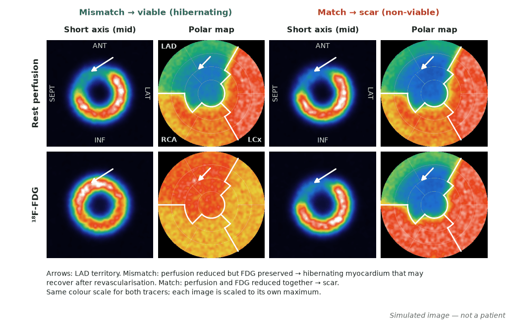
In ischaemic left-ventricular dysfunction, FDG PET identifies hibernating (viable) myocardium — regions with reduced perfusion but preserved glucose metabolism (a perfusion–metabolism mismatch). Viable segments may recover function after revascularisation, whereas a matched perfusion–metabolism defect indicates scar. FDG PET is widely regarded as the reference standard for viability, although randomised trials have not shown that viability testing selects patients who benefit from revascularisation.
Chronically hypoperfused but living myocardium down-regulates contraction while switching to glucose metabolism. Pairing a perfusion study with FDG reveals this metabolism–perfusion dissociation.
When to image
- Ischaemic cardiomyopathy with reduced EF being considered for revascularisation.
- Deciding whether dysfunctional segments are likely to recover.
- Clarifying viability where other tests are equivocal.
How to read it
- Perfusion–FDG mismatch (reduced perfusion, preserved/increased FDG) = hibernating, viable.
- Concordant reduction (match) = scar, non-viable (non-transmural if the reduction is only mild).
- Quantify the extent of viable myocardium to inform revascularisation benefit.
Protocol
- Rest perfusion study (SPECT or PET) paired with FDG PET.
- Metabolic preparation: glucose loading ± insulin (or clamp) to drive myocardial FDG uptake.
- Compare perfusion and metabolism maps segment by segment.
Pitfalls
- Inadequate metabolic preparation degrades FDG images.
- Diabetes complicates glucose handling — may need a clamp.
- In STICH and REVIVED-BCIS2, viability did not identify patients who benefit from CABG or PCI; the extent of scar, not of viable myocardium, predicted outcome in REVIVED — integrate with the whole picture.
Evidence & guidelines
- FDG PET is the reference standard for hibernating myocardium.
- PARR-2: FDG PET-assisted management did not significantly reduce cardiac events overall (HR 0.78, p=0.15); benefit was confined to adherent and post hoc subgroups.
- Interpreted within heart-team decision-making.
In depth
- Thallium-201 (K⁺ analogue via Na/K-ATPase) has ~80–85% first-pass extraction, a 73-hour half-life and 68–80 keV X-rays; uptake >50–60% of normal segments indicates viability.
- A severe fixed defect on thallium redistribution imaging suggests predominantly non-viable myocardium, but reinjection shows improved uptake in about half of apparently irreversible defects.
- Hibernating myocardium shifts from fatty-acid to glucose metabolism, so it remains FDG-avid despite reduced perfusion (perfusion–metabolism mismatch), whereas scar shows a matched defect.
- Late redistribution imaging at 18–24 h recovers additional fixed-appearing defects, but some viable segments only redistribute after reinjection.
- IV glucose/insulin loading for non-diabetic patients with fasting glucose <110 mg/dL (one sample protocol): prime with 5 U regular insulin and 10 g dextrose, infuse insulin 1.5 mU/kg/min with glucose 10 mg/kg/min for 60 min, give FDG once glucose is 100–200 mg/dL (preferably <150), then continue 20% dextrose during acquisition.
- In diabetic patients (or fasting glucose >110 mg/dL), one sample protocol primes with 10 U regular insulin if glucose >140 mg/dL or 6 U if <140, then infuses insulin with 20% dextrose adjusted to hold glucose at 80–140 mg/dL for 20–30 min before FDG.
- In the CASS registry (ejection fraction <0.36), surgery prolonged survival compared with medical therapy, most clearly when ejection fraction was <0.26 (5-year survival 63% vs 43%); this predates viability imaging, and STICH later showed that viability testing did not identify who benefits from surgery.
Sources: ASNC imaging guidelines (Dilsizian, J Nucl Cardiol) · PMID 2362606 · PMID 2362606 (Dilsizian 1990, NEJM) · ASNC PET guideline 2009 (Dilsizian, J Nucl Cardiol) · PMID 3263995 (Kiat 1988) · ASNC PET guideline 2009, appendix 1 (protocol A) · PMID 6352078 (Alderman 1983) · PMID 21463153 (STICH viability, Bonow 2011)