Captopril (ACE-Inhibitor) Renography
Snapshot
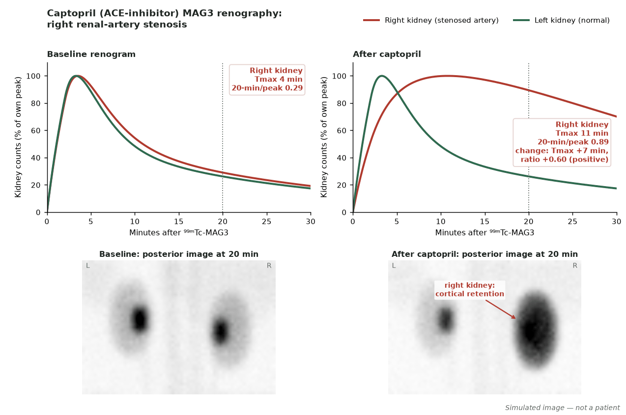
Captopril renography screens for renovascular hypertension. ACE-inhibition removes angiotensin-II-mediated efferent arteriolar constriction, so a kidney supplied by a haemodynamically significant renal-artery stenosis shows a worsened renogram after captopril compared with baseline — indicating a functionally relevant stenosis.
In renal-artery stenosis, angiotensin II maintains glomerular pressure by constricting the efferent arteriole. Blocking it unmasks the physiological significance of the stenosis on the renogram.
ACE-inhibitionUnmasks stenosis
FunctionalNot just anatomical
Curve changePost-captopril worsening
When to image
- Selected patients with suspected renovascular hypertension — its role has diminished now that Doppler, CT and MR angiography detect stenosis.
- Assessing the functional significance of a known renal-artery stenosis.
How to read it
- Post-captopril deterioration (delayed uptake/excretion) versus baseline suggests a functionally significant stenosis.
- A normal captopril study makes renovascular hypertension unlikely (<10% probability); sensitivity and specificity are ~90% when creatinine is <1.7 mg/dL and lower in azotaemia.
- Interpret both kidneys’ curves.
Protocol
- Withhold captopril 3 days and longer-acting ACE-inhibitors 5–7 days (stop ARBs too); stop chronic diuretics if possible; hydrate (e.g. 7 mL/kg water 30–60 min before); no solid food within 4 h of oral captopril.
- Captopril 25–50 mg orally 1 h before tracer, or enalaprilat 40 µg/kg IV (max 2.5 mg) ≥15 min before; 1-day (baseline then ACE-I) or 2-day (ACE-I first) protocol.
- Monitor blood pressure during the study.
Pitfalls
- Bilateral stenosis and renal impairment reduce accuracy.
- Dehydration and ongoing ACE-inhibitor therapy confound results.
- Interpret alongside anatomical angiography.
Evidence & guidelines
- SNMMI procedure guideline for diagnosis of renovascular hypertension v3.0 (Taylor et al., 2003) and ACE-inhibitor renography consensus (Taylor et al., J Nucl Med 1996;37:1876–1882).
- A functional test complementary to CT/MR angiography.
In depth
- ACE-inhibitor renography tests for renovascular hypertension (not merely stenosis), most useful in moderate-to-high-risk patients (abrupt/severe or resistant hypertension, bruit, worsening function on ACEI/ARB).
- Hydrate the patient (for example 7 mL/kg water orally at least 30 and preferably 60 min before) and avoid a solid meal within 4 h if oral captopril is used; place an IV line for prompt saline infusion in high-risk patients and those receiving enalaprilat.
- Withhold short-acting ACE inhibitors such as captopril for 3 days and longer-acting ones for 5–7 days; angiotensin II receptor blockers should also be stopped because they may reduce sensitivity similarly, and chronic diuretics should be stopped several days before if possible.
- Captopril 25–50 mg orally with tracer 60 min later; or enalaprilat 40 µg/kg (max 2.5 mg) IV over 3–5 min with tracer 15 min later.
- ⁹⁹ᵐTc-MAG3 is preferred over ⁹⁹ᵐTc-DTPA when creatinine is raised because of its higher extraction; furosemide 20 mg with the tracer is optional and may improve detection of cortical retention, but it is not essential.
- A normal ACEI study makes renovascular hypertension unlikely, so the baseline study may be omitted.
- High probability (>90%) is a marked ACEI-induced change from baseline: for MAG3, unilateral parenchymal retention (20-min/peak ratio rise ≥0.15, or Tmax prolonged by ≥2–3 min or 40%); for DTPA, a fall in relative uptake >10% or in ipsilateral GFR >10%, or marked unilateral parenchymal retention.
- Bilateral symmetrical post-ACEI change usually does not indicate renovascular hypertension.
- Safety: record BP and pulse before ACEI and every 5–15 min thereafter (every 5 min with enalaprilat), with sitting and standing BP before discharge; do not discharge until standing mean BP is at least 70% of baseline and the patient is asymptomatic when standing.
Sources: SNMMI procedure guideline for diagnosis of renovascular hypertension v3.0 (2003, reviewed 2022) · SNMMI procedure guideline for diagnosis of renovascular hypertension v3.0 (2003)