Renovascular Hypertension
Snapshot
Renovascular hypertension (RVH) is high blood pressure driven by reduced renal perfusion — almost always renal-artery stenosis — which activates the renin–angiotensin–aldosterone system (RAAS). It is an important correctable cause of secondary hypertension (primary aldosteronism is more common). Anatomical imaging confirms the stenosis (Duplex ultrasound first-line, then CT or MR angiography); ACE-inhibitor (captopril) renography is a functional test with a narrower, selective role today.
RAASRenin-driven
ARAS ~90%Commonest cause
Duplex USFirst-line imaging
ACE-I renographyFunctional test
Reference values
- ⁹⁹ᵐTc-MAG3 (tubular; preferred if creatinine is raised) or ⁹⁹ᵐTc-DTPA (glomerular filtration); 1-day protocol: ~40 MBq baseline, then 200–400 MBq for the ACE-I study.
- ACE-inhibition: captopril 25–50 mg orally 1 h before tracer (fasting for absorption); or enalaprilat 40 µg/kg IV (max 2.5 mg) over 3–5 min, image ~15 min later.
- Stop ACE-inhibitor/ARB before the study (captopril 3 days; longer-acting agents 5–7 days); stop chronic diuretics if possible; hydrate; monitor blood pressure.
- Duplex US: peak systolic velocity ≥180–200 cm/s (criteria vary by laboratory) suggests ≥60% stenosis; resistive index >0.8 predicts poor revascularisation response.
- Anatomical accuracy: CT angiography and gadolinium-enhanced MR angiography performed best in meta-analysis, better than ultrasound and captopril renography (Vasbinder et al., Ann Intern Med 2001).
- ACE-I renography: sensitivity and specificity ~90% when creatinine is <1.7 mg/dL, lower in azotaemia; a normal study means <10% probability of RVH.
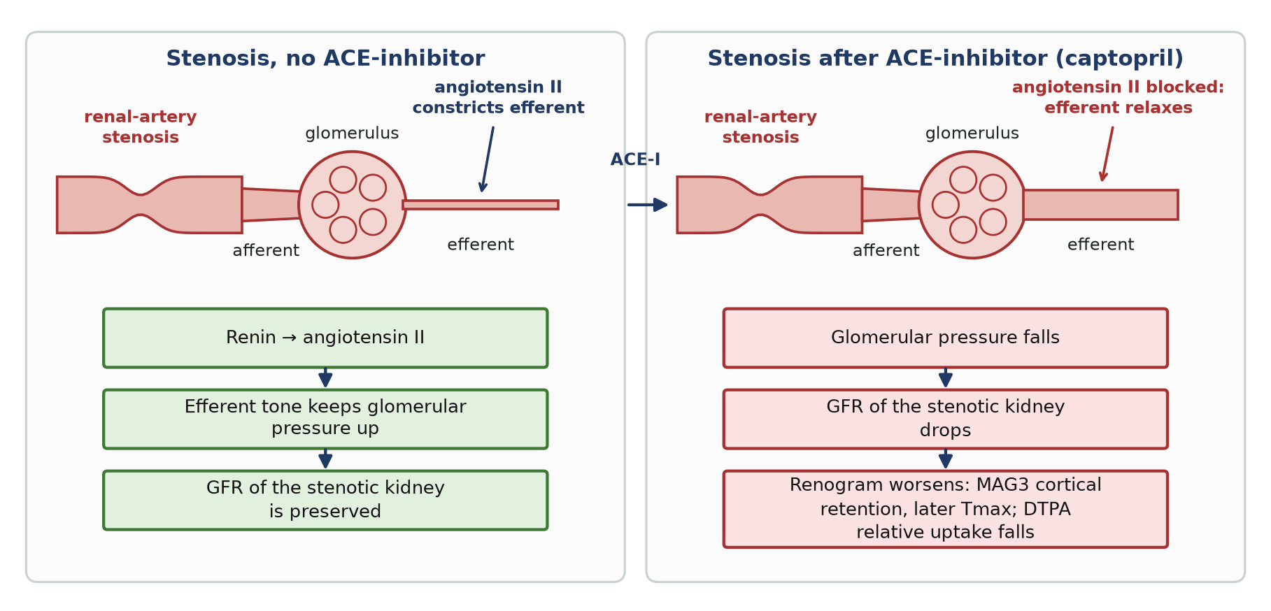
- High-probability ACE-I renogram = ACE-I-induced deterioration of the affected kidney: MAG3 unilateral parenchymal retention (20-min/peak ratio rise ≥0.15, Tmax prolonged ≥2–3 min or ≥40%, or worsened curve grade); DTPA relative uptake falls >10%.
Definition
- Hypertension caused by a haemodynamically significant reduction in renal blood flow, usually from stenosis of the main renal artery (or a branch).
- ‘Renal-artery stenosis’ is the anatomical lesion; ‘renovascular hypertension’ is the clinical syndrome it produces — the two are not synonymous (many stenoses are incidental and not causing the hypertension).
- It is an important potentially reversible cause of secondary hypertension (primary aldosteronism is more common).
Pathophysiology
- Renal-artery stenosis lowers perfusion pressure to the affected kidney.
- Juxtaglomerular cells release renin → angiotensin II (vasoconstriction) and aldosterone (sodium and water retention) → the RAAS raises blood pressure.
- Angiotensin II constricts the efferent arteriole to preserve glomerular filtration in the stenotic kidney — the physiological basis of ACE-inhibitor renography (blocking it drops that kidney’s GFR and worsens its renogram).
- Unilateral disease is renin-dependent and potentially curable; with bilateral disease or a stenosis to a solitary kidney, ACE-inhibitors/ARBs can precipitate acute kidney injury.
Causes
| Cause | Frequency / typical profile | Features |
|---|---|---|
| Atherosclerotic renal-artery stenosis (ARAS) | ~90% of RVH; older (>55–65), cardiovascular risk factors | Ostial/proximal stenosis; progressive; often bilateral |
| Fibromuscular dysplasia (FMD) | ~10%; young women (15–50) | Mid/distal artery, classic “string of beads”; often curable by angioplasty |
| Other (uncommon) | Any age | Vasculitis (Takayasu), dissection/thrombosis/infarction, radiation fibrosis, extrinsic compression, mid-aortic syndrome, transplant renal-artery stenosis |
When should renovascular hypertension be suspected?
- Hypertension onset before ~30 years (suggests FMD) or new/accelerating after ~55 years (suggests ARAS).
- Resistant hypertension — uncontrolled on ≥3 agents including a diuretic — or accelerated/malignant hypertension.
- A rise in serum creatinine >30% after starting an ACE-inhibitor or ARB.
- Recurrent ‘flash’ pulmonary oedema or unexplained heart failure.
- Abdominal or flank bruit; unexplained hypokalaemia with metabolic alkalosis.
- Asymmetric kidney size / an unexplained small kidney; unexplained renal impairment; widespread atherosclerosis.
Tests available & investigation of choice
Diagnosis is confirmed anatomically; functional testing is selective. Figures are cohort-dependent.
| Test | Sensitivity | Specificity | Role |
|---|---|---|---|
| Duplex ultrasound | High | High | First-line screen; PSV ≥180–200 cm/s (lab-dependent) suggests ≥60% stenosis; resistive index >0.8 predicts poor response to revascularisation |
| CT angiography | High | High | Anatomical investigation of choice when US is equivocal; needs iodinated contrast and radiation |
| MR angiography | High | High | Anatomy without radiation; tends to over-estimate stenosis; with eGFR <30 use a group II gadolinium agent only when needed (very low NSF risk) |
| Catheter angiography (DSA) | Gold standard | — | Definitive; used when non-invasive tests conflict, or at the time of intervention |
| ACE-inhibitor (captopril) renography | ≈90% (lower if azotaemic) | ≈90% | Functional test / lateralisation — selective role; less sensitive, radiation, time-consuming |
Where captopril renography stands in the pathway
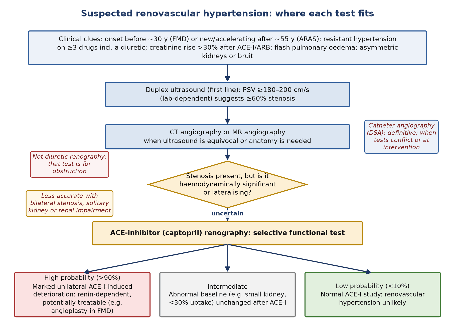
- It is no longer a first-line test — anatomical imaging (Duplex → CT/MR angiography) has largely replaced it for detecting stenosis.
- Practice shifted after the ASTRAL (2009) and CORAL (2014) trials, which showed renal-artery stenting adds little to optimal medical therapy in atherosclerotic disease — so proving ‘functional significance’ is less often decisive.
- Selective current roles: judging the haemodynamic significance and lateralising renin production when anatomy is borderline; helping decide whether a stenosis is worth treating (e.g. angioplasty in FMD, or before revascularisation); and when iodinated/gadolinium contrast is contraindicated.
- Note: for renovascular hypertension the nuclear test is ACE-inhibitor (captopril) renography — not diuretic renography, which is used for suspected obstruction.
Tracers, doses & protocol
- ⁹⁹ᵐTc-MAG3 is preferred (high tubular extraction, better in impaired function); ⁹⁹ᵐTc-DTPA (filtration) is an alternative.
- Give the ACE-inhibitor: captopril 25–50 mg orally 1 h before the tracer (patient fasting), or enalaprilat 40 µg/kg IV (max 2.5 mg) over 3–5 min with imaging ~15 min later.
- Withhold ACE-inhibitors/ARBs beforehand, stop diuretics, and hydrate well; monitor blood pressure (risk of hypotension after ACE-I).
- Two-day protocol: ACE-I study first, with a baseline study only if it is abnormal. One-day protocol: low-activity (~40 MBq) baseline first, then the ACE-I study (200–400 MBq).
Interpretation — criteria for a positive scan
- Compare the post-ACE-I renogram with baseline: a functionally significant stenosis shows ACE-I-induced deterioration of the affected kidney.
- ⁹⁹ᵐTc-MAG3 (tubular): worsening of the renogram curve grade; prolonged time-to-peak (Tmax) — an increase of ≥2–3 min or ≥40%; a rise in the parenchymal 20-min/peak ratio of ≥0.15 — unilateral parenchymal retention is the key criterion.
- ⁹⁹ᵐTc-DTPA (filtration): a fall in the affected kidney’s relative (split) function of >10% after ACE-inhibition.
- Standardised probability: high (>90%) = marked unilateral ACE-I-induced change; low (<10%) = normal ACE-I study; intermediate = abnormal baseline (e.g. small kidney with <30% uptake) unchanged after ACE-I.
- A normal ACE-inhibitor renogram makes renovascular hypertension unlikely; a high-probability study supports a renin-dependent, potentially treatable lesion.
Common pitfalls
- Bilateral stenosis, a stenosis to a solitary kidney, and significant renal impairment reduce sensitivity and cause false negatives.
- Ongoing ACE-inhibitor/ARB therapy slightly lowers sensitivity; dehydration or chronic diuretics lower specificity (bilateral symmetrical changes); poor captopril absorption (solid food within 4 h) can invalidate an oral study.
- The test reflects functional significance, not anatomy — always correlate with angiographic imaging.
- Watch for symptomatic hypotension after ACE-inhibition — monitor and hydrate; use caution in volume-depleted patients.
Conclusion
- Renovascular hypertension is an important correctable cause of secondary hypertension — suspect it from the clinical clues above.
- Confirm the stenosis with Duplex ultrasound first, then CT or MR angiography; catheter angiography is definitive.
- ACE-inhibitor (captopril) renography is now a selective functional test — most useful to show whether a stenosis is haemodynamically significant or lateralising, particularly when revascularisation (e.g. angioplasty for FMD) is being considered.
Sources
- Regalla DKR et al. Renovascular Hypertension. StatPearls (NCBI Bookshelf NBK551587).
- ACR Appropriateness Criteria® Renovascular Hypertension. J Am Coll Radiol 2017;14:S540–S549.
- Taylor AT et al. SNM procedure guideline for diagnosis of renovascular hypertension, v3.0 (2003); Taylor A et al. Consensus report on ACE inhibitor renography. J Nucl Med 1996;37:1876–1882.
- ASTRAL Investigators. N Engl J Med 2009;361:1953–1962; Cooper CJ et al. (CORAL). N Engl J Med 2014;370:13–22; Vasbinder GB et al. Ann Intern Med 2001;135:401–411; Radermacher J et al. N Engl J Med 2001;344:410–417; 2024 ESC hypertension guidelines, Eur Heart J 2024;45:3912–4018.