Radionuclide GFR Measurement
Radionuclide GFR uses a filtered tracer (⁹⁹ᵐTc-DTPA or ⁵¹Cr-EDTA) with timed blood samples to give an accurate, reproducible glomerular filtration rate — more precise than creatinine-based estimates. It is used where accuracy matters, notably before and during nephrotoxic chemotherapy and before kidney donation.
A known amount of a purely filtered tracer is injected; its plasma clearance (from timed samples) equals GFR. This avoids the biases of creatinine-based equations.
When to measure
- Before and during nephrotoxic chemotherapy (e.g. carboplatin dosing).
- Living kidney-donor assessment.
- When accurate GFR is needed and creatinine-based estimates are unreliable.
How it is done
- Inject a filtered tracer (⁹⁹ᵐTc-DTPA or ⁵¹Cr-EDTA).
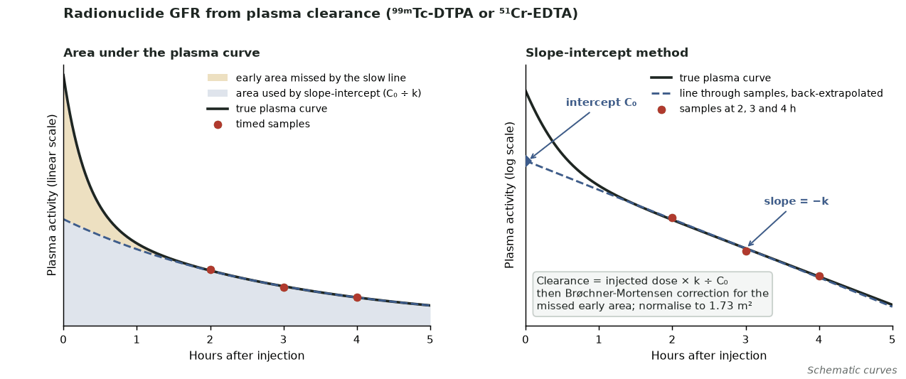
- Timed venous samples: slope-intercept (2–4 samples, ~2–5 h) with Brøchner-Mortensen correction, or a single sample timed to expected GFR (2, 3 or 4 h) in adults without oedema/ascites.
- Normalise to 1.73 m² body surface area (use absolute mL/min for carboplatin dosing).
Pitfalls
- Accurate injection and sample timing are essential (avoid extravasation).
- Oedema/ascites (expanded extracellular fluid) invalidate single-sample methods — use multi-sample methods.
- Follow a validated sampling protocol; in severe renal impairment extend sampling (up to 24 h).
Evidence & guidelines
- BNMS GFR guidelines (Fleming et al., Nucl Med Commun 2004;25:759–769; updated 2018).
- More accurate than estimated GFR for dosing decisions.
In depth
- ⁵¹Cr-EDTA (not available in the USA) and ⁹⁹ᵐTc-DTPA are the usual radionuclide GFR agents, both with low radiation doses; plasma-sampling clearance has replaced urinary collection methods, which are prone to collection and bladder-emptying errors.
- Among plasma methods, full multi-sample characterisation of the clearance curve (area under the curve) is the reference, but it is impractical routinely; the true gold standard, constant-infusion urinary clearance, is a research technique.
- The slope-intercept method uses 2–4 samples taken roughly 2–5 h after injection; because it ignores the early fast exponential it overestimates GFR, so a Brøchner-Mortensen-type correction is applied, and the result is separately normalised to 1.73 m² body surface area.
- The 2018 BNMS guideline recommends the single-sample method (Fleming formula) for routine GFR measurement in adults without ascites or oedema, with the sample time chosen according to the expected GFR.
- In severe renal impairment, delayed sampling (up to 24 h) is needed because measurement error rises steeply at low GFR, and single-sample methods are invalid with expanded extracellular fluid (ascites, effusions, oedema), which requires multi-sample methods.
- Camera-based GFR (Gates method, no blood sampling) is convenient but less accurate than plasma-sampling methods, so it should not be relied on when an accurate absolute GFR is required.
- Serial GFR results must be interpreted against biological and measurement variability, which increases sharply at low GFR (especially below about 25 mL/min/1.73 m²), so small changes may not be significant.
Sources: Taylor, J Nucl Med 2014 · 55:608-15 (PMID 24549283) · BNMS GFR guideline, Fleming et al. Nucl Med Commun 2004 (PMID 15266169) · Taylor, J Nucl Med 2014 (PMID 24549283) · Murray et al. J Nucl Med Technol 2013 · 41:67-75 (PMID 23658207) · BNMS GFR guideline (Fleming et al. 2004, PMID 15266169) · BNMS GFR guideline 2018, as summarised in McMeekin et al. EJNMMI Phys 2022